Idiopathic intracranial hypertension (IIH, pseudotumor cerebri) often affects obese and young women of childbearing age.1 Idiopathic intracranial hypertension is characterized by increased intracranial pressure (ICP), which leads to visual disturbances, tinnitus, and chronic headaches.1 Although factors such as cerebrospinal fluid (CSF) dynamics, hormonal changes, and cerebral vein obstruction are implicated in the pathophysiology of IIH,1 the exact cause, by definition, remains uncertain.
An elevated ICP may result in visual disturbance due to papilledema (optic disc swelling). However, the origin of the chronic headaches in IIH remains unclear.2 Diagnosing IIH involves obtaining a thorough medical history, physical examination, and comprehensive diagnostic testing including with lumbar puncture, visual field testing, neuroimaging, and blood tests.3 There is also a significant association between IIH and preexisting migraine, so differentiating between chronic migraine headaches and IIH-related headaches can be difficult.2
Liposuction was the most common cosmetic surgical procedure in 2021, with more than 1.9 million procedures performed worldwide.4 Despite a low complication rate, there are rare descriptions of visual loss after liposuction, potentially due to ischemia, hypovolemia, and acute blood loss.5 Here we present the case of a patient who underwent circumferential truncal liposuction and subsequently experienced delayed headaches and visual changes indicative of IIH.
METHODS
A literature search of the US National Library of Medicine National Institutes of Health (PubMed) database was conducted using the keywords “liposuction,” “idiopathic intracranial hypertension,” “ischemic optic neuropathy,” “vision loss,” and “papilledema.” Publications in all languages were included, provided that English translations were available. For this review, we selected case reports that focused on visual changes or headaches following high-volume liposuction.
CASE REPORT
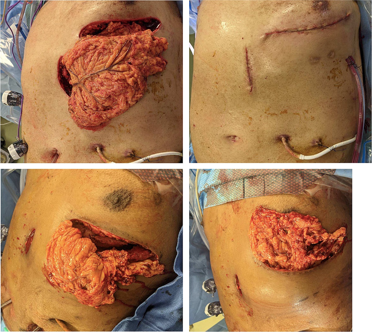
A 31-year-old African American woman (height, 163 cm; weight, 89.8 kg; body mass index, 33.8 kg/m2) underwent circumferential liposuction of the trunk with fat transfer to the hips and superior buttocks and bilateral breast reduction. The patient had a history of migraines and myopia, for which she used corrective lenses. There was neither history of papilledema, optic disc swelling, nor other visual changes. Throughout her consultation and preoperative visits, she did not experience any migraines; nor did she take any medication that could potentially trigger migraines. Furthermore, she did not report any headache immediately before the surgery.
Preoperatively, her blood pressure was 161/90 mm Hg; pulse rate, 70 beats/min; oxygenation, 100%; fasting blood sugar level, 5.4 mmol/L; hemoglobin, 11.5 g/dL; hematocrit, 33.8%; and glomerular filtration rate, 119 mL/min per 1.73 m2. Intraoperatively, she remained hemodynamically stable, with systolic blood pressure of 110 to 150 mm Hg and diastolic blood pressure of 60 to 90 mm Hg. The procedure was performed under general anesthesia for 5 h 29 min, during which the patient was in the prone position for less than 45 min. During liposuction, 3,100 mL of total tumescent fluid was infiltrated, with removal of 5,000 mL of aspirate, of which 1,200 mL was injected into her hips and buttocks. Additionally, during bilateral breast reduction, 202 and 260 g of tissue were excised from her left and right breasts, respectively. Following surgery, her vital signs were stable in the recovery room, with a blood pressure of 169/100 mm Hg and a pulse rate of 80 beats/min. She did not report headache or visual changes in the immediate postoperative period.
The patient recovered uneventfully until her second postoperative visit at 10 days, when she reported headache radiating to the back of her head. She was advised to take nonsteroidal anti-inflammatory drugs, which slightly improved the headaches; however, within 48 hours, she again experienced severe headache. Consequently, she was examined in the emergency room (ER), where a head computed tomography was performed, which was unremarkable. She was discharged from the ER with prescriptions for nonsteroidal anti-inflammatory drugs and lorazepam.
Three weeks after surgery, the patient left the country and returned home to Montreal, Canada. One week later, she presented to the ER again because of severe headache and visual disturbance characterized by blurry vision. She was evaluated, discharged from the ER, and asked to follow up with an ophthalmologist to monitor the visual changes. After visual field testing, venography, and magnetic resonance imaging, the ophthalmologist eliminated the possibility of cerebral venous sinus thrombosis and other space-occupying lesions. The final diagnosis was papilledema. Subsequently, she returned to the ER, where a lumbar puncture was performed, revealing a CSF pressure and opening pressure of 30 cm H2O (reference range, 6–25 cm H2O) and 29 cm H2O (reference range, 6–25 cm H2O), respectively. Excess CSF was successfully drained to alleviate the high ICP, and the patient was prescribed 0.5 g of acetazolamide, which effectively resolved her symptoms. The patient was diagnosed with IIH according to the modified Dandy criteria.6
RESULTS
The literature review revealed that IIH and visual loss are rare complications associated with liposuction. Previous cases of nonarteritic ischemic optic neuropathy occurring after liposuction are detailed in Table 1.
TABLE 1 -
Cases of Ischemic Optic Neuropathy Following Liposuction
Authors
Age, y
Sex
Preoperative Medical Condition
Clinical Outcome
Minagar et al
7
47
Female
N/A
Unilateral AION
Sibgatullah et al
8
36
Female
N/A
Unilateral AION
Foroozan and Varon
9
30
Female
N/A
Bilateral AION and transverse sinus thrombosis
Moura et al
5
49
Female
N/A
Bilateral AION
Rath et al
10
43
Female
N/A
Unilateral posterior ischemic optic neuropathy
Ahmad and Pelak
11
38
Female
N/A
Bilateral AION
Abri Aghdam et al
12
30
Female
Optic disc drusen
Unilateral AION
Ribeiro Monteiro et al
13
30
Female
IIH
AION
Our literature review identified 8 women aged between 30 and 49 years who developed ischemic optic neuropathy following liposuction, most commonly anterior ischemic optic neuropathy (AION) but posterior ischemic optic neuropathy in 1 case.10 Of these 8 cases, 6 were most likely triggered by acute blood loss during surgery. Other factors were implicated in some of the cases. Moura et al5 presented the case of a patient who also had acute blood loss during liposuction but who also had small, crowded optic discs, a risk factor for AION. Abri Aghdam et al12 presented a case of a patient with preexisting optic disc drusen, characterized by the presence of crystalline deposits or calcifications on the optic disc, who subsequently developed unilateral AION following liposuction. Finally, Ribeiro Monteiro et al13 reported the first case of a patient with preexisting IIH who developed bilateral optic nerve–related visual loss after undergoing liposuction.
DISCUSSION
We present the novel case of a 31-year-old woman who underwent a 360° liposuction procedure and subsequently developed worsening headaches and visual disturbances. To our best knowledge, this is the first report of IIH developing after liposuction. Although other reported cases revealed visual disturbances after liposuction, these were ischemic cases, mainly secondary to surgery. This cause was highly unlikely in our case: our patient was in the prone position for less than 45 minutes during the operation, and ION typically occurs immediately postoperatively and is associated with prolonged intraoperative hypotension, anemia, dehydration, or prolonged time spent in the prone position, often exceeding 90 minutes.12,14,15 The only other reported case of IIH was preexisting and did not develop after surgery.13 Consistent with liposuction triggering IIH in our case, the condition developed after several days and presented with delayed-onset, persistent postoperative headache and visual changes, distinguishing the patient from those with surgery-related ION who developed visual changes in the immediate postoperative period. Although our patient described previous migraine, this was likely true migraine and not IIH present before surgery, as she did not experience any migraine headaches throughout her consultation or preoperative visits. Although the pathogenesis of the development of IIH is unclear, we propose that the surgery triggered the development of IIH in this case.
Elevated ICP is commonly associated with papilledema, as observed in our patient, which in IIH can then progress to ischemia and severe visual impairment. Possible causes of papilledema include high blood pressure, tumors, infections or inflammation, cerebral venous thrombosis, hemorrhage, and IIH. Based on the unremarkable laboratory values and presence of papilledema in our patient, she was diagnosed with IIH.16
Abdominal surgery and combined breast surgery pose a high risk of complications, the primary complications being hematomas, followed by infections, venous thromboembolism, and respiratory issues rather than IIH.17
The diagnosis of IIH was based on the modified Dandy criteria, that is, symptoms of elevated ICP such as papilledema and headache without any abnormal neurological findings. In addition, both the magnetic resonance imaging and venogram results were normal, with no signs of venous obstruction. A lumbar puncture confirmed high ICP greater than 25 cm H2O without any other CSF abnormality. Furthermore, the patient remained awake and alert during her ER visit.18
This case report emphasizes the importance of having a high index of suspicion for IIH as a complication of liposuction. In our case, rapid medical intervention, including performing a lumbar puncture and prescribing acetazolamide, prevented worsening of the visual loss. Surgeons must be aware of the patient's medical history, particularly in relation to headaches and visual changes, and in patients describing symptoms, assess for papilledema before performing large-volume liposuction. In addition, a detailed preprocedural health assessment is always warranted to rule out the presence of IIH, as the demographics of patients with IIH and those who elect to undergo liposuction often overlap.1 Given that liposuction is an extremely common procedure and the subsequent development of IIH is so rare, screening every patient may not be cost-effective. Nevertheless, surgeons should still suspect intracranial hypertension in patients who present with persistent headaches and/or visual changes following liposuction.
CONCLUSIONS
Surgeons should be vigilant about comorbidities and be aware of the potential impact of procedures that may alter ICP. This case report adds to the existing literature by highlighting the association between liposuction and development of IIH and emphasizing the importance of thorough preoperative evaluation before the procedure and consideration of rare associated conditions.
REFERENCES
1. Markey KA, Mollan SP, Jensen RH, et al. Understanding idiopathic intracranial hypertension: mechanisms, management, and future directions. Lancet Neurol. 2016;15:78–91.
2. Raoof N, Hoffmann J. Diagnosis and treatment of idiopathic intracranial hypertension. Cephalalgia. 2021;41:472–478.
3. Jensen RH, Radojicic A, Yri H. The diagnosis and management of idiopathic intracranial hypertension and the associated headache. Ther Adv Neurol Disord. 2016;9:317–326.
4. Surgery ISoAP. The latest global survey from ISAPS reports a significant rise in aesthetic surgery worldwide. Available at:
https://www.isaps.org/discover/about-isaps/global-statistics/reports-and-press-releases/global-survey-2021-full-report-and-press-releases/. Accessed date February 7, 2024.
5. Moura FC, Cunha LP, Monteiro ML. Bilateral visual loss after liposuction: case report and review of the literature. Clinics (Sao Paulo). 2006;61:489–491.
6. Friedman DI, Jacobson DM. Diagnostic criteria for idiopathic intracranial hypertension. Neurology. 2002;59:1492–1495.
7. Minagar A, Schatz NJ, Glaser JS. Liposuction and ischemic optic neuropathy. Case report and review of literature. J Neurol Sci. 2000;181(1–2):132–136.
8. Sibgatullah M, Kupersmith MJ, Zerykier A, et al. Ischemic optic neuropathy after liposuction: case report and review. Neuroophthalmology. 2009;29:91–93.
9. Foroozan R, Varon J. Bilateral anterior ischemic optic neuropathy after liposuction. J Neuroophthalmol. 2004;24:211–213.
10. Rath EZ, Falick Y, Rumelt S. Posterior ischemic optic neuropathy following breast augmentation and abdominal liposuction. Can J Ophthalmol. 2009;44:346–347.
11. Ahmad M, Pelak V. Vision loss in a young, healthy woman following liposuction. Am J Cosmet Surg. 2016;33:96–101.
12. Abri Aghdam K, Aghajani A, Soltan Sanjari M, et al. Anterior ischaemic optic neuropathy following liposuction in a patient with optic disc drusen. Neuroophthalmology. 2021;45:261–264.
13. Ribeiro Monteiro ML, Moura FC, Cunha LP. Bilateral visual loss complicating liposuction in a patient with idiopathic intracranial hypertension. J Neuroophthalmol. 2006;26:34–37.
14. Rupp-Montpetit K, Moody ML. Visual loss as a complication of non-ophthalmic surgery: a review of the literature. Insight. 2005;30:10–17.
15. Buono LM, Foroozan R. Perioperative posterior ischemic optic neuropathy: review of the literature. Surv Ophthalmol. 2005;50:15–26.
16. Whiting AS, Johnson LN. Papilledema: clinical clues and differential diagnosis. Am Fam Phys. 1992;45:1125–1134.
17. Winocour J, Gupta V, Ramirez JR, et al. Abdominoplasty: risk factors, complication rates, and safety of combined procedures. Plast Reconstr Surg. 2015;136:597e–606e.
18. Shaw GY, Million SK. Benign intracranial hypertension: a diagnostic dilemma. Case Rep Otolaryngol. 2012;2012:814696.


留言 (0)