Gaucher disease (GD) is the most prevalent lysosomal storage disease that leads to the accumulation of glucocerebroside in reticuloendothelial cell, thus haematological, skeletal and abdominal organs. It is an autosomal recessive disease secondary to a deficit in glucocerebrosidase. Its incidence rate is 1/50000 in the general population but could reach 1/800 among Ashkenazi Jews [2]. Three major clinical sub-types are described depending on the absence (Type I) or presence of neurologic involvement (Type II and III). Type 1 of GD is the most frequent, especially within the Ashkenazi, evolving from adult-onset disorders with major haematological and bony manifestations to childhood disorders, with mainly visceral and bony involvement. Type II is the infant subtype, seen within the first year of life, with acute neurologic involvement and severe prognosis [3]. GD Type III is another form of GD with a subacute neurological involvement associated with visceral findings.
Concerning cardiac involvement, it is rare and mainly reported in type IIIc GD, a subtype described in Spanish and Arab populations, in patients with D409H mutation. It is characterized by extensive calcification of the aorta and leaflets valve [1, 4]. Constrictive calcific pericarditis is a rare cardiac manifestation resulting from intrapericardial haemorrhage related to thrombocytopenia seen in GD [5]. Coexistence of Gaucher disease with restrictive cardiomyopathy is rare [6].
It is important to evaluate pulmonary artery hypertension, which is rare but well-described in type I GD [7] with an incidence rate of up to 20%, based on Doppler echocardiographic assessment [8]. According to the European Association of Cardiovascular Imaging criteria to assess the probability of PAH, our patient presented mild pulmonary hypertension with an estimated systolic PAP (sPAP) of 40mmH. Similar results were reported in a large Romanian cohort with mean sPAP of 41mmHg, found in 13% of patients [9]. Risk factors of symptomatic PAH in GD are female sex, splenectomy, non-N409S GBA mutation, angiotensin-converting enzyme gene polymorphism and family history. None of those is present in our case.
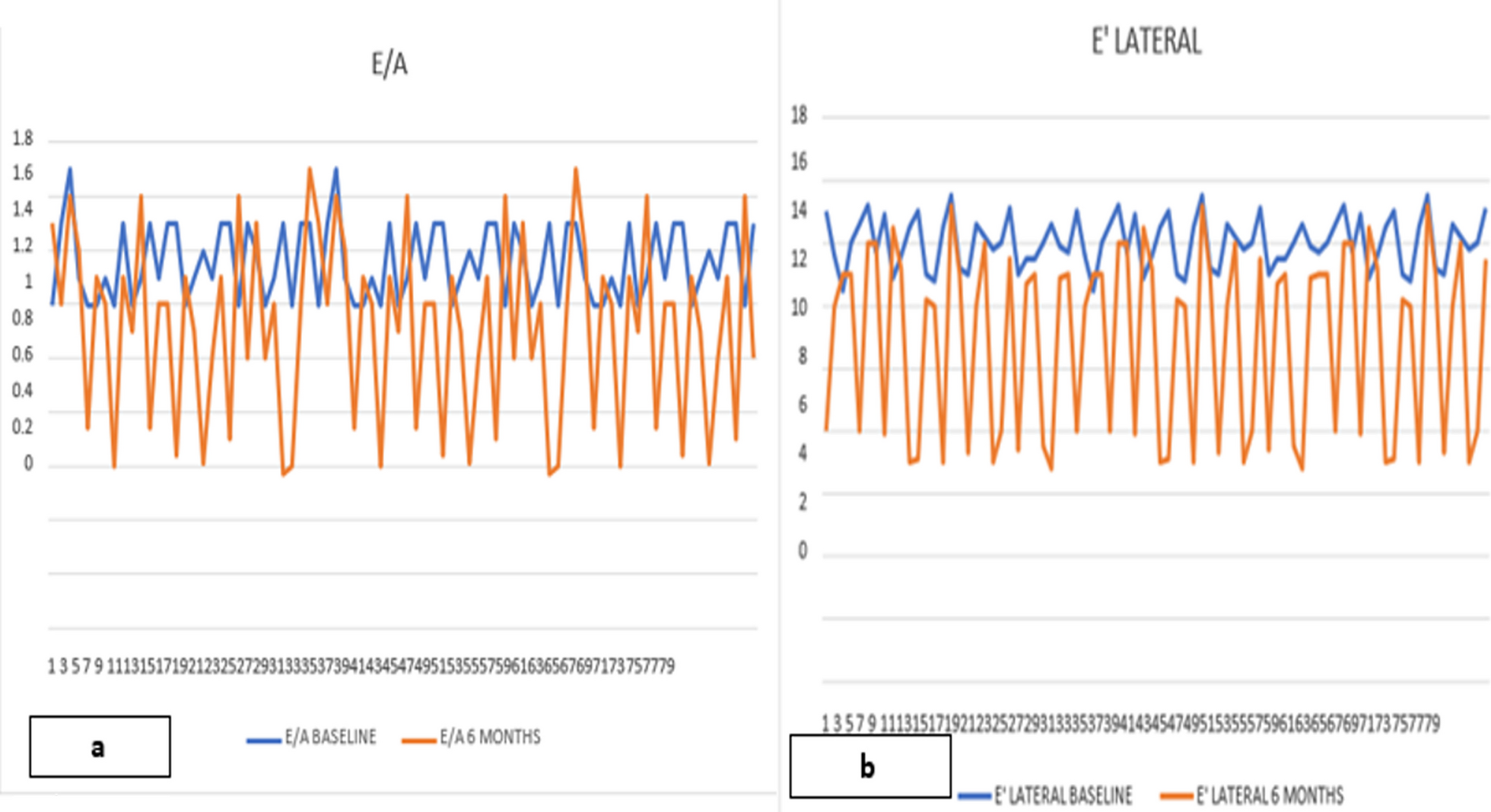
The diastolic function may be compromised due to infiltrative processes leading to left ventricular hypertrophy and patchy interstitial fibrosis [10]. In our study, Doppler assessment found a grade II diastolic dysfunction with elevated filling pressure; however, E/e’ ratio was normal. Neither E/A ratio nor A mitral wave could be studied due to Atrial arrhythmia.
Only two studies were interested in evaluating diastolic function in Type 1 GD. Koželj and al. reported a tendency towards dysfunction with no statistically significant difference between patients and the control group [11]. However, Lo Iudice investigated LV geometry and function in 18 GD patients compared to hypertensive and normal subjects. A prolonged deceleration time of E wave velocity, greater atrial filling fraction and normal E/e’ ratio were observed. There is a pattern of impaired relaxation with normal filling pressure [12]. Worthy of note, all GD were under enzyme replacement therapy, which partly explains the absence of LV hypertrophy.
We hypothesize that our patient had an impaired diastolic function, recently decompensated by the onset of tachyarrhythmia.
Besides LV hypertrophy and bi-atrial enlargement, our patient presented a global hypokinesis with a regional wall motion abnormalities in basal and middle infero-septal and inferior segments. Alizad described similar findings in adult GD patients, with increased LV mass and a septal muscular prominence [13].
CMR was performed in order to study myocardial infiltration. Recently, Solanich assessed the GD cardiac involvement of a 62-year-old type I GD patient with no cardiac symptoms using CMR. LGE showed multiple foci consistent with interstitial infiltrative fibrosis in the basal and middle inferior and inferolateral segments, in addition to bi-atrial enlargement [14]. Similar findings are seen in our case, even if LGE was only located in the basal infero-septal segment. Without an objective histological examination, the focal interstitial fibrosis may correspond to a local reaction in response to sphingolipid infiltration.
In a larger cohort of patients with type I GD, no interstitial fibrosis was reported neither in patients on ERT nor at diagnosis. Atrial enlargement is present even in the absence of structural heart disease. An accurate assessment of diastolic function and interstitial fibrosis is warranted by using a multi-modal approach with the aid of CMR imaging and echocardiography evaluation [8].
Enzyme replacement therapy is the cornerstone for treating GD with a positive outcome regarding anaemia, thrombocytopenia, and organomegaly [3]. Concerning cardiac involvement, Spada reported an improvement of LV systolic and diastolic function and normalization of T-wave within months of a high dose of ERT, and complete normalization of echocardiogram after 3 years [15].
Considering the absence of myocardial infiltration in GD on ERT in the cohort of Roghi et al. [8], ERT may have a significant role in preventing cardiac manifestations. Nonetheless, its effect in treating valve calcification is debatable. As reported by previous cases, it did not seem to stop calcification progression [1, 16].


留言 (0)