Cardiomyopathy (CMP) in children has a diverse clinical as well as genetic characteristics. It is associated with significant morbidity and mortality. The precise incidence remains uncertain, with reported annual rates ranging between 0.65 and 1.24 per 100,000 children. Dilated cardiomyopathy (DCM) and hypertrophic cardiomyopathy (HCM) are the predominant types, accounting for 50 to 58% and 25 to 42%, respectively [2, 14].
The accurate incidence in our society remains uncertain due to inadequate registration of patients with cardiomyopathy (CMP) and the existence of undiagnosed cases. PSCC-Qassim is recognized as the sole referral cardiac center in the region, catering to a population of approximately 1.2 million. In our study, 70% of our patients were diagnosed with dilated cardiomyopathy (DCM), while 26% had hypertrophic cardiomyopathy (HCM).
Both HCM and DCM exhibit a familial pattern, marked by notable genetic heterogeneity. The predominant inheritance pattern is autosomal dominant, although autosomal recessive, X-linked, and mitochondrial inheritance patterns have also been documented [10, 15]. Many patients in our cohort had consanguineous parents and a positive family history of cardiomyopathy, particularly prevalent in those with hypertrophic cardiomyopathy (HCM).
Disease-associated mutations have been identified in over 50 genes, with some overlap between categories. Genetic causes are more prevalent in familial HCM, accounting for up to 65% of patients, and less frequently in other types of cardiomyopathies (e.g., 50% in arrhythmogenic right ventricular dysplasia, and 30% in dilated cardiomyopathy). To address the significant genetic heterogeneity and uncover novel genes, we employed whole-exome sequencing (WES) as a primary diagnostic test in our patients, particularly those with HCM, following the approach adopted by other researchers in the study of cardiomyopathy [10, 16].
In our cohort, the most prevalent genetic abnormalities identified were ELAC2 mutations and very long-chain acyl-CoA dehydrogenase deficiency (VLCADD). Within our cohort, all patients with ELAC2 mutations and 44% of patients with VLCADD experienced mortality. Notably, it has been reported that nearly one-third of patients with ELAC2 mutations succumb during the initial presentation [17].
According to data from the Pediatric Cardiomyopathy Registry published in 2003, about one-third of children diagnosed with cardiomyopathy had a confirmed cause. The documented causes encompassed myocarditis (16% in dilated cardiomyopathy—DCM), metabolic factors (4% in DCM, 9% in hypertrophic cardiomyopathy—HCM), syndromic conditions (1% in DCM, 9% in HCM), and neuromuscular causes (9% in DCM, 9% in HCM) [18].
Infants and children diagnosed with heart failure are currently assessed using the Ross heart failure class, which grades symptoms related to respiration, feeding, and weight gain. Although widely adopted, it is important to note that this classification lacks formal validation against outcomes. Moreover, patients with hypertrophic cardiomyopathy (HCM), restrictive cardiomyopathy (RCM), and noncompaction cardiomyopathy (NCM) may display signs of heart failure with preserved ejection fraction [19].
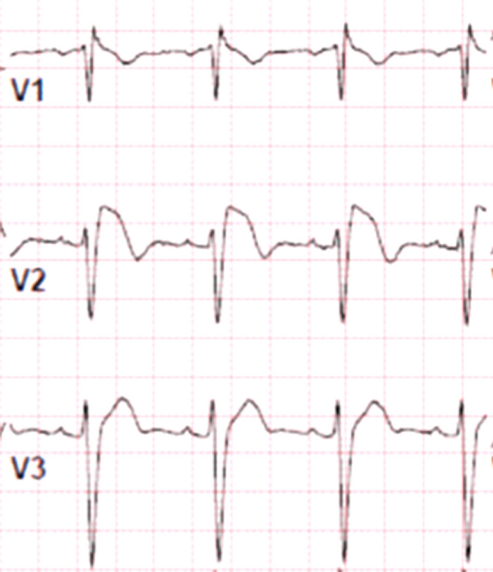
Echocardiography serves as the primary diagnostic tool for establishing the diagnosis and cardiomyopathy (CMP) phenotype. Following the diagnosis, serial echocardiography is conducted during the acute setting and in the follow-up period. In children with dilated cardiomyopathy (DCM), the extent of ventricular dilation and dysfunction stands as a robust predictor of mortality or transplantation. For patients with hypertrophic cardiomyopathy (HCM), the thickness of the septal wall has been linked to the risk of sudden death [19, 20].
Cardiac MRI proves valuable in identifying inflammatory processes, aiding in the definition of the underlying cause and contributing to the management and risk stratification of cardiac conditions [6].
In dilated cardiomyopathy (DCM), children often exhibit elevated cardiac biomarkers, including B-type natriuretic peptides [21].
Dilated cardiomyopathy
Dilated cardiomyopathy (DCM) comprises two-thirds of cardiomyopathy cases in our cohort. DCM serves as the ultimate common pathway for various pathological processes that result in left ventricular (LV) dilation and systolic dysfunction, often presenting with signs and symptoms of congestive heart failure. In roughly two-thirds of patients, the underlying cause of DCM remains idiopathic. Potential contributors to DCM include antecedent viral myocarditis, generalized myopathies, and abnormalities in sarcomeric, sarcolemmal, cytoskeletal, or nuclear proteins [22].
Due to the associated risks with endomyocardial biopsy (EMB), the initial diagnosis of myocarditis typically relies on clinical presentation. Moreover, EMB is not widely available and is not routinely performed [23]. Globally, similar to our society, the true prevalence of pediatric myocarditis remains unknown. Myocarditis is reported to contribute to 17% of sudden cardiac deaths in children younger than 16 years old [14]. In our cohort, none of the individuals underwent endomyocardial biopsy (EMB) or cardiac MRI.
The diagnosis of myocarditis was established based on a history of viral infection along with clinical and laboratory evidence of myocardial cell injury. Although this represents a limitation in our study, the focus remains on potential genetic causes and the outcomes associated with various types of cardiomyopathy. It is worth noting that EMB is not recommended for infants weighing less than 10 kg or for hemodynamically unstable patients. The general recommendation is to perform EMB only if confirming the clinical diagnosis of myocarditis would have a clear impact on the patient’s treatment plan, such as listing for transplantation [24].
Cardiac MRI has emerged as the preferred noninvasive test for diagnosing myocarditis. The MRI Lake Louise Criteria (LLC) diagnostic criteria for myocarditis rely on the identification of tissue edema, hyperemia, and necrosis, forming the foundational features observed in both acute and chronic stages [6].
The clinical presentation of children with dilated cardiomyopathy (DCM) varies from asymptomatic cases to acute decompensated heart failure and cardiogenic shock. According to literature, a considerable number of children with DCM necessitate hospitalization, with 54% receiving inotropic support, 41% requiring mechanical ventilation, 13% needing extracorporeal membrane oxygenation (ECMO) support, and 11% undergoing urgent transplantation [25, 26]. In our cohort, 70% of our patients needed hospitalization, with 54% requiring admission to the intensive care unit (ICU) at the initial presentation. Notably, none of the patients in our cohort underwent extracorporeal membrane oxygenation (ECMO) support.
The rate of complete spontaneous recovery in pediatric myocarditis/dilated cardiomyopathy (DCM) following disease onset ranges from 22 to 37% of patients during the follow-up period [4, 27]. Notably, almost 35% of our patients with DCM experienced normalized cardiac function.
The risk factors for death and transplantation in children varied depending on the etiology of dilated cardiomyopathy (DCM). Some reports indicate that predictors of a poor outcome include an initial left ventricular ejection fraction (LVEF) less than 30%, age less than 2 years at presentation, a prolonged course of the disease, and a high level of N-terminal pro-brain natriuretic peptide [27]. On the other hand, younger age at presentation and a lower left ventricular end-diastolic dimension (LVEDD) z-score were reported as independent predictors of normalization [4]. In comparison with idiopathic dilated cardiomyopathy (IDCM), familial dilated cardiomyopathy (FDCM) was reported to have a lower cumulative incidence of death (P = 0.04; hazard ratio 0.64, P = 0.06), with no significant difference in the risk of transplantation or the combined death or transplant outcome [22, 28].
In this descriptive study, results of multivariate survival analysis for patients with dilated cardiomyopathy (DCM) reveal that most deaths occur in the first 2 years after diagnosis. Risk factors for adverse outcomes include an ejection fraction (EF) at presentation of 20% or less (p-value < 0.001), admission to the hospital (and intensive care unit (ICU)) at the first presentation, consanguineous parents, and a positive family history of cardiomyopathy. Patients who received inotropic medications had a lower mortality rate (P-value 0.022). Additionally, patients with DCM and positive genetic studies, except for ELAC2 mutation, have a better prognosis than those with unknown/idiopathic DCM.
However, organ shortage poses a risk, particularly for children on waiting lists. Furthermore, a strong and reliable social support system is crucial for the long-term success of HTx [29]. In addition to the lifelong use of immunosuppressant, acute graft rejection, and chronic graft failure are important challenges with HTx. In our cohort, only two patients underwent heart transplantation. The long-term survival rate of children with cardiomyopathy after HTX in experienced centers is high. Morbidity and mortality were higher in patients with systemic diseases than in those with cardiac-specific conditions [30].
The effectiveness of Beta-Blockade therapy, such as Carvedilol, metoprolol, or bisoprolol, in children, including those with a structurally normal heart, remains unclear. These medications may be initiated in the treatment of moderate to severe systolic dysfunction of a systemic left ventricle.
Intravenous immunoglobulin G (IVIG) is not recommended as a routine treatment for myocarditis. Although some case reports and retrospective studies have suggested a potential benefit of IVIG in the treatment of myocarditis, there is a lack of higher-quality studies with low bias risk and larger sample sizes to support its routine use [31]. In our study, IVIG administration showed no significant effect in reducing mortalities (p-value 0.074).
Corticosteroids are not recommended as a routine treatment for myocarditis, particularly in the absence of robust randomized controlled trial evidence.
Hypertrophic cardiomyopathy
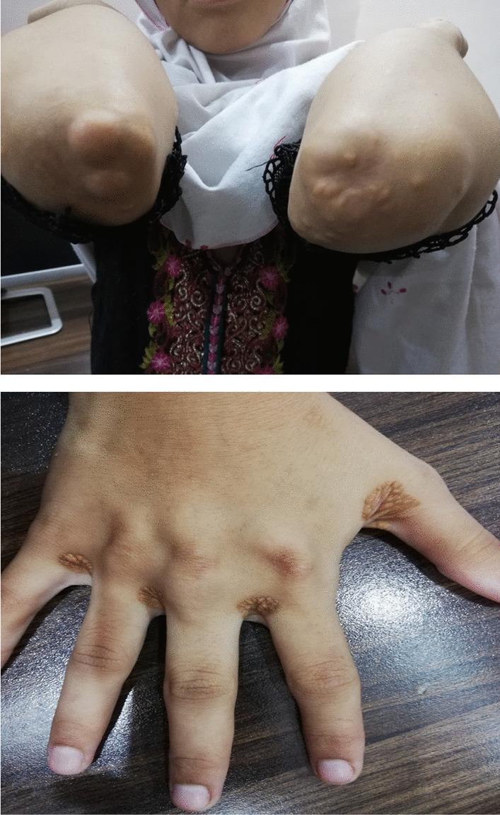
More than 1400 mutations in various sarcomeric genes have been identified in approximately 70% of patients with hypertrophic cardiomyopathy (HCM) [32]. Infants with hypertrophic cardiomyopathy (HCM) stemming from malformation syndromes and inborn errors of metabolism commonly exhibit symptoms in infancy and, often linked to neurological and musculoskeletal issues. The mortality rate for these infants is notably elevated, reaching 30% within a two-year period, surpassing that of older children. Poor prognostic factors include mixed phenotypes, presentation at 1 year of age or younger, low weight, the presence of signs and symptoms of heart failure, lower left ventricular fractional shortening, and increased left ventricular posterior wall septal thickness at the time of diagnosis [33].
Patients with the infantile form of Pompe disease might respond to enzyme replacement therapy, which can be effective in reducing the progression of left ventricular hypertrophy if initiated early. Enzyme replacement therapy or bone marrow transplantation is currently the treatment of choice in some patients with specific types of lysosomal storage diseases, namely Mucopolysaccharidoses I, II, IV, and VI [34, 35].
For patients with restrictive cardiomyopathy (RCM), the 5-year mortality rate is high and has been reported to be 68% from the time of diagnosis [2].
At least 5% of children diagnosed with cardiomyopathy have some form of left ventricular noncompaction (LVNC). This condition may occur in isolation (23%) or be associated with dilated or hypertrophic cardiomyopathy (59% and 11%, respectively). There is a tendency for both under- and over-diagnosis of LVNC, possibly stemming from the lack of consensus on diagnostic criteria for LVNC [19].
Noncompaction of the left ventricle is a common finding in Barth syndrome, which is an X-linked recessive disorder caused by a mutation in the tafazzin (TAZ) gene on chromosome Xq28 [36]. LVNC can also be associated with other inborn errors of metabolism and certain congenital heart diseases, including septal defects and pulmonary stenosis [36].
Children with noncompaction cardiomyopathy (NCM) who have normal cardiac dimensions and systolic function are at a very low risk for adverse outcomes. However, those with NCM mixed with other phenotypes, such as dilated cardiomyopathy (DCM) or hypertrophic cardiomyopathy (HCM), have a worse prognosis with an increased incidence of death or transplantation (18–25%) [19].


留言 (0)