
記住我

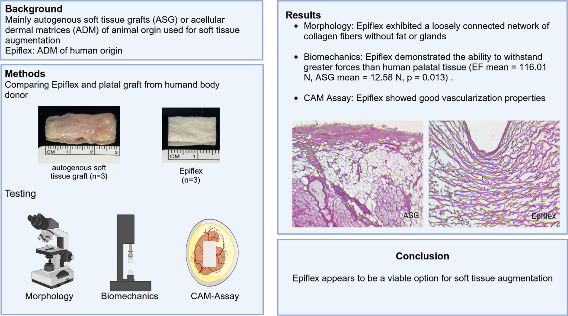


In this project we compared autogenous soft tissue grafts (ASG) to Epiflex (EF; Deutsches Institut für Zell- und Gewebeersatz gGmbH, Berlin, Germany). EF is harvested postmortem from screened donors and later decellularized, sterilized and preserved. For testing it is rehydrated with 0.9% saline solution (B. Braun Melsungen AG Melsungen, Germany) according to the manufactures protocol (Fig. 1).
Fig. 1
a Epiflex before testing; b autologus soft tissue graft after harvesting, before testing
ASG was gathered from two body donors’ hard palate as connective tissue graft (30 × 15 mm, n = 4) using standard surgical protocols considering the anatomy of the palatine artery and nerve and the distance to the upper molars.
In accordance with common donation procedures for Anatomical Institutes in Germany, the bodies were donated for medical education and research. After the death and transfer to the Institute for Anatomy, the bodies were stored frozen at − 20° C and later defrosted for collection of the ASG. The thickness of the ASG was controlled by a sliding calliper. The ASG was used without any further processing. Both ASG and EF were cut in a rectangular shape (15 mm × 10 mm).
Biomechanical testingASG (n = 3) and EF (n = 3) biomechanical properties were evaluated with a material test machine (model 5942; Instron, Pfungstadt, Germany) and the software BlueHill (version 2.25; Instron). Each graft was put between the opposing brackets and draged uniaxial with a constant strain rate of 0.5 mm/s under displacement control until rupture. The maximum load (ML, Newton [N]) and the expansion (E, [mm]) were measured [7].
CAM-assayTo analyse angiogenetic properties the CAM-Assay was used to histologically evaluate vessel growth and proliferation. Epiflex was cut in a rectangular shape (10 mm × 20 mm) and placed on the chicken embryo for up to 24, 72 and 120 h. Afterwards the membrane was harvested and prepared for histology. ASG was not used due to its body donor origin and in literature well demonstrated healing properties. Since grafts from body donors were obtained post mortem and start to decompose shortly after thawing, it was impossible to perform CAM-Assays with them.
HistologyNative ASG, EF and CAM-Assay samples were fixed in buffered formaldehyde solution (4%) and embedded in paraffin. Staining of native ASG and EF was performed with hematoxylin and eosin (H&E), Heidenhain’s Azan, Sirius red and Weigert’s elastic according to standard protocols. Briefly, sections were deparaffinized in xylol and rehydrated in ethanol of decreasing concentrations. H&E staining was performed with hematoxylin (Sigma-Aldrich, St. Louis, Missouri, US) and eosin (Sigma-Aldrich) after flushing in distilled water. For Heidenhain’s Azan staining, a staining kit was used (Cat No. 12079, MORPHISTO Ltd., Offenbach am Main, Germany). Sirius red staining was conducted using Sirius red F3B (Chroma, Waldeck GmbH & Co. KG, Münster, Germany), and picric acid (Sigma-Aldrich). For Weigert’s elastic staining resorcin-fuchsin (Chroma) and nuclear fast red (Sigma-Aldrich) were used. After staining, a dehydration of ethanol in increasing concentration and treatment in xylol followed. CAM-Assay samples were H&E stained as mentioned above. Vascular endothelium was stained with an immunoperoxidase reaction. Briefly, 7 μm thick sections were deparaffinized in xylol and rehydrated in ethanol of decreasing concentrations. After flushing in distilled water, slices were protein blocked and incubated with a polyclonal mouse antibody against CD31 in an 0.015 mol/L sodium azide overnight at 4 °C, followed by incubation with biotinylated anti-mouse IgG (1:200 in PBS/5% BSA) for 30 min at room temperature. The Vectastain® Elite ABC kit for peroxidase (Vector laboratories, Burlingame, California) was used, according to the protocol of the manufacturer, for signal enhancement. Detection was carried out with 3,3′diaminobenzidine (DAB, Sigma, St. Louis, Missouri) as chromogen. Nuclear staining was performed with hematoxylin (Roth, Karlsruhe, Germany). After staining, slides were dehydrated in ethanol of raising concentrations and embedded in Eukitt® (Sigma). Negative controls were performed by omitting the primary antibody on consecutive sections.
Histological sections were imaged with a Leica MS 5 tripod (Leica Microsystems, Germany) and a JVC KY-F75U C mount digital camera (JVC, Yokohama, Japan).
Scanning electron microscopyAfter tensile strength measurement ASG and EF were prepared for scanning electron microscopy (SEM). The samples of both grafts were fixed in 2.5% buffered glutaraldehyde solution, washed in phosphate buffered saline (PBS), dehydrated in ethanol, freeze–dried, mounted on specimen holders, sputtered with gold in an argon atmosphere, and visualized with a scanning electron microscope (SEM; ESEM XL-30, Philips, Eindhoven, Netherlands).
StatisticsStatistical analysis was performed with IBM SPSS Statistics 27 (Armonk, USA) using the Mann–Whitney-U-test. P-values below or equal to 0.05 (p ≤ 0.05) were deemed statistically significant.
留言 (0)