
記住我


We retrieved 1464 articles (after removing duplicates) but finally included 28 articles which met the inclusion criteria. The PRISMA flowchart showing the inclusion of studies is presented in Fig. 1.
Fig. 1
The PRISMA flowchart showing the inclusion of studies
Characteristics of included studiesThe characteristics of the included studies are shown in Table 1 [8,9,10,11,12,13,14,15,16,17,18,19,20,21,22,23,24,25,26,27,28,29,30,31,32,33,34,35]. All studies were single-centre with most of them being cross-sectional in nature. Overall we included a total of 534 renal biopsies and 107 renal autopsy findings. Supplementary appendix 1 shows PRISMA-ScR Checklist for the reported studies Two studies, [22, 31] were exclusively done on children with a mean age (± SD) in years of 11.52 ± 2.88 years and 5.8 ± 1.0 years respectively. In the majority of studies, AKI was caused by Russell’s viper, however, in some studies, Echis carinatus and sea snakes were also identified to be associated with kidney involvement [15,16,17, 27, 29].
Table 1 Characteristics of included studies Timing and indication of renal biopsyIn the majority of studies, kidney biopsies were done after 2–3 weeks from the onset of AKI, The most common indication for renal biopsies was persistent renal dysfunction or dialysis dependency. Pathological changes varied depending upon the time lag from the onset of AKI to kidney biopsy. Histopathological findings in Kidney biopsies of included studies are shown in Table 2. Kidney tissue obtained during the early diuretic phase of acute tubular necrosis (ATN) revealed epithelial degeneration in tubules, tubular vacuolation, desquamation, and severe intertubular interstitial oedema with regenerative changes developing at the later stages of AKI (after 3 to 8 weeks) [9, 30, 31]. Interstitial haemorrhage was more common in the 1st week of the bite [15]. The nature of the tubular casts also changed with the time lag between the bite and kidney biopsy. Hyaline casts with degenerating cellular-granular casts were more commonly observed in earlier stages whereas red blood cells (RBC) casts appeared later.
Table 2 Kidney biopsy findings chartRenal Biopsy findingsGlomerular lesionsChanges in the glomerulus were largely underreported. Glomerular changes occurring with acute cortical necrosis (ACN) were the most commonly reported finding. However, thrombotic microangiopathy (TMA) and focal and diffuse mesangial proliferation have also been reported [30, 32]. Studies also reported necrosis of glomerular tuft and isolated glomerular thrombosis [9]. Specific glomerular changes like ballooning and dilatation in the glomerular capillary loops, focal proliferation of mesangial cells, endothelial cell swelling, splitting of glomerular capillary basement membrane were reported by Mittal et al. and Acharya et al. [15, 17].
Tubulointerstitial changesATN was the most frequently encountered finding in kidney biopsy tissue. The incidence varied from 30 to 100% [8, 9, 11, 12, 14,15,16, 18, 23, 25,26,27, 32, 34]. Hyaline, granular, or pigment casts were frequently seen along with dilated tubules lined with flattened epithelium and desquamation of necrotic cells [12, 28]. Acute interstitial nephritis (AIN) has been reported non uniformly [25,26,27, 32, 34]. In a series by Priyamvada et al., 5.7% of snakebite-induced AKI were reported to have AIN on kidney biopsy [26]. The kidney biopsy demonstrated mixed infiltrate of predominantly lymphocytes and variable proportions of other cells like neutrophils, eosinophils, and occasional plasma cells. Neutrophil cast was reported in one patient. The prevelance of AIN was slightly higher in other series; out of 85 biopsies, 20 (23.5%) patients had AIN [27]. Marked infiltration of eosinophils and lymphocytes with substantial tubular injury was reported. Golay et al. observed extensive interstitial inflammation, with a predominant lymphocytic infiltration [20]). In viperine envenoming, “hemorrhagic interstitial nephritis” characterized by haemorrhages in the interstitium with tubular necrosis and RBC congestion in the tubular lumen was reported [10, 17]. One study reported features of pyelonephritis complicating ATN [17]. Patchy and diffuse areas of hemorrhagic necrosis in the cortex and widespread medullary areas have also been reported in in Russell viper’s envenomation [9].
Pigment induced nephropathySakthirajan et al., in their analysis of pigment-induced nephropathy, found snake envenomation as the most frequent etiology of rhabdomyolysis [28]. Out of the 26 patients with rhabdomyolysis, 10 were caused by snakebite envenomation. All biopsies revealed features of acute tubular injury and pigment casts. In an another series, among a cohort of 56 patients diagnosed with hemoglobin cast nephropathy, the second most prevalent etiology, following drug-induced cases, was attributed to snake envenomation-induced hemolysis. This particular cause was observed in 16 patients, accounting for approximately 28.4% of all patients. Positive myoglobin immunostaining was observed in two patients who had suffered snake bites envenomation [35].
Vascular changesVasculitis-like changes including necrotizing arteritis, thrombophlebitis, and vessel wall necrosis have been described in Russell’s viper bite cases [9, 13, 15]. Severe congestion of large intrarenal vessels along with venules and veins and crowding of neutrophils in intertubular capillaries were reported by Chugh et al. [16].
Thrombotic microangiopathyRao et al. described a series of TMA in AKI induced by snake-bite envenomation [30]. In this study, out of 103 patients post snake-bite envenomation AKI, 19 (18.5%) had clinical features of TMA. However, renal biopsy was done in only 2 patients which showed features of TMA such as fibrin thrombi in glomerular capillary lumen and arterioles with patchy cortical necrosis. Priyamvada et al. also reported chronic TMA in patients who developed CKD following snakebite envenomation [29].
Acute cortical necrosis (ACN)Following ATN, ACN was the second most common finding reported in patients with snake-bite AKI. Its incidence varies between 5 and 100% of biopsies reporting it [10,11,12,13, 15,16,17,18, 21, 22, 24, 31, 32, 34]. Fibrin and platelet thrombi were found predominantly in lobar and sublobar arteries. In an analysis by Chugh et al. including 113 cases of ACN, viperine snake-bite envenomation was one of the major causes responsible for 16 (14.2% ) of total ACN cases [18].
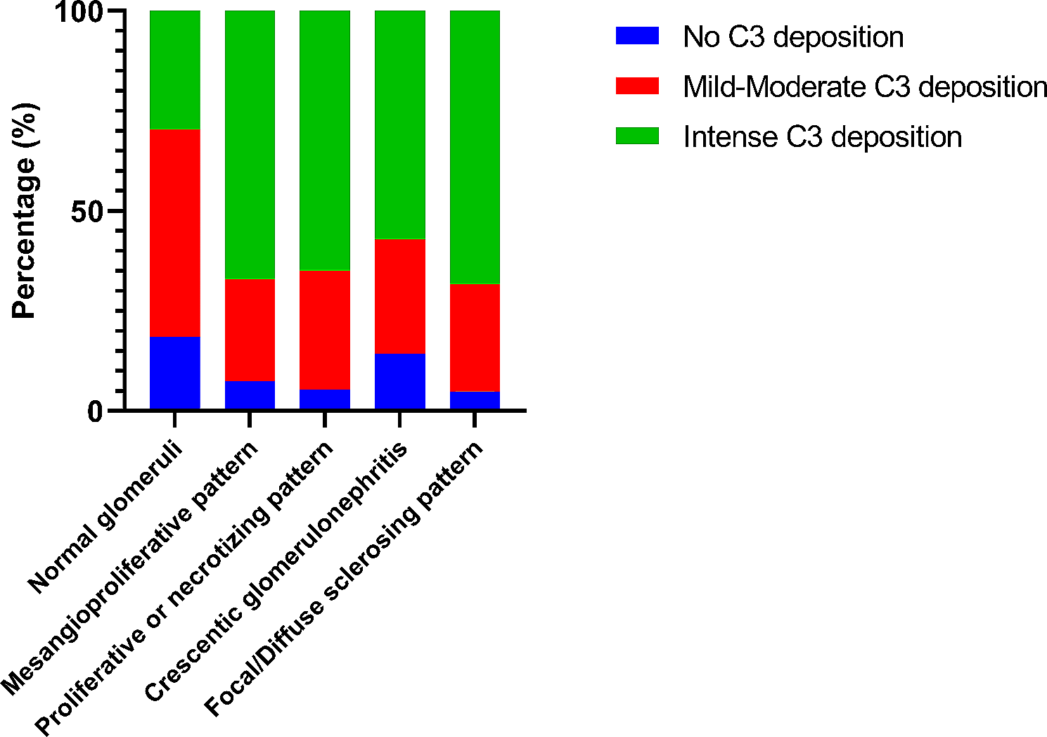
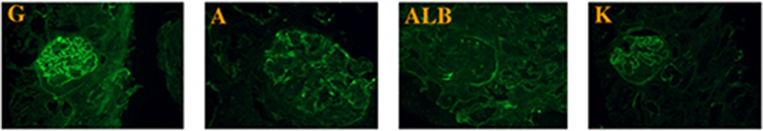
Immunofluorescence (IF)Only a few studies reported IF findings [13, 17, 20, 26, 27]. A study showed dense C3 deposits in the afferent and efferent arteriolar walls in cases of necrotizing arteritis [16]. Mittal et al. demonstrated IF results in 7 patients and reported a weak positivity for IgG and IgM, along with C3 positivity in the mesangial area [17]. Priyamvada et al. found a weak C1q deposition in the mesangium [26]. Other studies failed to demonstrate any deposits in IF [20, 27].
Electron microscopyStudies describing electron microscopic findings were scanty [12, 14]. Date et al. provided a detailed description of ultrastructural findings. The authors reported swollen cytoplasm of bowman’s capsule epithelium with visceral epithelium showing blebs, microvilli, patchy foot process fusion, and intracytoplasmic lipid vacuoles. The basement membrane of the glomerular capillaries was thick and wrinkled. In blood vessels, endothelial cells were swollen and cytoplasmic protrusions were seen to be protruding into the lumen. Infiltration of inflammatory cells was found in the interstitium. Intracytoplasmic bodies were seen in the proximal tubules representing degenerating organelles.
Renal biopsy findings and outcomesThe kidney outcomes in AKI following snakebite envenomation varied from partial or complete recovery of kidney functions to progression to end-stage kidney disease (ESKD) resulting in dialysis dependence. Studies reported that snakebite envenoming patients who did not recover their kidney functions had diffuse cortical necrosis and TMA as the predominant pathological findings. Patients with acute tubular injury were reported to respond well to conservative management and dialysis contrary to those with ACN who only responded partially or not at all [10]. Supplementary Table 1 shows renal and patient outcomes in biopsied patients. Sarangi et al. demonstrated that the clinical presentation and prognosis of the patients were directly proportional to the severity of renal histopathological lesion on the kidney biopsy [11]. Lower survival rates were reported in ACN. 8 out of 10 patients (80%) who had bilateral renal cortical necrosis, and 4 out of 23 patients with less severe acute tubular lesions died (P < .001). Other series reported a mortality rate of up to 100% in patients with ACN [8].
Studies also reported that as compared to non-TMA cases of AKI, TMA cases were associated with more advanced azotemia at presentation with an almost universal requirement for dialysis. These patients required a longer duration of renal replacement therapy (RRT), and hospitalization and had higher chances of progressing into chronic kidney disease (CKD) with higher mortality insinuating a poor prognosis [29, 30]. Golay et al. reported worse clinical outcomes while comparing cases with and without AIN [23].
留言 (0)