A unique case of a typical pancreatic ductal adenocarcinoma that initially presented with a cystic component but underwent morphological changes
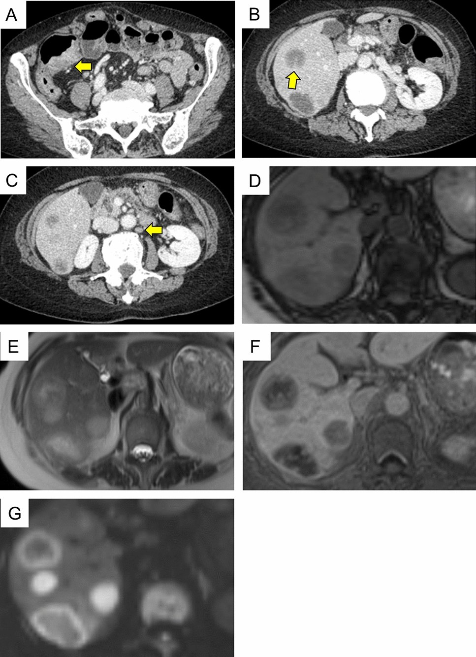
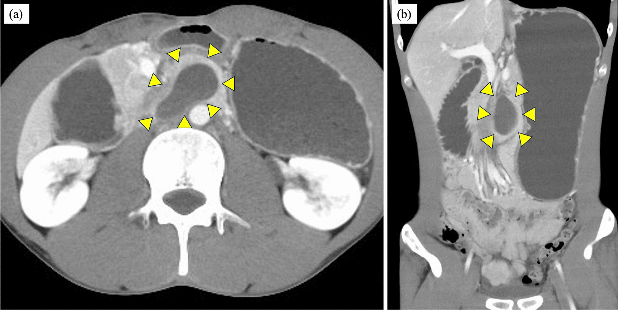
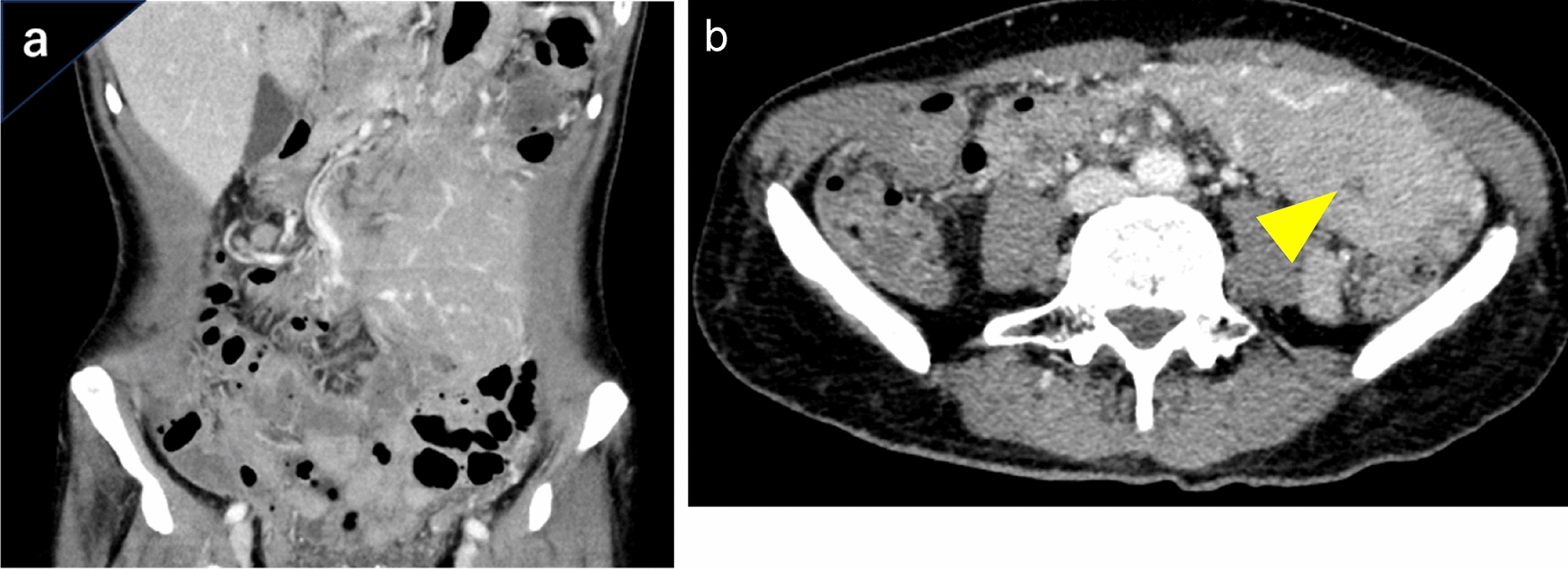
A 66-year-old man was initially suspected of having a microcystic serous cystic neoplasm based on magnetic resonance imaging findings of a multifocal mass measuring 46 mm in the pancreatic head, with a cystic component showing a high signal on T2-weighted images. The tumor marker levels were within normal limits. However, contrast-enhanced computed tomography revealed thick cyst walls with delayed staining, which was atypical for serous cystic neoplasms; therefore, the patient was followed up closely. Twenty-two months later, the delayed contrast area was enlarged, carbohydrate antigen 19–9 levels were elevated, and 18 F-fluorodeoxyglucose-positron emission tomography revealed increased accumulation, indicating a potentially malignant lesion. Pancreatoduodenectomy was performed and histopathological examination confirmed the diagnosis of normal-type pancreatic carcinoma with predominantly poorly differentiated cells. Based on the pathological findings and a literature review, it is highly likely that this case represents pancreatic ductal adenocarcinoma with a cystic structure from the beginning. While distinguishing pancreatic ductal adenocarcinoma from other pancreatic cystic tumors, such as serous cystic neoplasms, is critical owing to differing treatments and prognoses, caution is warranted as they may exhibit similar imaging features, as observed in our patient.


留言 (0)