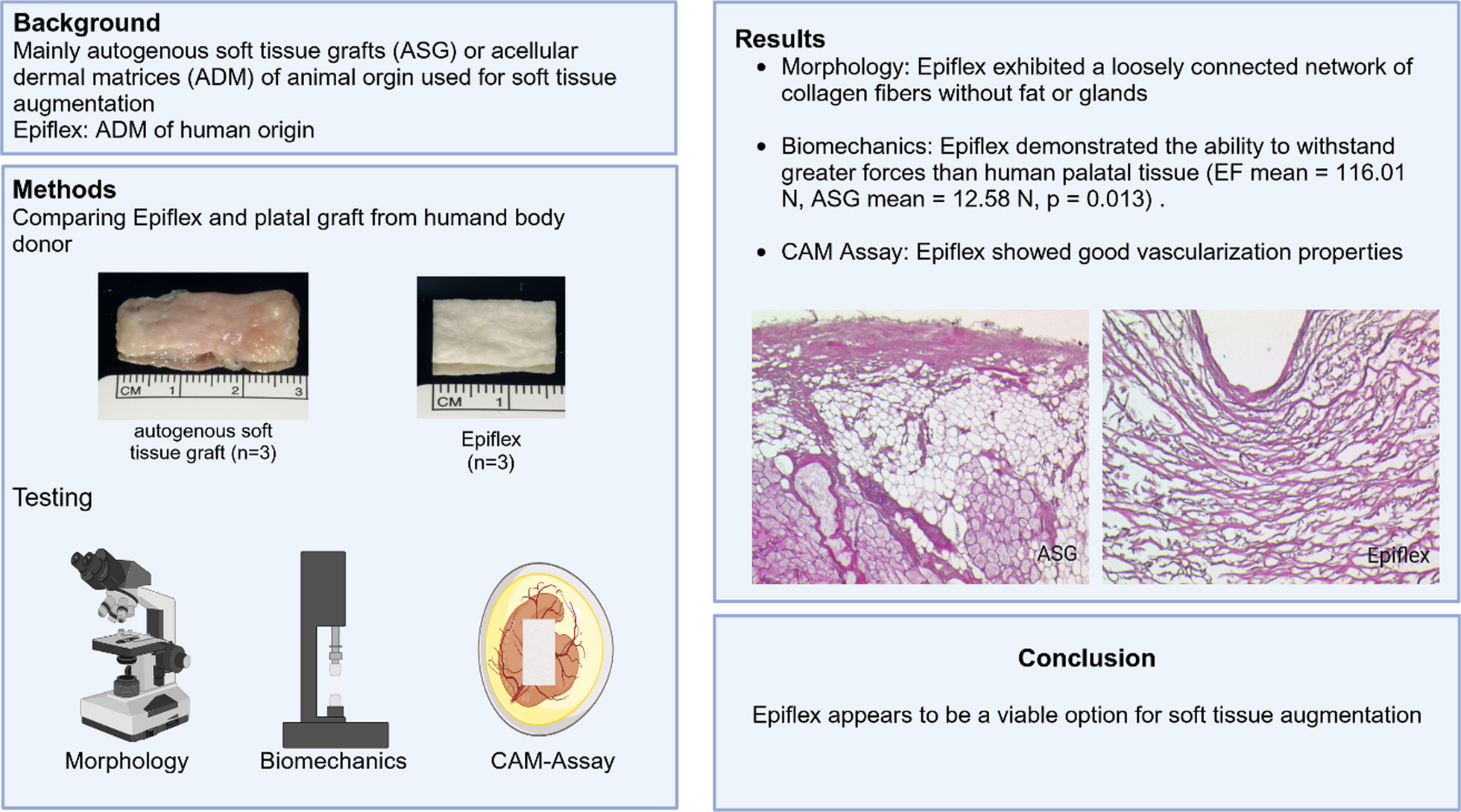
As part of the iterative process in patient care, innovations or clinical applications have to be critically analyzed. Therefore, this work aimed to investigate the outcome of individualized hybrid zirconia abutments for cemented zirconia bridges after switching from prefabricated titanium abutments. Despite many obvious reasons why this change should be beneficial for the quality of dental restorations and, consequently, patient satisfaction, it is crucial to perform critical clinical investigation and quality control.
The “implant supracrestal complex” is a recently introduced term that summarizes the implant-abutment-prosthesis complex, comprising factors such as the emergence profile, emergence angle, and implant abutment junctions [21]. But even though the authors postulated that these factors could influence the short- and long-term clinical outcome in terms of peri-implant tissue health, they could not report on corroborating evidence that the prosthetic abutment or its material (zirconia vs. titanium) had a relevant impact on the risk of peri-implantitis [22]. In the present study, design features of the two abutment types diverged significantly, such as implant shoulder-to-crown margin, ellipse circumference, shell surface, and abutment angle, as illustrated in Table 1. Individualized shapes, at least in theory, should lead to improved, more natural gingival esthetics by supporting soft tissues, maintain gingiva thickness and lead to a more favorable anatomic shape and EP. Still, in contrast to the emergence profile, none of these properties could be singled out as an individual risk factor for peri-implantitis. Whereas the abutment angle in ihZiA was significantly smaller than in the pTiA group, the shoulder-to-crown margin, ellipse circumference, and emergence profile/shell surface were significantly bigger. Nevertheless, the aim was to optimize the individual anatomical shape of the abutment, not to minimize or maximize any of these technical abutment-specific parameters.
Nevertheless, individualized abutments facilitate an advantageous concave emergence profile with an angulation tailored to the specific anatomical situation [19, 23]. This could improve accessibility for oral hygiene measures and provide an approximation to a favorable emergence angle of less than 30° [19, 23, 24]. Nevertheless, the measurements of Katafuchi et al., that lead to the recommendation of an EA of < 30°, were done on restorations without platform switching [19]. In this study on the other hand all implants were restored taking advantage of platform switching. Notably individualized ihZiA were restored with a uniformly designed adhesive base, which made measurements more difficult resulting in an initial EA of 30° from the implant collar border to the upper border of the adhesive base. For that reason, angles for the ihZiA group were measured from the adhesive base such as the implant collar border as depicted in Fig. 4. Notwithstanding the method of measurement, the impact of EA and EP was similar, when compared to ihZiA. The evaluation of the EP in this study provided further evidence that, especially in crowns emerging convexly, higher EA values are especially detrimental and lead to increased BL. To a lesser but still significant degree, this can also be stated for pontics and splinted implants and should also be considered for the design of milled bars. Due to the limited number of observations and the consideration of implant clustering in single patients, no statistically significant difference could be observed for free-end sites. Since these are explicitly located distally, only a tendency toward an impact of shape (p = 0.067) could be observed when evaluating distal sites.
Nevertheless, this fact and Fig. 4a leave room for speculation that studies with higher sample sizes of implants positioned in free-end locations might also show an impact of the EP in these situations. The analysis of different combinations of EA and EP shows that shape is essential when the EA is larger than 30° since no significant impact could be found when comparing EA smaller than 30° regarding sites with a neighboring tooth or pontic, as well as free-end positions. Therefore, the shape of individualized abutments should allow for the compensation of clinical situations that lead to higher EA. Interestingly, the relevance of the EP for splinted sites and those facing pontics was remarkable. In these cases, EA of less than 30° and with a concave shape was correlated with significantly less bone loss than EA of more than 30° and a convex profile. Of course, EA cannot be designed infinitely small, and convexity is also limited to the clinical situation. Especially in the esthetic zone, compromises like “black triangles” can hamper patient satisfaction significantly, which might be why various recommendations for pontic design exist [25, 26]. From the implant’s perspective on the other hand, the presented results lead the authors to the conclusion that the EA should be as small as possible, but at least smaller than 30°, and combined with a convex profile. In the clinical context, the EP should therefore be convex and of an EA as low as reasonably achievable, which coincides with prioritization of esthetic factors in the esthetic zone and functional/constructional parameters in the molar region [27, 28]. These suggestions are backed by the publications by Katafuchi et al. and Soulami et al. [19, 29]. Yi et al. additionally found that splinting of implants could be considered a relevant risk factor for peri-implantitis [23]. Our study also provides evidence that the mentioned parameters for EP and EA facing pontics can be regarded as relevant risk factors. A detrimental crown and pontic design with consecutive BL is illustrated in Fig. 2c, and beneficial alterations are highlighted.
In a recent review of peri-implant soft tissue phenotype modification and its impact on bone loss, Tavelli et al. argue that low supracrestal tissue height and gingiva thickness are associated with higher marginal BL [30]. This statement is backed by observations by Linkevicius as well as Berglundh & Lindhe et al. and affects implants placed at bone level in particular, and consensus exists that a peri-implant gingiva thickness of less than 2 mm coincides with early bone loss [31,32,33,34]. This is in line with the present study’s results, which demonstrate a significant correlation between gingiva thickness and BL, further underlining the necessity to evaluate this parameter in advance and consider soft tissue augmentation, which can improve peri-implant tissue health and reduce marginal BL [30, 35]. It has to be stated though that in the patient cohort examined, thinner mucosa also led to higher EA, resulting in two factors potentially augmenting the negative effect on BL.
Proper contouring of the prosthesis was shown to improve clinical outcomes for treating peri-implant mucositis, potentially preventing peri-implantitis [24]. This might explain why bone-loss without active inflammation was significantly higher in the prefabricated pTiA group when adjusted for time, even though the values for plaque or bleeding index overall did not differ significantly between the two abutment types. On explanation might be considered an abutment associated initial bone loss, since this significant difference could only be observed for bone loss without BoP of ≤ 2 mm (Table 2). The documented bone loss could also be a sign of peri-implant disease that could be controlled with regular recalls and professional cleaning. After all, the impact might be limited since abutment type was not a significant influencing factor for the other definitions of more severe peri-implantitis, especially considering BoP as an indicator for current inflammation [36]. Bone loss of ≥ 2 mm was significantly more frequent in the maxilla when compared to the mandible. This was especially the case for distal sites (p < 0.001) in the metric analysis, as demonstrated in Table 3. The literature on whether maxillary implants are generally more prone to bone level changes is inconsistent, providing evidence for both scenarios [37, 38]. The fact that especially upper distal sites were affected in the present study might indicate that accessibility to regular dental hygiene measures could be the cause. Sex, on the other hand, was found to be a significant factor only for distal sites resulting in more bone loss in men. Considering this isolated result, the clinical relevance is somewhat limited, but according to a population-based, cross-sectional study by Varela-Centelles and colleagues, being female was associated with good oral hygiene habits [39]. Still, some studies indicate higher bone resorption in women, which aside from (distal) location, further emphasizes the potential influence of dental hygiene in this study [14, 39, 40]. All the more important is an abutment design that facilitates an easily cleanable contour of the dental prosthesis. Interestingly, while PD did correlate significantly with bone resorption in pTiA, this was not the case for ihZiA, which might result from the lower bacterial adhesion to zirconia [3, 41]. Even though it is hard to account for specific anti-bacterial surface alterations, this could indicate a lower bacterial load in the peri-implant sulci [3]. Still, overall bone loss in both groups was considerably below 1 mm after roughly five to six years, which is in line with the literature. [42, 43]
To evaluate overall treatment success, the esthetic outcome and patient satisfaction also have to be considered, as they influence the patient’s QoL [44, 45]. Aside from the esthetic and natural shape of the individualized ihZiA, leading to a more natural scalloped look of the gingiva, pTiA in this study showed significantly more visible titanium (p = 0.0008). Of course, the immediate visibility of minor recessions in pTiA influences this. In restorations with ihZiA, the darkish titanium coloration, which stands in stark contrast to the ceramic crowns, becomes visible only after the recession leads to the exposure of the entire abutment. Furthermore, bone resorption did not correlate with PD to the same extent as with pTiA, potentially leading to a more stable gingival margin. While abutment type alone did not impact OHIP scores, the amount of visible titanium in the esthetic zone group significantly influenced OHIP-14 scores in the pTiA group. Taken together, this demonstrates a measurable but minor effect on patients’ QoL, considering that this was not the case for OHIP-49 scores.
Nevertheless, this emphasizes that patient expectations have risen and visible metal components are considered unacceptable, ultimately affecting patient satisfaction [45]. Figure 6 underlines this assumption. Nelson et al. and Hu et al. showed that gingival display in Caucasians can be relevant even in the first molar region [27, 28]. While this was the case for elderly patients, it was especially relevant in the younger population, who showed papillary display of over 90% in the first premolar and 85% in the second molar region [28]. Therefore, optimization of the gingival contour as well as a natural coloration without metal show is a crucial criterion for esthetically pleasing restorations, which ultimately influence the patient’s self-perception and self-confidence.
Despite the prevailing argument that ceramic restorations are more prone to chipping or fractures, none of these events occurred in this study, even though most abutments were located in the molar and premolar region known to be subjected to two to three times higher biting forces [5, 46]. This is consistent with the findings of Klongbunjit and colleagues, who found in vitro that hybrind and titanium abutments had comparable stability and strength when subjected to bending and torque fatigue tests [47]. Similarly Al-Zordk et al. did observed high fracture resistance of zirconia hybrid abutments, which exceeded the maximum masticatory forces in molar teeth by a wide margin [48]. Waltenberger and Wolfart even described a concept, that utilizes a custom-made, adhesively bonded zirconia abutment secured to a titanium base [49]. This abutment is digitally designed and placed upon implant placement, enabling immediate loading with a provisional PEEK crown [49].
A potential limitation of this study arises when comparing prefabricated with individualized abutments and is inherent in the presented setting after changing the standard of care and documenting the respective outcomes. Another limitation is the evaluation via panoramic x-ray, which bears limitations in the evaluation of bone loss in the anterior region. However, only radiographs of sufficient quality were included. Furthermore, the annual bone loss is mathematically calculated and therefore varying dynamics of tissue inflammation over time are not reflected in this figure.
According to the results of the present study, the outcome of individualized ihZiA was similar to that of pTiA. They both provide a viable option for anatomically correct and esthetically pleasing implant-based dental restorations. Furthermore, early-stage peri-implantitis was less frequently encountered in ihZiA, and OHIP-14 scores in patients of the pTiA group were significantly impacted by visible titanium. Therefore, ihZiA, if properly designed, could be superior in terms of peri-implantitis prevention and QoL. Still, further studies need to verify this assumption since the true impact of the material can be evaluated only if the same design features are applied. Therefore, this study is limited to the comparison of these specific pTiA and ihZiA in cemented zirconia FDP and the results cannot be transferred to other platforms without careful consideration and constraints. This is due to the great variety of abutment and fixation systems, which bear specific strengths, weaknesses and limits the generalizability of this study. Nevertheless, the present study highlights the clinical relevance of the supracrestal complex. Moreover, abutment features alone insufficiently reflect the interproximal situation. Thinner peri-implant soft tissue at the time of the prosthetic restoration had a significantly negative impact on annual bone loss. A convexly shaped EP should be avoided in all circumstances, and the EA should be designed as low as reasonably achievable.


留言 (0)