Chemicals and reactants
Shanghai Yuanye Bio-Technology Co., Ltd (Shanghai,China) provided GLA (CAS No. 79498-31-0, Batch No. B20698, 98% purity). The chemical structure of GLA is illustrated in Fig. 1A. To obtain the original solution (50 mM), we dissolved the GLA in dimethyl sulfoxide and kept it at − 80 °C. Pancreatic enzyme, Dulbecco’s Modified Eagle Media: Nutrient Mixture F-12 (DMEM/F12), radio-immunoprecipitation assay (RIPA) buffer, 4, 6-diamidino-2-phenylindole (DAPI) solution and toluidine blue staining solution were purchased from Beijing Solaybao Technology (Beijing, China). Betulinic acid (Batch No. S360301, CAS No. 472-15-1, 99.88% purity) was purchased from selleck (Shanghai, China). Horseradish peroxidase-conjugated goat anti-rabbit and goat anti-mouse antibodies were purchased from Boster Biological Technology (Wuhan, China). Primary antibodies against Primary antibodies against (GAPDH), COL2A1, MMP13, SOX9, iNOS, COX-2 were obtained from Abcam (Shanghai, China). Primary antibodies against extracellular signal-regulated kinase (ERK), c-Jun N-terminal kinase (JNK), NF-κB p65, phosphorylated-ERK (p-ERK;Thr202/Tyr204),phosphorylated-JNK (p-JNK;Thr183/Tyr185), phosphorylated-NF-κB p65 (p-P65;Ser536) were from Cell Signaling Technology (Danvers, MA, USA).
Cell culture and grouping
A chondrogenic ATDC5 cell line was purchased from Riken Cell Bank (Ibaraki, Japan) and was used as a reference for the analysis. ATDC5 cells were grown at 37 °C in a cell incubator with 5% carbon dioxide in DMEM/F12 media supplemented with 10% fetal bovine serum and 1% penicillin/streptomycin. For control experiments, ATDC5 cells were divided into four groups. To observe the GLA in IL-1β stimulation cartilage cells play a role. (1) Sham group, GLA and IL-1β were not administered to the ATDC5 cells; (2) IL-1β group, In the case of ATDC5 cells, only IL-1β (10 ng/ml) was used; (3) GLA group, Only the GLA (0.5 μM) was applied to the ATDC5 cells; (4) GLA+IL-1β group, we treated ATDC5 cells with IL-1β (10 ng/ml) and GLA (0.5 μM). GLA is dissolved in DMSO. In this experiment, the concentration of DMSO in all cell culture media was kept below 0.05%, and there was no significant effect on the growth of cells.
Cell-viability assay
CCK-8 reagen was used to detect the effect of GLA on chondrocyte cell viability. ATDC5 cells (6 × 10³ cells/well) were planted in 96-well plates and cultivated for 24 h. ATDC5 cells then underwent treatment with GLA at various doses (0, 0.1, 0.3, 0.5, 1, 3, 5 μM) during 24 h, 48 h, or 72 h. Following treatment, 10μl CCK-8 solution was added into each well and it was incubated at 37 °C for 1–4 h. Samples were assayed for absorbance at the 450 nm band level using an ELX800 microplate reader (Bio-Tek Instruments, Inc, Winooski, VT, USA). All experiments were carried out independently three times.
RNA extraction and quantitative reverse transcription PCR
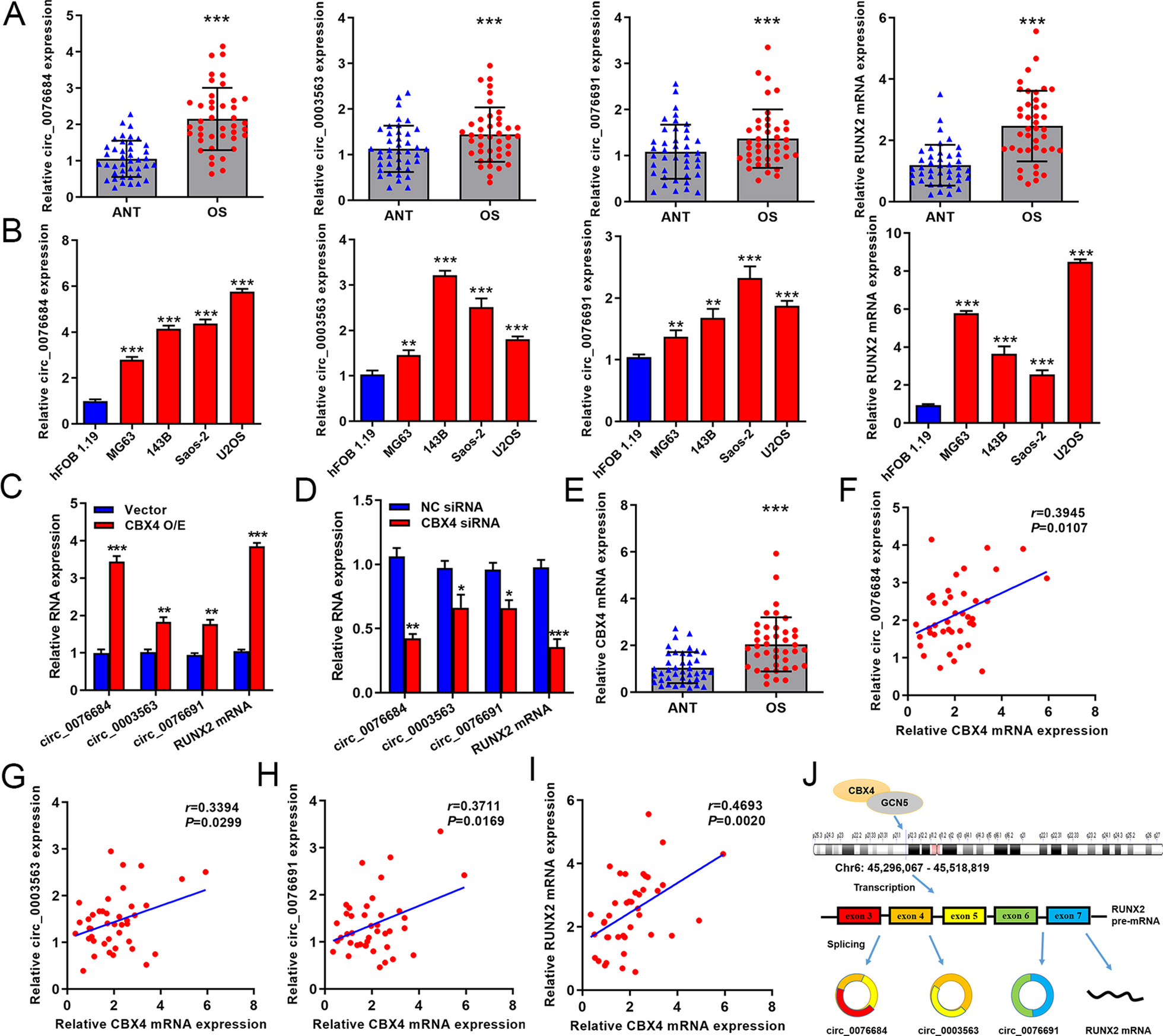
ATDC5 cells were separated into four groups and injected in 6-well plates (1 × 10⁵ cells/well). The cells were treated for 48 h with IL-1β or GLA, and total RNA from chondrocytes in each of the groups was isolated with TRIzol (Beijing Tianen Biotechnology Co., Ltd., Beijing, China). We took RNA (1 μg) from each chondrocyte group and synthesized complementary Dna using reverse transcriptase according to the manufacturer’s protocol (TaKaRa Bio, Otsu, Japan). We performed real-time PCR using SYBR PreMix Ex Taq Kit (Takara Bio) and a LightCycler 96 PCR machine (Roche, Germany). All reactions were carried out in triplicate. The specific primers employed are as follows:
INOS Forward primer 5′-CTGGCAAGCCCAAGGTCTAT-3′
Reverse primer 5′-TCCCCGCAAACATAGAGGTG-3′;
COX-2 Forward primer 5′-CGG TGAAACTCTGGCTAGACAG-3′
Reverse Sequence 5′-GCAAACCGT AGATGCTCAGGGA-3′;
MMP13 forward primer 5ʹ-GACCCCAACCCTAAGCATCC-3′
Reverse primer 5ʹ-CCTCGGAGACTGGTAATGGC-3′;
COL2A1 forward primer 5′-CTCAAGTCGCTGAACAACCA-3′
reverse primer 5′-GTCTCCGCTCTTCCACTCTG-3′;
SOX9 forward primer 5′-GCAGGCGGAGGCAGAGGAG-3′
reverse primer 5′-GGAGGAGGAGTGTGGCGAGTC-3′;
GAPDH forward primer 5′-ACCCAGAAGACTGTGGATGG-3′
reverse primer 5′-CACATTGGGGGTAGGAACAC-3′.
Western blot analysis
We washed the ATDC5 cells twice with cold PBS after treating them with IL-1β or GLA, and then extracted the total cellular proteins from the ATDC5 cells using RIPA lysis solution mixed with protease and phosphatase inhibitors (Sigma-Aldrich, Rockford, USA). The lysate was kept on ice for 10 min before centrifuge at 12,000 rpm for 10 min at 4 °C. The required total protein was present in the supernatant. The target protein quantity was evaluated using a Bicinchoninic Acid (BCA) Protein assay kit (Beyotime, Nanjing, China), and the protein was quantified. Sodium dodecyl sulfate–polyacrylamide gel (SDS-PAGE) was used to electrophorese protein (20 μg/well), which was then transferred to polyvinylidene fluoride membranes (0.45 μm, Millipore, Bedford, MA, United States). The polyvinylidene fluoride (PVDF) membrane has been blocked with 5% skimmed milk for 2 h at ambient temperature before being treated with the matching primary antibody overnight at 4 °C. The PVDF membrane was washed three times with Tris-buffered saline-Tween 20 for ten minutes each time before being incubated with a horse-radish peroxidase (HRP) conjugate secondary antibody for one hour at ambient temperature. Finally, we detected the protein bands using the Odyssey V3.0 image scanner (LiCOR Biosciences, Lincoln, NE, USA) and analyzed them using ImageJ software. In this experiment, GAPDH was used as a source of internal control.
Enzyme-linked immunosorbent assay (ELISA)
ATDC5 cells have been grown on 6-well plates, and their supernatant was collected following treatments. The enzyme-linked immunosorbent assay kit (ELISA; R&D System) was used to measure the quantity of IL-6 in the supernatant.
Immunofluorescence microscopy
ATDC5 cells were seeded in 24-well plates and exposed to either IL1β or GLA for 48 h. For 30 min, the cells were submerged in 4% paraformaldehyde (PFA) at the ambient temperature. For 15 min, the cells were treated with 0.1% Triton-100 (Solarbio, Beijing, China). The primary antibody was incubated at 4 °C overnight after blocking with 1% bovine serum albumin (Sigma Aldrich, Germany) for 30 min. The cells were rinsed three times with PBS before being incubated at room temperature for two hours with goat anti-rabbit Ig G antibody (1:400). After that, the nucleus had been stained for 5 min using DAPI staining buffer. Under a confocal microscope (Leica, Germany), cells are viewed and photographed.
High density culture and toluidine blue staining
We used this method to measure chondrocyte ECM level. Each well of a 24-well plate was seeded with 10 μL of ATDC5 cells suspension (107 cells), and each group was seeded three times. ATDC5 cells were grown for 1 h in a cell incubator until they stuck to the wall. After cell adhesion, for a total of 24 h, each well received 700 μL of DMEM/F12 medium for cell cultivation with a concentration of 10% fetal bovine serum and 1% penicillin/streptomycin. Then different quantities of IL-1β (10 ng/ml) and GLA (0, 0.3, 0.5 μM) were added. Every 2–3 days, the culture media was replaced. After 5–8 days of cell culture, ATDC5 cells were fixed for 30 min in 4% PFA before being stained with the color toluidine blue. ImageJ (National Institutes of Health, Bethesda, MD, United States) was used to determine the staining intensity of cells in each group.
Animal model
All experiments involving animals in the present study have been carried out in conformity with Nanchang University’s Animal Ethics Committee (No.: DM20210912). The animal model we established this time is the DMM model [18]. C57BL/6 mice (6 weeks old, n = 24) were anesthetized with a 50 mg/kg intraperitoneal dose of pentobarbital sodium. Under the microscope, the attachment point between the medial meniscus and the tibial plateau of the right knee (medial meniscus-tibial ligament) was severed with a surgical knife, and the injury of other ligaments around the medial meniscus should be avoided during the operation. The experimental animals were randomly divided into 4 groups: Sham group, DMM group, DMM + 10 mg/kg GLA group (low concentration GLA group), DMM + 20 mg/kg GLA group (high concentration GLA group). Only the right knee joint was incised in the Sham group, without the medial meniscus-tibial ligament being severed. The mice in the low-concentration GLA group were intraperitoneally injected with GLA 10 mg/kg, and the mice in the high-concentration GLA group were given GLA 20 mg/kg intraperitoneally on two separate days for 8 weeks. Mice in the sham surgery and DMM groups received the same quantity of phosphate buffered saline (PBS) intraperitoneally. During the experiment, mice were free to obtain food and water. After 8 weeks, pentobarbital sodium was injected intraperitoneally. Following anesthesia, each of the mice were euthanized, and samples of knee tissue from the joints were taken for further research.
Histological scoring
Six mice in each group had their right knee joints frozen in 4% paraformaldehyde for 24 h before being dissolved with a 10% ethylenediaminetetraacetic acid (EDTA) decalcified solution for one month. The mice’s knee joints were implanted in paraffin, and the knee joints containing the mice were cut into sections of 5 μm thickness from the sagittal position. Hematoxylin–eosin (HE) staining, safranin O-fast green staining and IHC staining (p-p65, MMP13, COL2A1, COX-2) were performed on the sections. In this study, the pathological changes of joints were analyzed by the International Association for the Study of OA (OARSI) scoring system [19]. Xylene was used to dewax the paraffin sections of the knee joint, and the related staining was performed after gradient ethanol hydration. Under a microscope, the cartilage damage of the joint in the knee and the manifestation of associated indicators were seen.
Statistic analysis
All results were shown as mean ± standard deviation (N ≥ 3). For statistical analysis, we utilized GraphPad Prism 9 software (GraphPad Software, California, USA). To examine the variations between two groups or three or more groups, all statistical analyses were performed using the t-test or a one-way analyses of variance (ANOVA). In statistical terms, P 0.05 was considered meaningful.


留言 (0)