
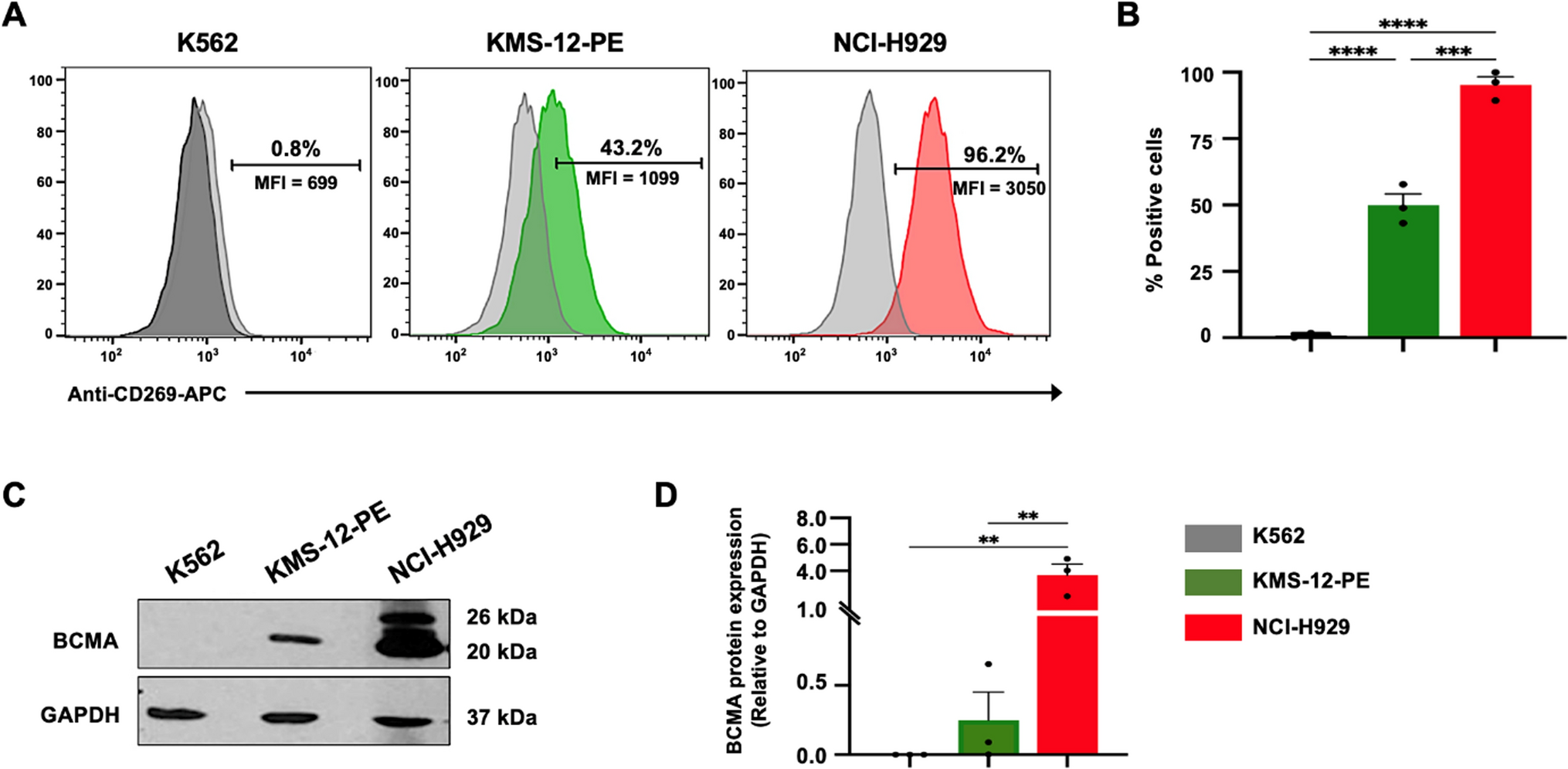
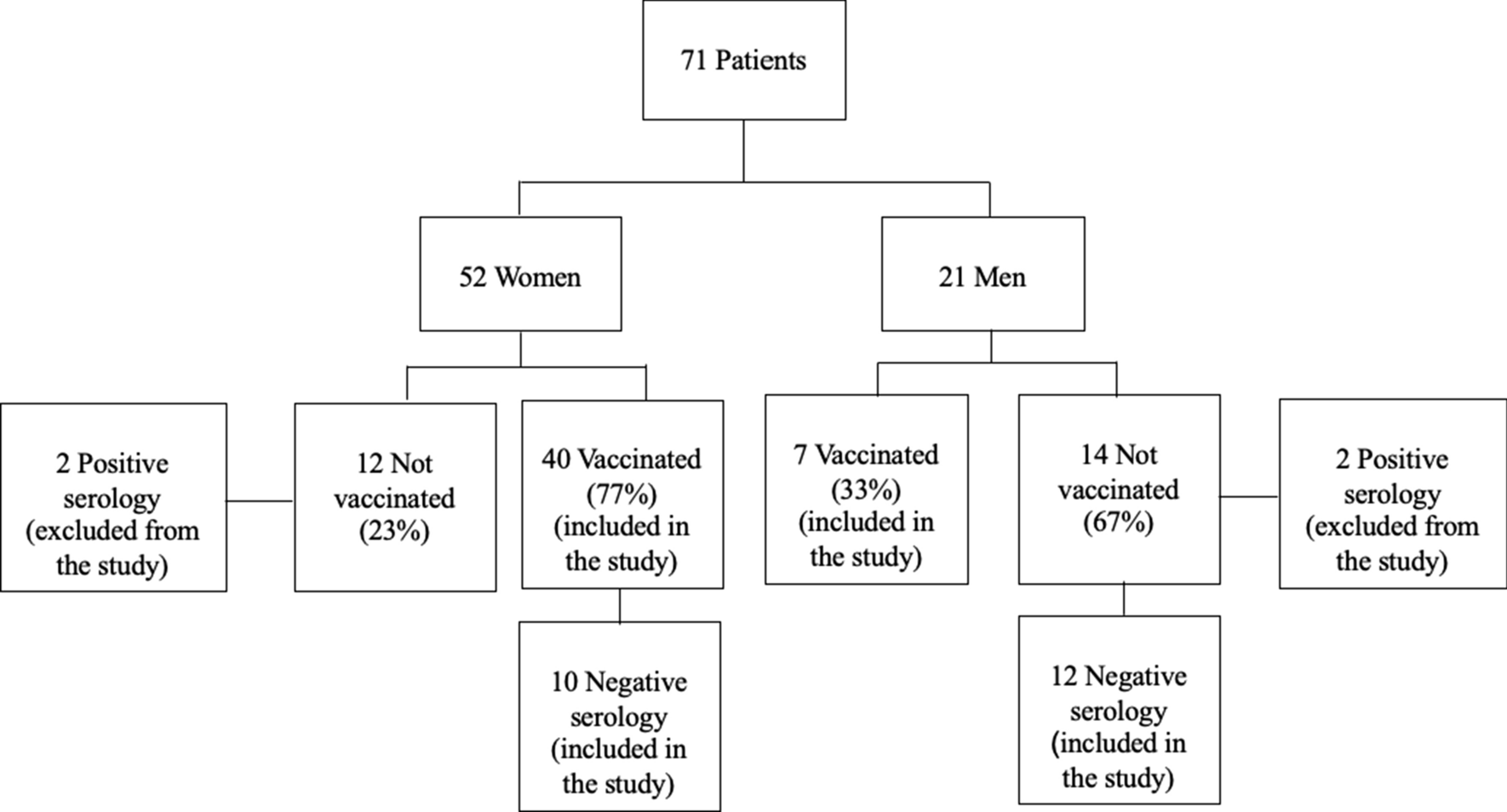
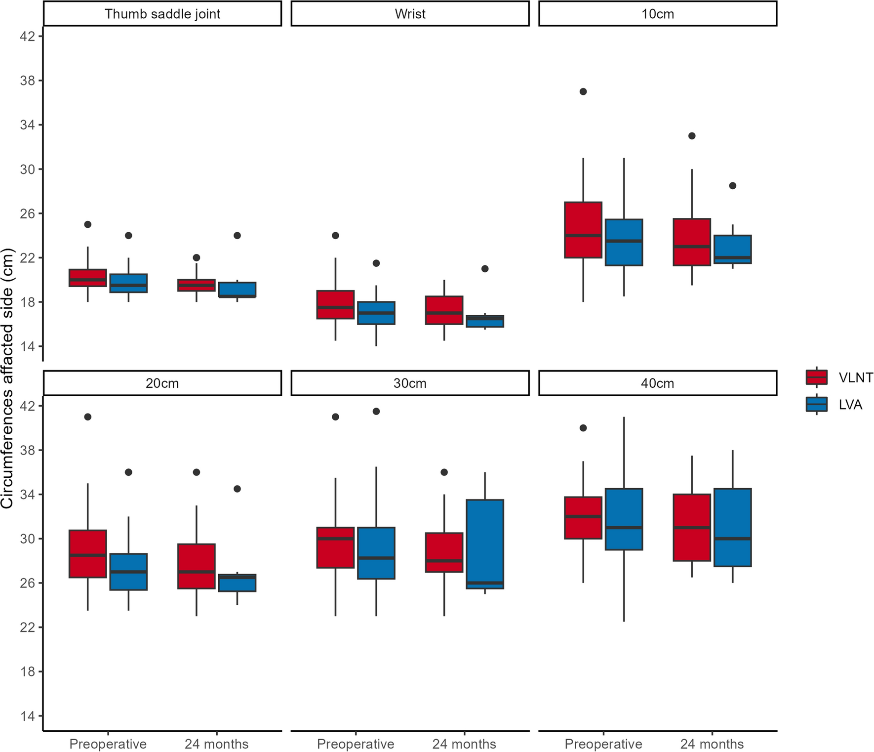
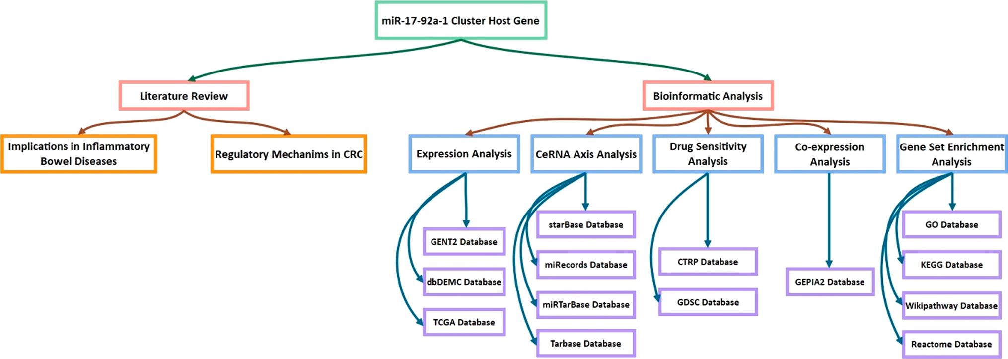
記住我

Clinicopathological analysis was performed on 182 patients receiving immunotherapy combined with platinum-based chemotherapy or monotherapy according to PD-L1 TPS or ECOG status. These patients were randomized in a 1:1 ratio into a training cohort and a validation cohort (Table 1). The median follow-up for the training cohort was 34.6 months, with a median OS of 26.3 months (95% confidence interval (CI) 18.5–34.1); the 1-, 3-, and 5-year OS rates were 78%, 33%, and 12%, respectively. The median PFS was 7.6 months (95% CI 5.9–9.4). The median follow-up for the validation cohort was 34.2 months, with a median OS of 32.5 months (95% CI 25.5–39.5); the 1-, 3-, and 5-year OS rates were 88%, 44%, and 3%, respectively. The median PFS was 7.7 months (95% CI 6.5–8.8). In summary, the demographics, tumor characteristics and treatment regimens were highly comparable in the two cohorts, except for the higher proportion of patients with PD-L1≧ 1% in the training cohort (80.2% vs. 64.8%) compared to the validation cohort.
Table 1 Baseline characteristics of patientsEstablishment of the NTRSX-tile software was used to identify the best cutoff values for 15 indicators, including neutrophils, lymphocyte, platelets, NLR, PLR, HCT, β2-microglobulin, ALB levels, ALP levels, FIB levels, LDH levels, HDL-C levels, and CYFRA21-1 levels, SCC, and CEA. The results suggested that in the training cohort, the optimal cutoff values for ALP, HDL-C, and CYFRA21-1 levels were 162 U/L, 0.7 mmol/l and 6.3 ng/ml, respectively (Fig. S1A, B, C). Thus, three independent predictors of poor OS, namely a pooled cohort with ALP, HDL-C, and CYFRA21-1 were included in NTRS (Fig. S2). One point was scored for each factor and the total score (0–3) was calculated. For patients distinguished by the best cutoff value for the other indicators, there was no distinction between the groups with high and low best cutoff values in terms of OS or P-values that were statistically significant.
Identification of the NTRS with OS in the training and validation cohortUnivariate and multivariate Cox regression analyses in Table 2 and Table S1 demonstrated the relationship between clinicopathological variables and OS and PFS in the training and validation cohorts. In the univariate analysis of the training and validation cohorts, PD-L1 expression, maximum diameter, LDH levels, CYFRA21-1 levels and NTRS were associated with OS; NTRS was associated with PFS. These variables were subsequently included in multivariate analyses.
Table 2 Univariate and multivariate analyses for predictors of overall survival and progress-free survival in the training cohortComparison of the NTRS in prognosis abilityIn the training cohort, time-dependent ROC analysis showed that the prognostic value of NTRS for OS was significantly better than PD-L1 expression (Wilcoxon matched pairs P = 0.012, 0.004) (Fig. 1A, B). The AUC of NTRS was significantly greater than PD-L1 expression at the 1, 2, 3, 4, 5-year time point. For example, in the training cohort (Fig. 1A), the C-index for 4-year OS probability prediction in NTRS was 0.767 (95% CI 0.636–0.859), which was significantly higher than PD-L1 expression (C-index 0.608, 95% CI 0.477–0.728); in the validation cohort (Fig. 1B), the C-index for 4-year OS in NTRS was 0.728 (95% CI 0.589–0.841), which was also greater than PD-L1 expression (C-index 0.635, 95% CI 0.492–0.763). However, the NTRS did not show better predictive value for PFS than PD-L1 expression in the training (P = 0.755) and validation groups (P = 0.503). In the training cohort, the 2-year PFS C-index was 0.644 (95% CI 0.546–0.768) for NTRS and 0.712 (95% CI 0.597–0.810) for PD-L1 expression; in the validation cohort the NTRS was 0.640 (95% CI 0.521–0.747) and PD-L1 expression was 0.542 (95% CI 0.424–0.657) (Fig. 1C, D). These results suggested that the NTRS was a better predictor of OS than PD-L1 expression.
Fig. 1
Time-dependent ROC curve analysis to compare the effect of NTRS, PD-L1 expression, tumor size, ALP, CYFRA21-1, SCC, HDL-C, NLR, PLR, ALB, FIB, HCT, and LDH in predicting A, C overall survival and B, D progression-free survival in A, B training cohort and C, D validation cohort. The vertical axis shows the corresponding area under the ROC curve at different time points, and the horizontal axis represents the time (months). PD-L1 programmed death ligand 1, ALP alkaline phosphatase, SCC squamous epithelial cell carcinoma-associated antigens, HDL-C high-density lipoprotein cholesterol, NLR neutrophil–lymphocyte ratio, PLR platelet-lymphocyte ratio, ALB albumin, FIB fibrinogen, HCT hematocrit, LDH lactic dehydrogenase, NTRS non-tumor-related scores
OS and PFS assessed by the NTRS and PD-L1 expressionFigures 2 and S3 showed OS and PFS for different scores (0–3) of PD-L1 expression and NTRS stratification in the training and validation cohorts were shown. Survival curves for NTRS were more discriminatory compared to PD-L1 expression.
Fig. 2
A–D OS and E–H PFS in A, E PD-L1 (< 1%, 1–49%, ≧ 50%) expression and different risk score (score = 0, 1, 2, 3) in B, F PD-L1 < 1% group, C, G PD-L1 1–49% group and D, H PD-L1 ≧ 50% group in training cohort. The number and P-value can be seen clearly. A P-value of ≦ 0.05 was considered significant. PD-L1 programmed death ligand 1, OS overall survival, PFS progress-free survival
In the training cohort, mOS was 8.6, 26.3 and 33.2 months for patients with PD-L1 expression < 1%, 1–49% and ≧ 50%, respectively. mOS was 64.9, 21.1, 12.4 and 6.3 months for patients with NTRS of 0–3, respectively (Figs. 2A, S3A). In patients with PD-L1 1–49%, mOS was statistically different at 33.5, 20.3, 12.4 and 6.3 months when score = 0, 1, 2, and 3, respectively (P < 0.001) (Fig. 2C), but no survival differences was seen among scores of PD-L1 < 1% and ≧ 50% (Fig. 2B, D). mPFS for PD-L1 expression was 4.4, 7.6 and 8.8 months and mPFS for NTRS were 9.8, 5.9, 4.4 and 3.2 months, respectively (Figs. 2E, S3B). Differences in PFS were seen in patients with PD-L1 1–49% when scores = 0, 1, 2, and 3 (P = 0.012), while no differences were seen in patients with PD-L1 < 1% and ≧ 50% (Fig. 2F–H).
In the validation cohort, mOS was 31.7, 29.1 and 64.6 months for patients with PD-L1 expression < 1%, 1–49% and ≧ 50%, respectively, and 43.6, 44.0, 18.7 and 11.1 months for patients with scores 0–3 (Figs. 3A, S3C). In the stratification of PD-L1, OS was difference when risk score = 0, 1, 2, 3 (P < 0.001, 0.01, and 0.003, respectively) (Fig. 3B–D). mPFS for PD-L1 expression was 8.3, 6.9 and 10.3 months; while, mPFS for NTRS was 8.3, 7.9, 4.2, and 2.5 months, respectively (Figs. 3E, S3D). When risk score = 0, 1, 2, 3, there was a difference in PFS in patients with PD-L1 1–49%, while no difference was seen in patients with PD-L1 < 1% and ≧ 50% (Fig. 3F–H).
Fig. 3
A–D OS and E–H PFS in A, E PD-L1 (< 1%, 1–49%, ≧ 50%) expression and different risk score (score = 0, 1, 2, 3) in B, F PD-L1 < 1% group, C, G PD-L1 1–49% group and D, H PD-L1 ≧ 50% group in validation cohort. The number and P-value can be seen clearly. A P-value of ≦ 0.05 was considered significant. PD-L1 programmed death ligand 1, OS overall survival, PFS progress-free survival
In pooled cohort, OS was statistically different when PD-L1 expression was stratified at < 1%, 1–49% and ≧ 50% (mOS by score = 0, 1, 2, 3. mOS at PD-L1 < 1%: 32.5 m, 18.0 m, 30.1 m, 10.2 m, P = 0.013; mOS at PD-L1 1–49%: 32.5 m, 23.5 m, 13.2 m, 13.6 m, P = 0.001; mOS at PD-L1≧ 50%: 64.6 m, 33.2 m, 17.6 m, 23.1 m, P = 0.006). In summary, among patients with PD-L1 1–49%, the higher the score, the worse the PFS and there was a statistical difference (P = 0.407 for PD-L1 < 1%; P = 0.011 for PD-L1 1–49%; P = 0.618 for PD-L1≧ 50%) (Fig. S4).
Thus, even though PD-L1 had statistically different OS in the < 1%, 1–49%, and ≧ 50% groups, there was no PFS difference. Moreover, both OS and PFS were statistically different in the PD-L1 1–49% stratification in each NRTS score (0–3) in both the training and validation cohorts.
NTRS showed higher efficiency than PD-L1 in predicting responseNTRS demonstrated a higher efficiency than PD-L1 expression as a biomarker for immunotherapy. When response was evaluated with RECIST1.1, NTRS were more different between the DCB and NDB groups than PD-L1 expression (Fig. 4A–D), but there was no significant difference between the BOR and non-BOR groups (Fig. 4E–H). The proportion of NTRS and PD-L1 was 90% (N = 81) and 90% (N = 81) for non-PD (PR + SD) patients in the training cohort and 92% (N = 83) and 91% (N = 82) for non-PD patients in the validation cohort (Fig. 4I–L).
Fig. 4
Comparisons between PD-L1 expression and NTRS e in predicting response to ICIs in NSCLC in A, B, E, F, I, J training cohort and C, D, G, H, K, L validation cohort. Differential analyses of A, C NTRS and B, D PD-L1 expression among patients with durable clinical benefit. Differential analyses of E, G NTRS and F, H PD-L1 expression among PR/SD/PD patients. Number of patients with PR/SD/PD responses among I, K PD-L1 expression and J, L NTRS in I, J training cohort and K, L validation cohort. PD-L1 programmed death ligand 1, NTRS non-tumor-related scores, ICIs immune checkpoint inhibitors, NSCLC non-small cell lung cancer, PR partial response, SD stable disease, PD progressed disease
ROC analysis demonstrated that NTRS significantly enhanced the predictive efficiency of PD-L1 expression (Fig. S5). When BOR was assessed in the training cohort, the AUC of NTRS was 0.650, which was the trend of assessing prognosis than 0.522 of PD-L1 (Figs. S5A, 5E). In evaluation of DCB in the training cohort, the AUC of NTRS was higher, 0.644, than 0.572 for PD-L1 (Fig. S5B, 5E). When BOR was assessed in the validation cohort, the AUC for NTRS was 0.584, significantly lower than 0.637 for PD-L1 (Figs. S5C, 5E). In evaluation of DCB in the validation cohort, the AUC of NTRS was highest at 0.665, which was higher than that of 0.547 for PD-L1 (Fig. S5D, 5E). Taken together, the combination of PD-L1 expression and NTRS had a greater AUC and higher predictive prognosis value to improve the effectiveness of PD-L1 expression as an immunotherapy biomarker.
Distribution and classification characteristics of NTRS and PD-L1 expression in metastatic sites and tumorigenic factorsThe distribution of PD-L1 expression in tissue samples from different metastatic sites was moderately different depending on organ differences (Fig. S6). Among metastatic sites, the proportion of patients with high PD-L1 expression was greatest in lung (PD-L1 (≧ 50%) = 29%) and smallest in pleural (PD-L1 (≧ 50%) = 13%), and the proportion of patients was greatest in the adrenals (scores (1 + 2 + 3) of 66%) and smallest in the lung (scores (1 + 2 + 3) of 29%) (Fig. S6A, B). Differences in the distribution of PD-L1 expression and NTRS at different metastatic sites were further explored. PD-L1 status (PD-L1≧ 1% vs. < 1%) was predictive of ICI treatment response in brain metastases, but no significance was found between metastasis site and treatment response in the risk score (Fig. S6C, D).
Categorical and regression analyses were used to assess the prognostic factors of NTRS and PD-L1 expression in long-term survival. Table S2 provided the tumor correlates of NTRS and PD-L1 expression. In PD-L1 expression, the level of the tumor marker SCC was identified as the most important correlate, while in the NTRS, LNMs status was determined as the most important correlate. And the critical ranges of LNMs and SCC levels were identified as N2 and 2.5 ng/ml. Specifically, regression analysis of critical values in the PD-L1≧ 50% group suggested worse PFS when SCC levels exceeded 2.5 ng/ml (mPFS: 8.4 vs. 2.4 months, P < 0.001), but no difference was found in OS (Fig. S7). In addition, in the NTRS, LNMs suggested survival differences in OS and PFS (Fig. 5). In particular, N2 staging resulted in shorter OS and PFS in this group of patients with NTRS ≧ 1 (mOS: 42.6 vs. 14.2 months, P < 0.001; mPFS: 10.37 vs. 4.17 months, P = 0.001). These results implied longer OS in patients with PD-L1 ≥ 1% brain metastases, but there was no correlation between NRTS and metastatic site. In a further, patients with NTRS (≧ 1, host factor) and LNMs (N2, tumorigenic factor) had worse OS and PFS; patients with PD-L1 (≧ 50%) and SCC levels (> 2.5 ng/mL) had shorter PFS.
Fig. 5
A Kaplan–Meier curve model comparing the hierarchical association of prediction factors associated with OS and PFS among patients with LNMs. OS overall survival, PFS progress-free survival, LNM lymph node metastases
Subgroup analyses of OS and PFS based on NTRS and PD-L1 expressionPatients with NTRS scores (0 vs. ≧ 1) (Fig. 6) and PD-L1 expression (0 vs. ≧ 1) (Fig. S8) were examined for differences in OS and PFS in the training and validation cohorts, respectively. When risk scores ≧ 1, OS was significantly shorter for both risk factors (female: P = 0.01; size < 86 mm: P < 0.025) (Fig. 6A, C), but PFS was not uniform for these factors in the training and validation cohorts (Fig. 6B, D). Furthermore, in the five factors of male, size < 86 mm, ECOG 0–1, N2-3, and SCC level less than 2.5, OS was statistically prolonged despite the absence of positive expression of PD-L1 in the training and validation cohorts (Fig. S8). To summarize, OS shortened with increasing PD-L1 levels and NTRS, but no similar results for PFS. Also, there are 5 factors (male, size < 86 mm, ECOG 0-1, N2-3, SCC level < 2.5) that indicated an extended OS.
Fig. 6
Subgroup analysis using univariate Cox regression was performed to assess the ability of NTRS to discriminate A, C OS and B, D PFS in patients with different clinical characteristics in A, B training cohort and C, D validation cohort. OS overall survival, PFS progress-free survival, CI confidence intervals, ECOG Eastern Oncology Collaborative Group, TKI tyrosine kinase inhibitors, LUAD Lung adenocarcinoma, LUSC Lung squamous cell carcinoma, CEA carcinoma embryonic antigen, SCC squamous cell carcinoma antigen, PD-1 programmed cell death protein 1, TPS tumor proportion score, NTRS non-tumor-related scores
留言 (0)