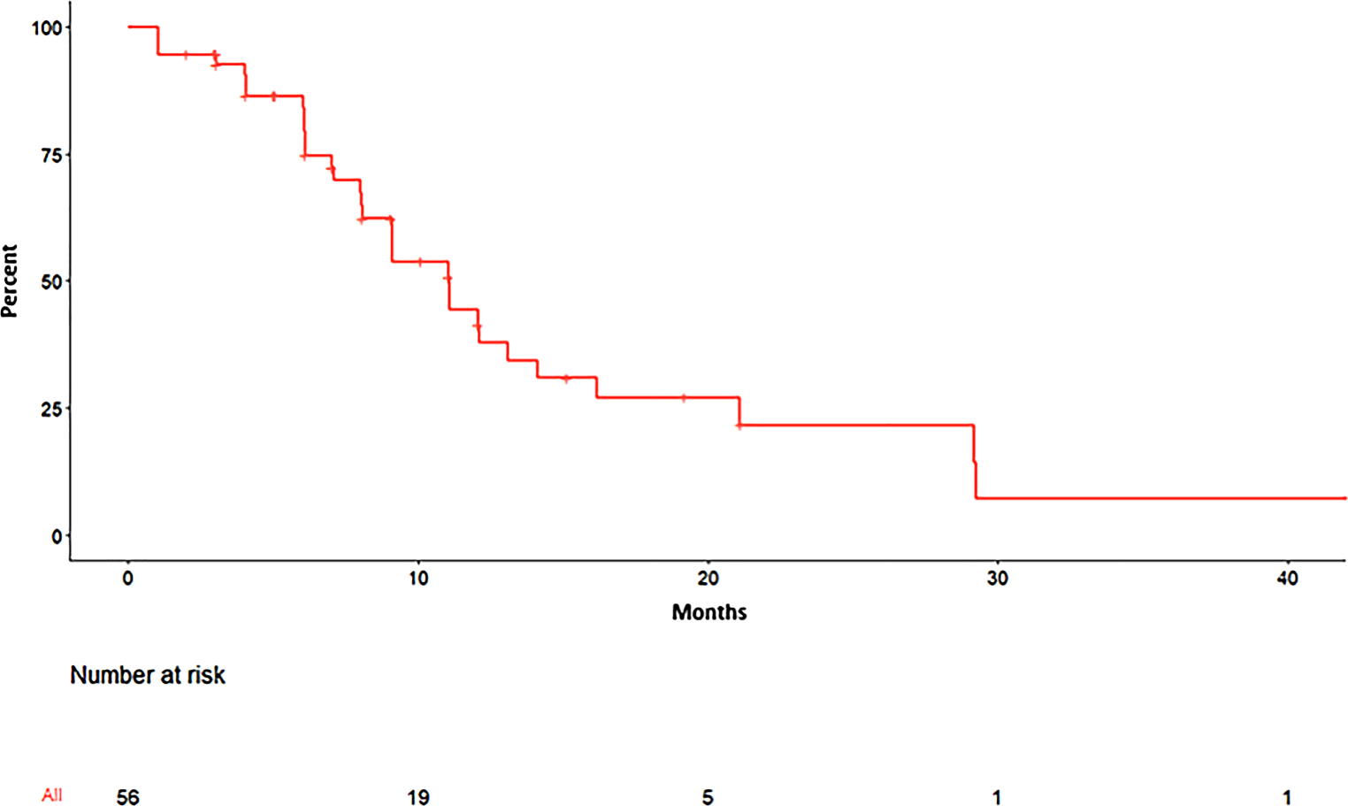
In this study, we assessed the calculated margin using the Van Herk formula in the context of CBCT scans of patients with early glottic cancer. The determined margins were 1.8 mm for the LR direction, 2.2 mm for the AP direction, and 5.3 mm for the SI direction. Moreover, considering instances where movements exceeded these calculated margins, we proposed an adjusted margin strategy of 3 mm for the AP and LR directions and 5 mm for the SI direction. To validate this approach, we employed ViewRay real-time cine MRI, revealing an overlap ratio of 0.87. Importantly, this ratio was notably higher in sessions without swallowing events (averaging 0.92) compared to sessions with swallowing events (averaging 0.77).
Several reports have been published regarding hypofractionated treatment of early glottic cancer [4, 7]. Two prospective studies have been reported, with conflicting results [6, 10]. Sher et al. observed that among 29 patients, dose-limiting toxicities were encountered in two active smokers, while the rest tolerated up to 42.5 Gy for 5 fractions [6]. On the contrary, Kang et al. reported early closure of their prospective dose-escalation trial due to two cases of grade 3 laryngeal inflammation with a SIB dose of 55 Gy and 40.7 Gy over 11 fractions [10]. Discrepancies between these results may be attributed to differences in manipulated target volumes. Sher et al. defined the CTV as the ITV plus 2 mm, while Kang et al. applied an SIB approach, encompassing the total larynx in the RT field. Larger treatments are related to higher doses to normal tissues, which can consequently lead to laryngeal toxicities [8]. Additionally, Sher et al. employed fiducial markers for respiratory tracking with four-dimensional CT to account for movements of vocal cord. These variations potentially contribute to the divergent findings witnessed in these contemporaneous clinical trials. In fact, we are currently trying to conduct a prospective multi-institutional trial for SBRT of early glottic cancer. A critical step in our preparatory phase involved the execution of this study, aimed at assessing the potential margins for the forthcoming trial. Notably, our investigation unveiled significant movement along the SI axis. This finding gains particular relevance given our institution’s adherence to a 3-mm PTV margin in all spatial directions.
Similar works using CBCT to evaluate the motion of vocal cords have been reported previously [19, 20]. Kwa et al. reported that the estimated margin for intrafractional motion using CBCT in 42 patients was 1.6 mm for LR, 4.3 mm for SI, and 2.2 mm for AP [20]. Additionally, the applied margin of 3 mm for AP and LR and 5 mm for SI was adequate, as all patients had at least 94% of the prescribed dose. Perillo et al. evaluated 23 patients with early glottic cancer who received 36 Gy in 3 fractions. Their study demonstrated similar results, i.e., 2.4 mm for LR, 5.1 mm for SI, and 2.2 mm for AP. We also report that the calculated margin was 1.78 mm for the LR, 2.16 for the AP, and 5.33 mm for SI direction, which were comparable to previous studies. Additionally, our study supports the use of margin of 3 mm for the AP and LR directions and 5 mm for the SI direction, which were commonly used margins in previous studies [6, 19, 20].
A notable limitation in using CBCT for evaluating vocal cord motion lies in its lack of real-time imaging capability, preventing an assessment of motion induced by swallowing. While multiple CBCT sessions can help capture potential intrafractional vocal cord motion, the dynamic influence of swallowing remains unaccounted for. On the contrary, cine MRI offers a noninvasive and ionizing radiation-free approach, comparable to gold standard methods like fluoroscopy or endoscopy for swallowing evaluation [15, 17]. Notably, Bradley et al. highlighted larynx motion of up to 3 mm in the SI direction even during rest, a factor that can be overlooked in CBCT analyses [15]. Addressing this limitation, we bolstered our study by corroborating the CBCT-derived margin using findings from participants undergoing real-time cine MRI. Through the combined application of CBCT and real-time cine MRI, our evaluation revealed the acquired margin to be satisfactory. However, as higher discrepancies between ITV and PTV were observed when swallowing, strategies aimed at reducing the impact of swallowing on treatment accuracy should be explored, to ensure the precision and efficacy of RT.
The larynx is known as a mobile organ, motion which is caused by breathing and swallowing. Although the thermoplastic mask is known to reduce possible setup errors and general movement, motion of the glottis cannot be prevented, especially in the SI direction. It is known that swallowing can cause severe vocal cord displacement of more than 2 cm [12, 16, 21]. Some efforts to reduce the effect of swallowing during RT have been reported [6, 11, 12, 19]. One potential approach involves providing patients with instructions to refrain from swallowing during the course of RT. Notably, such instructions have been shown to decrease the frequency of swallowing events compared to situations where no specific guidance was provided [12]. This parallels the findings of a study conducted by Perillo, which similarly incorporated instructions against swallowing during RT, and reported a 5-mm margin requirement for the SI direction [19]. An alternative strategy involves use of a gating system to mitigate potential mistargeting of the vocal cord induced by swallowing [6, 11]. Sher et al. initially employed an internal fiducial-based respiratory tracking system employing three fiducials. Due to challenges in tracking all fiducials consistently, they introduced additional skin fiducials to enhance accuracy [6]. Additionally, the same group proposed a surface-based gating technique, wherein real-time calculations of surface discrepancies are made using cameras [11]. Notably, our study aligns with their findings, revealing significant mistargeting of the vocal cord attributed to swallowing even after applying a 5-mm SI margin. In light of these results, it becomes imperative to consider measures aimed at mitigating the influence of swallowing in future prospective trials.
One of the limitations notably stems from the imaging and deformation capabilities of the ViewRay system. Due to its use of a 0.35‑T MRI for cine purposes, the attained image quality is inherently restricted. The process of deformation within the real-time MRI is also marred by errors attributed to the compromised image quality, potentially contributing to the observed reduction in the overlap ratio. Additionally, the intrinsic constraints of the real-time cine MRI, offering only a single sagittal image per simulation, restrict assessment of the LR direction. An additional limitation is the lack of representation of early glottic cancer patients due to the small sample size. While each methodology used in this study presents its unique limitations, the acquisition and validation of the margin for the vocal cord was successfully achieved through incorporation of two distinct methodologies.
In summary, the determined PTV margins for early glottic cancer were 1.8 mm for the LR, 2.2 mm for the AP, and 5.3 mm for the SI direction. Upon applying a 3-mm AP and 5‑mm SI margin, the observed overlap ratio proved satisfactory in sessions without swallowing events (average of 0.92), in contrast to sessions with swallowing events (average of 0.77). Given the impact of swallowing-induced movement, strategies to mitigate the influence of swallowing should be investigated in future studies.


留言 (0)