
記住我


This single-center study investigates the oncological and safety outcomes in a larger cohort of patients with biopsy-proven endophytic RCC treated with PCA. We demonstrate the use of PCA to treat endophytic RCC is associated with a promising primary efficacy rate of 86%. Notably, none of the patients in this cohort developed metastatic RCC during the almost three-year mean follow-up period. Furthermore, overall rate of major complications was low.
Only a few studies investigating oncological outcomes after PCA of endophytic RCC have been published. This study found Nelson–Aalen cumulative incidence estimates of primary local tumor recurrence, which were 12.8% at 3 years and 25.8% at 5 years, respectively. These findings are consistent with the results reported by Murray et al. [8], who observed comparable outcomes of 10% at 3 years and 25% at 5 years in their RCC sub-cohort (n = 25) . The cohort and technical execution of PCA in our study closely resemble those in the study by Murray et al. However, the tumors selected for our study were defined as endophytic according to the RENAL classification, while the study by Murray et al. defined endophytic tumors as those covered by the renal cortex, making them more central than those included in our study.
In a study by Autrusseau et al. [9], 14 patients with centrally placed RCC were treated with PCA, along with concomitant balloon occlusion of the renal artery. Among the patients, 14% (n = 2) experienced local tumor progression; one treatment was incomplete, and another had local recurrence with a median follow-up of 25 months [9]. Balloon occlusion of the renal artery might have a place as a standard practice of endophytic tumors in the future. However, it is important to note that this study includes a small cohort and has relatively short follow-up.
Lastly, in a study by De Marini et al., outcomes after MRI-guided PCA of RCC (n = 31) reported a primary local recurrence rate of 36% after five years of follow-up. In contrast, De Marini et al. showed a high rate of progression to metastatic disease 16% (n = 5), which is difficult to explain. However, the mean tumor size was larger in the cohort studied by De Marini et al. [10] compared to Murray et al. and this study, which could be one explanation for the higher rate of metastatic progression. Therefore, whether MRI guidance is beneficial for PCA is yet to be determined. In the study by Bhagavatula et al. [17], patients with RCC underwent PCA with either CT (n = 155) or MRI (n = 152) guidance. The study found no differences in key oncological outcomes. MRI-guided PCA of RCC should have a larger role for the treatment of endophytic RCC, given MRI’s excellent soft tissue contrast. However, MRI capacity is limited and this must be weighed against the practicality and lower cost of CT guidance.
From a practical standpoint, it is important to consider the secondary efficacy rate of PCA when evaluating its oncological outcomes. According to Okhunov et al. [18], repeated PCA is a feasible, safe, and less challenging option compared to primary PCA. In this study, repeated PCA was performed in five out of the eight cases of local tumor progression, due to either incomplete ablation or recurrence. Among these, four patients received an additional treatment, which raised the PCA efficacy rate from 86 to 93% and reduced the cumulative incidence estimates of local tumor progression (Fig. 2).
Late local recurrencies after three years were found in both this study and the study by Murray et al. [8], highlighting the importance of long-term follow-up for assessing the true oncological efficacy of PCA in treating endophytic tumors. The results presented exhibit relatively large statistical uncertainties due to the small study size, especially in long-term oncological outcomes. However, this study builds on other data in the literature in supporting the use of PCA for endophytic tumors due to relatively high rates of local control (Fig. 3).
Fig. 3
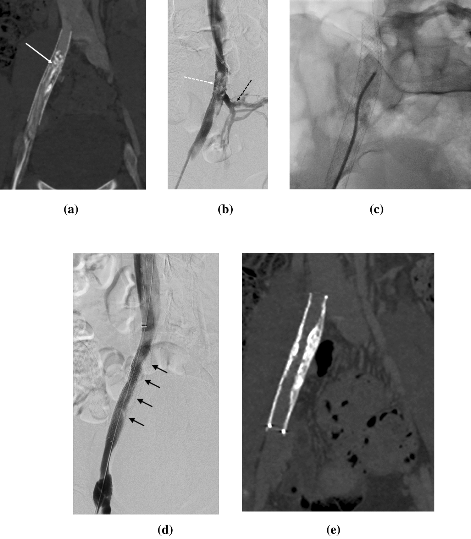
CT images from a 62-year-old male patient with a 38-mm endophytic biopsy-proven clear cell RCC in the left kidney where PCA was performed. A Preprocedural CT images with intravenous contrast, venous phase. The tumor is marked with a white circle. B Intraprocedural CT images with the patient in an oblique position. The image demonstrates the iceball zone. C Venous contrast CT imaging one year after primary PCA shows no residual tumor mass. The ablation zone is marked with a white circle. aComputed tomography (CT). bRenal cell carcinoma (RCC).cPercutaneous cryoablation (PCA)
The treatment of endophytic and central RCC has been associated with an increased risk of complications [12, 19].
In our study, we observed a low major complication rate of 5.4% (n = 3) according to the CDC system. Murray et al. [8] reported a 10% major complication rate in their cohort. De Marini et al. reported no major complications; however, 23% (n = 7) of patients experienced minor (grade ≤ 2) complications, graded by the CDC system.
In a recently published non-randomized prospective study that compared 190 patients with T1a RCC undergoing PCA or PN, both treatments showed similar overall complication rates of 23%, with major complication rates of 3% after PN and 10% after PCA. Notably, in that study, the PCA group treated 16 endophytic tumors, while only one endophytic tumor was treated with PN [4]. This discrepancy might indicate the challenges in treating endophytic RCC.
The complication rates in the literature are heterogeneous. This could be due to a substantial interobserver variability, different treatment modalities, and tumor types [20]. The CDC has been validated for use in the field of urology, but complication classifications related to procedures within interventional radiology, such as the CIRSE system, may better describe the complications after PCA [15, 21].
Ureteral protection during PCA is essential, and this can be done with hydro- or gas dissection, placement of double J-stent, or retrograde pyeloperfusion. At our institution, we do not use pyeloperfusion during PCA. Interestingly, an in vivo experimental study in pigs by Ahmad et al. showed that deep endophytic cryoablation did not affect the renal urothelium [22]. Marion et al. addressed the potential of retrograde pyeloperfusion during PCA. In their RCC cohort, 67 patients with increased risk of ureteral damage underwent pyeloperfusion during PCA and found an acceptable major complication rate given their cohort [23, 24]. With only few complications related to damage of the ureter, it seems that acceptable results can be achieved with the use of hydrodissection and double J-stents as is common practice in our institution.
The present study reflects the treatment of a subset of tumors that are sparsely described in the literature. The data are from a single center and reflect PCA procedural advancements in the period of inclusion. The relatively few tumors in this study do not allow for stratification of variables of interest. Due to the EAU guidelines for the treatment of RCC [3], patients chosen for PCA are often not eligible for extirpative surgery due to comorbidities and/or reduced renal function. Therefore, this study population is not necessarily representative of the average patient with RCC, making mortality and complication rates hard to compare. Furthermore, complications were not divided into early and late complications which could have given better insight in nature of complications after PCA.
留言 (0)