Biomechanical Evaluation of Zygomatic Implant Versus Pterygoid Implant in Atrophic Maxilla: An In vitro Finite Element Study
Background and Purpose
Dental implants are considered to be one of several treatment options that can be used to replace missing teeth. The objective of the study is to examine and compare the biomechanics of zygomatic and pterygoid implants planned on the atrophic maxilla with three different bone types.
Materials and Methods
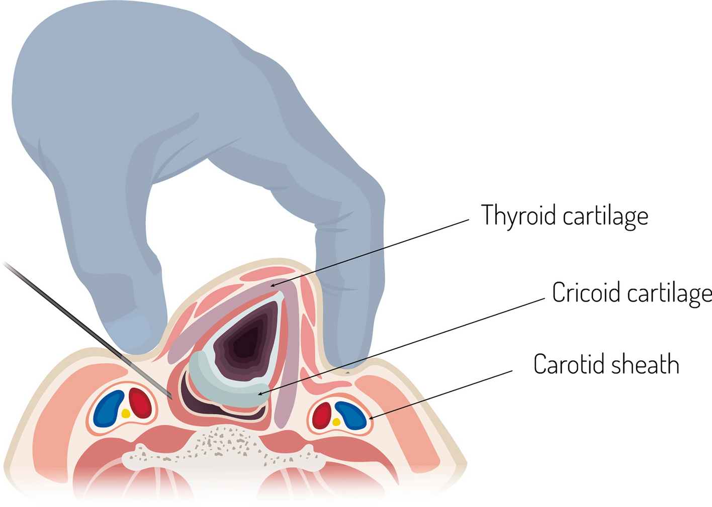
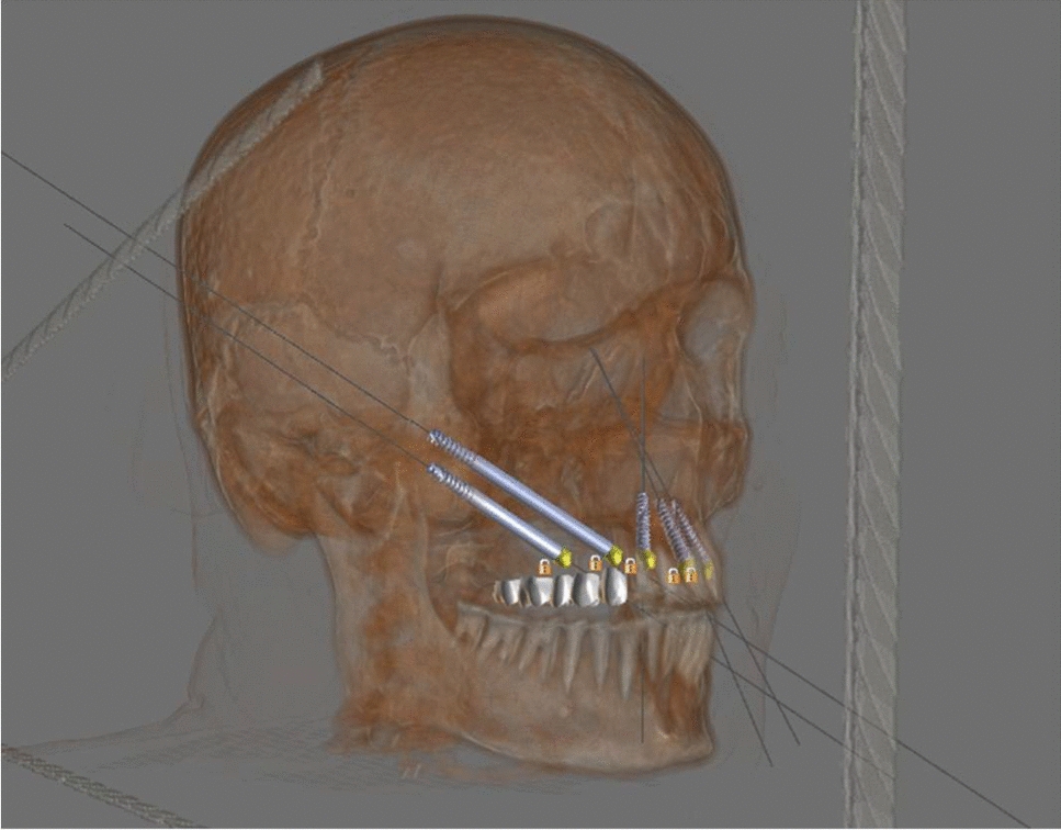
An in vitro finite element study was conducted on a three-dimensional model of zygomatic and pterygoid implants. In a total of 24 implants, two bilateral zygomatic and pterygoid implants with two anterior dental implants were inserted in models. 150 N vertical occlusal and 300 N load on masseter and medial pterygoid were simulated on the modeled prosthesis. The data were processed with ANSYS software. The stress on and deformations of the bones and implants were observed and compared.
Results
When comparing the D4, D3, and D2 bones in subgroup I with zygomatic implants, the D2 bone was subjected to less stress compared to D3 and D4. The smallest displacement (0.125784 mm) was seen in D4 followed by the largest displacement (0.74073 mm) in D2. Similarly, when comparing the D2, D3, and D4 bone in subgroup II with pterygoid implants, the D2 bone in the atrophic maxilla received the least amount of stress from the pterygoid implants compared to D3 and D4. Furthermore, the smallest displacement (0.030934 mm) was seen in D2, and the largest (0.046319 mm) in D4.
Conclusion
Results suggest firstly, that the overall stress was better distributed in D2 bone and secondly, the pterygoid implant showed higher stress concentration than the zygomatic implant.


留言 (0)