
記住我


Epilepsies are a group of heterogenous disorders affecting more than 50 million people worldwide ranking fifth among all neurological disorders in terms of standardized disability-adjusted life-years (DALYs) [1,2▪] with an overall annual cost estimated at $119.27 billion per year, globally [3]. Despite more than 30 antiseizure medications (ASM) one third of patients remain drug resistant, which carries a high risk of premature mortality due to sudden unexpected death (SUDEP), accidents, and suicide [4] with overall reduced life expectancy [5], high morbidity including cardio- and cerebrovascular diseases [6], mental comorbidities [7▪] and cognitive dysfunction, in particular in the young and the elderly population with epilepsies [8].
The aim of presurgical evaluation is to define the chance of complete seizure freedom and the likelihood of inducing new neurological deficits in each individual person with epilepsy (PWE).
Seizure-freedom provides better chances for access to education and work, driving, and personal development [9]. Even if surgery fails to achieve complete seizure-freedom, early epilepsy surgery can limit major developmental delay [10], and reduce mortality and morbidity [11,12▪].
Three randomized controlled trials and multiple open case series with long term follow up demonstrated that this interdisciplinary approach leads to improved seizure control, quality of life and more social participation [10,13–16]. In the very long term, epilepsy surgery though renders not more than 50% of individuals seizure-free depending on aetiology [17].
State-of-the-art epilepsy surgery programs require the interdisciplinary and multiprofessional collaboration of highly specialized neurology, clinical neurophysiology, neuropsychology, neuroradiology and neurosurgery departments. These requirements are met at a restricted number of sites, mainly in high income countries, leading to a substantial treatment gap [18▪▪]. Consensus based expert guidelines have been published but their scope was mainly national, rather than global [19–23].
 Box 1:
 no caption available
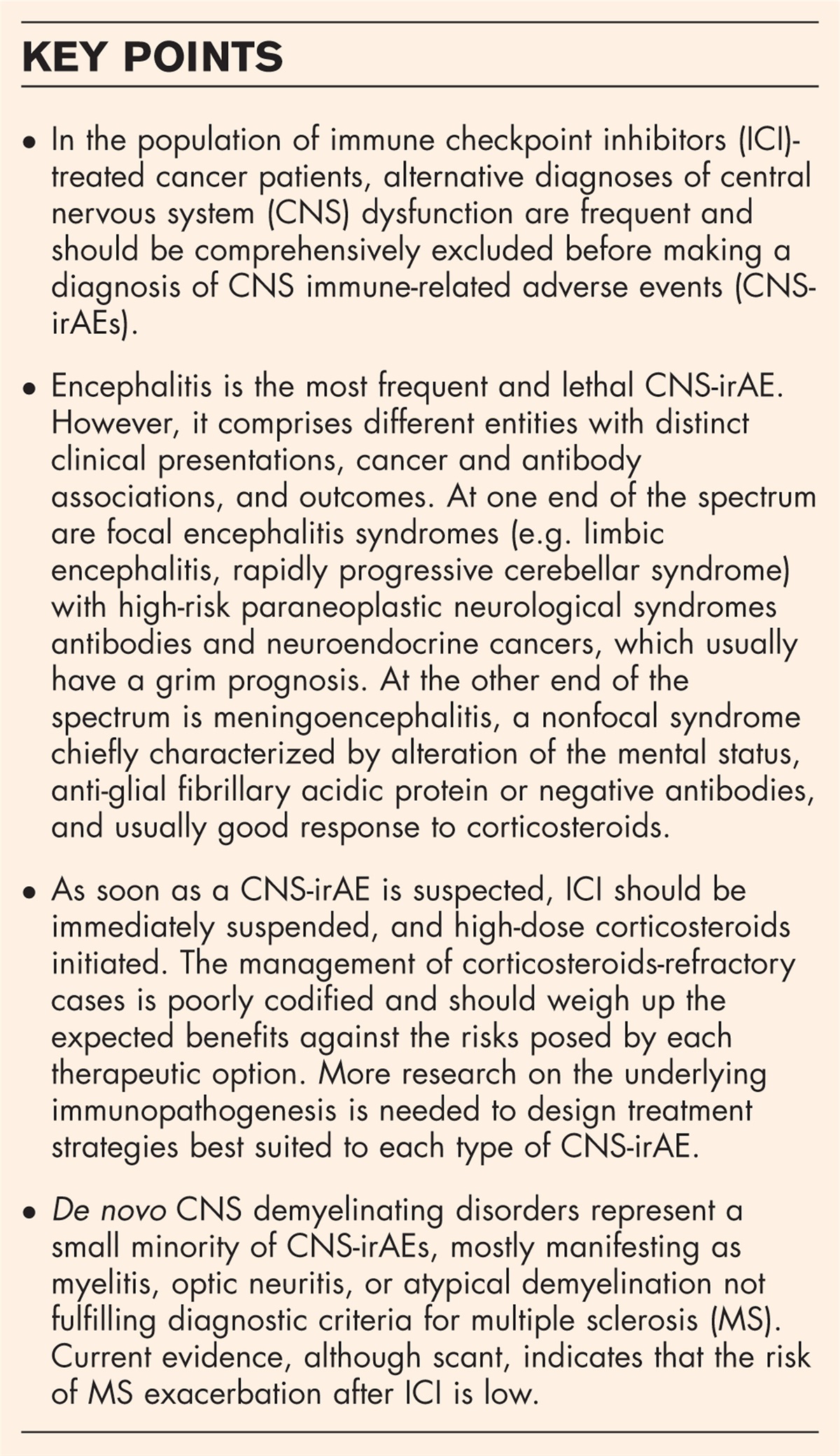
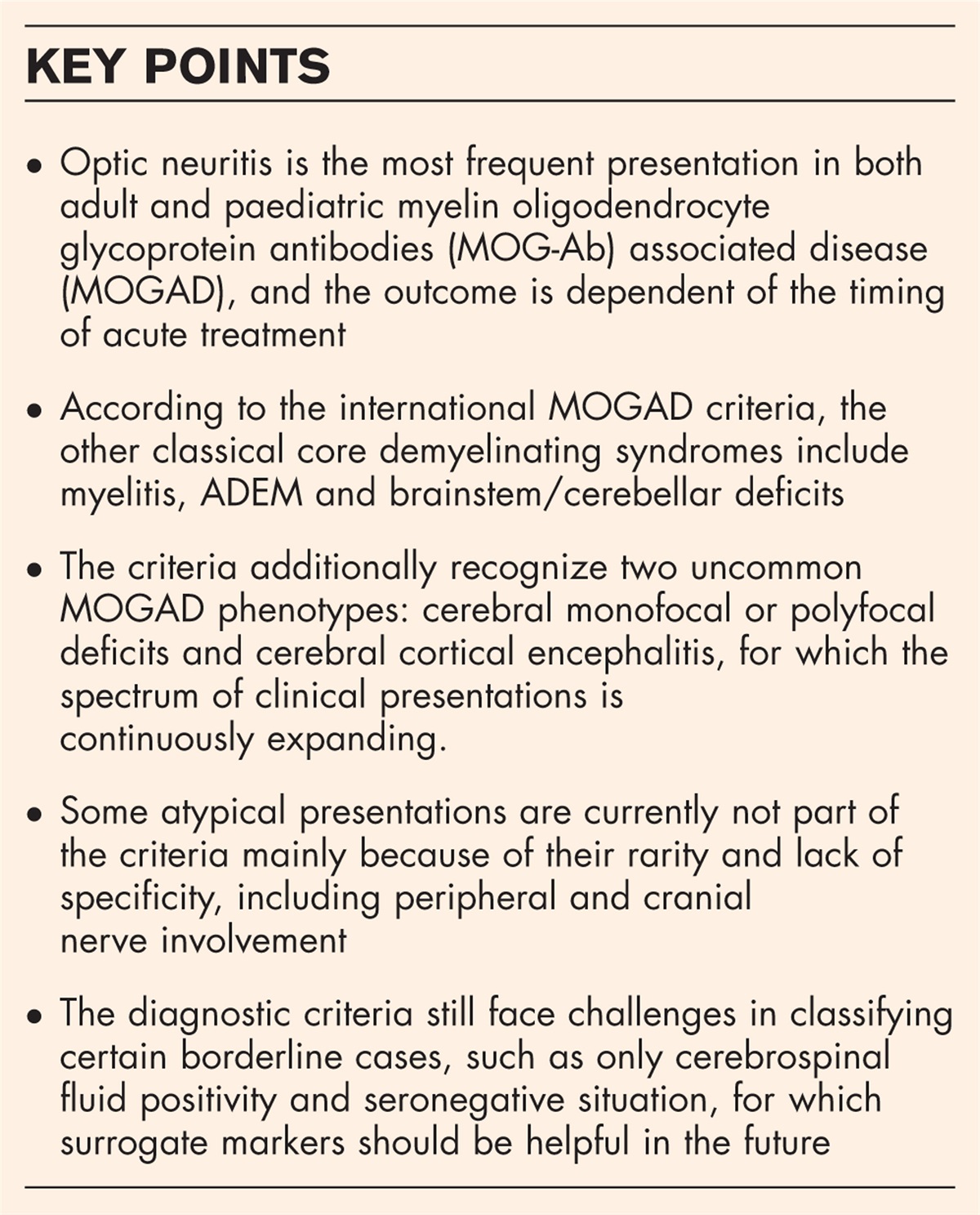
GENERAL FRAMEWORK OF PRESURGICAL EVALUATIONThe current framework of presurgical evaluation of drug resistant epilepsies is interdisciplinary and multiprofessional, but by no means evidence based, and different models may apply to different settings for instance in low to middle income countries (Figs. 1 and 2) [20–22,24].
 FIGURE 1:
 Pathways of noninvasive presurgical evaluation for epilepsy surgery.
 FIGURE 2:
 Pathways of invasive presurgical evaluation for epilepsy surgery.
In short, the presurgical evaluation starts with noninvasive investigations (so-called phase 1a and phase 1b) for all patients (Fig. 1), and if conclusive the patient may directly undergo surgery. If there are inconclusive or ambiguous results additional noninvasive investigations (phase 1b) may help localizing the epileptogenic zone or provide better information on the risk of potential postoperative deficits. The choice of phase 1b investigations depends on the clinical questions, which remain unanswered after the obligatory phase 1a, but mainly depends on the availability at the center and practices vary considerably (Table 1). There is currently no evidence on comparative utility, cost effectiveness, and ideal sequence of investigations. If phase 1b investigations yield contradictory results or the presumed epileptogenic zone is near an eloquent brain area, practices for phase 2 investigations vary similarly, with intracranial EEG with subdural electrodes or depth electrodes (Stereo-EEG; SEEG) including cortical stimulation to identify the seizure onset zone and the functional eloquent areas [25–27]. SEEG became the dominant paradigm and replaced subdural grid and strips almost completely notably without any randomized controlled trial [27,28]. At this stage, the phase 2 investigations are individualized treatment decisions, which represent both: personalized and precision medicine at the highest level (Fig. 2).
Table 1 - Additional Phase 1 investigations and type of studies available to support their use in presurgical evaluation Investigation Retrospective studies available Prospective studies available Systematic reviews with meta-analysis available MRI postprocessing techniques and MR tractography Yes Yes Yes Interictal and ictal high-resolution EEG (HD-EEG) Yes No No Interictal magnetoencephalography (MEG) Yes No No Interictal and ictal electrical source imaging (ESI) Yes Yes Yes Interictal magnetic source imaging (MSI) Yes Yes Yes Interictal EEG-functional MRI (EEG-fMRI) Yes No No Interictal 18F-fluoro-deoxyglucose (FDG) positron emission tomography (PET) Yes Yes Yes Ictal single-photon emission computed tomography (iSPECT) with MRI coregistration (SISCOM) Yes Yes No Functional MRI (fMRI) as well as Wada-test for language and/or memory lateralization and functional transcranial Doppler sonography (fTCD) Yes Yes YesEEG, electroencephalography; MRI, magnetic resonance imaging.
During long-term video-EEG it is often necessary to provoke seizures through withdrawal of ASMs, hyperventilation, sleep deprivation, and photic stimulation, alone or in combination. Although long-term monitoring is generally a safe procedure, it can pose a potential threat to patients’ safety including status epilepticus (SE), postictal psychosis, falls and physical injuries, complications associated with invasive electrodes, and even death [29–31].
There is a huge variety in practice patterns and at times lack of appropriate safety culture in Epilepsy monitoring units (EMUs) [32]. A survey among the 25 E-PILEPSY centers (https://epi-care.eu/therapeutics/8-surgery-e-pilepsy) [32] found that despite the risks, staff continuously observe patients in only 22 (81%) EMUs during, and even less frequently outside of regular working hours (17/25, 63%). Standardized Operating Procedures (SOP) for the treatment of seizure clusters and SE were available in only 16 EMUs (59%), with different safety measures (alarm seizure buttons: 21 (78%); restricted patient's ambulation in 19 (70%); guard rails in 16 (59%), and specially designed bathrooms in 7 (26%) [33].
A more recent development has been the ambulatory video-EEG technology [34], which has the advantage of lower cost and the possibility of more comfortable and stress-free home recordings, but with the disadvantage of lacking standardized periictal testing, and lack of safety measures including SUDEP prevention [35].
Current evidence for video-EEG long-term monitoring in the presurgical evaluation of complex drug-resistant epilepsiesVideo-EEG long-term monitoring is crucially important for documenting clinical events under investigation, drawing electroclinical correlations, and to assess the cognitive impact of seizures. Minimum standards and evidence-based guidelines have been developed [36▪▪]. In a systematic review to evaluate the current evidence on diagnostic accuracy and clinical value of noninvasive long-term video-EEG monitoring in defining the epileptogenic zone in patients who underwent epilepsy surgery for drug-resistant epilepsy [37], 44 studies including 3516 PWE (1271 women) the pooled sensitivity estimate was 0.70 [95% confidence interval (CI): 0.66–0.73] with a specificity estimate of 0.40 (95% CI: 0.33–0.45). In lesional temporal lobe epilepsy (TLE), sensitivity was 0.85 (95% CI = 0.81–0.89) and specificity −0.19 (95% CI = 0.13–0.28). In lesional extratemporal lobe epilepsy (ETLE), sensitivity was 0.47 (95% CI = 0.36–0.58) and specificity 0.35 (95% CI = 0.21–0.53). In lesional TLE, if monitoring was localizing and concordant with resection site, the seizure freedom rate was 247 of 333 (74%), whereas in lesional ETLE only 34 of 56 (61%). Based on these data, another systematic review [38] and a thorough analysis of the literature followed by a Delphi process, the joint task force of the International Federation of Clinical Neurophysiology (IFCN) and International League Against Epilepsy (ILAE) came to the conclusion, that: “long-term video-EEG monitoring must be used in the presurgical evaluation of patients with drug resistant epilepsies (strong recommendation)” [36▪▪].
Implementation of consensus-based practice and adherence to standardized protocols for ictal testing [39] and safety measures [29] is key to optimizing patient safety, clinical decision making that may eventually lead to better patient outcome and care experience [32,33], but remarkably little research has been done on patient's well being in EMUs. Enhancing comfort is a fundamental nursing goal in that for the patient stressful setting with a high level of discomfort and anxiety [40–42]. They feel that they are being observed and sense a lack of privacy. Recently an instrument [Epilepsy Monitoring Unit Comfort Questionnaire (EMUCQ)], based on Kolcaba's comfort theory [43] has been developed and validated in a multicenter study [44–47]. The experience of comfort is multidimensional, subjective and fluctuating. High school students older than 18 years seem especially vulnerable, but individual factors of the current life situation as well as factors related to organization, staff and environment of the EMU, influence comfort care [48▪▪].
Ambulatory video-EEG has been shown to be feasible detecting events, and it may help reduce the diagnostic gap in resource limited settings [49▪,50,51]. For some PWE, home telemetry might be more likely to record habitual seizures, which could be different from those provoked by ASM withdrawal, or different types of attacks, which might not all be observed during the more time-limited recordings in the laboratory environment of the EMUs. Evidence on the diagnostic utility and cost-effectiveness in the framework of presurgical evaluations is currently limited, and there are no comparative studies between the two approaches.
STRUCTURAL NEUROIMAGING Current practices of structural neuroimaging (magnetic resonance imaging)The aim of magnetic resonance imaging (MRI) is to identify an epileptogenic lesion which in turn increases the chances of postoperative seizure freedom significantly [16,17]. A dedicated protocol is mandatory and consensus-based recommendations have been published [26,52]. All published protocols include an anatomical 3D T1 weighted gradient-recalled-echo sequence, axial and coronal T2-weighted sequences, and a fluid-attenuated inversion recovery sequence (FLAIR) with similar slice orientations. For 3D T1 sequences voxel size should not exceed 1 mm. For T2 and FLAIR, slice thickness should not exceed 3 mm.
The E-PILEPSY network performed a survey on the clinical use of imaging, and postprocessing methods in the presurgical evaluation among the 25 centers [53]. Almost all (24/25) centers use standard MRI epilepsy protocols either at 3 Tesla (15/25) or 1.5 Tesla (9/25). Only six centers follow all guideline-recommended MRI sequences with the proposed slice orientation and slice thickness or voxel size. In total, 26 different MRI were reported by 22 centers. MRI postprocessing methods are used in 16 of 25 centers.
The large variation in the presurgical diagnostic workup among epilepsy surgery centers emphasizes the need for high-quality evidence-based recommendations. A first step towards harmonization was the consensus-based recommendation of the Neuroimaging Task Force of the ILAE task for a set of sequences, with three-dimensional acquisitions at its core (harmonized neuroimaging of epilepsy structural sequences; HARNESS) [54]. The recommended sequences are available on most MRI scanners and the protocol is applicable in most clinical setting and countries. The Neuroimaging Task Force also recommends computer-aided postprocessing methods of MRI.
Current evidence for MRI in the presurgical evaluation of complex drug-resistant epilepsiesThe influence of field strength and sequence selection for presurgical evaluation was analyzed by the E-PILEPSY consortium [55▪]. This analysis included 18 original research articles on diagnostic value of higher MRI field strength and 25 on guideline-recommended and additional MRI sequences in detecting an epileptogenic lesion. The lesion detection rate was used as a metric and articles were appraised on their risk of bias and their directness of evidence using QUADAS-2 [56]. In patients with normal MRI, 3T improved lesion detection rate by 18% compared to 1.5 T, and 7 T increased it further by 23% compared to 3 T. In patients with hippocampal sclerosis (HS), a higher field strength than 1.5 T did not lead to a higher detection rate. The use of epilepsy-specific MRI protocols yielded a detection rate of 83% for TLE. Dedicated MRI protocols and evaluation by an experienced epilepsy neuroradiologist increased lesion detection. For HS, 3DT1, T2, and FLAIR each had a lesion detection rate at around 90% [55▪]. The diagnostic yield of 7T for presurgical evaluation, assessed in a single center study on 41 patients, showed that a new epileptogenic lesion was detected in 19% of patients with a lesion-negative 3T MRI. In addition, in over 50% of patients the border zone of the epileptogenic lesion was better delineated [57▪].
FUNCTIONAL NEUROIMAGING (PET AND SPECT) Current evidence for PET and SPECT in the presurgical evaluation of complex drug-resistant epilepsiesOverall, the typically expected finding on interictal 18F-fluoro-deoxyglucose (FDG) positron emission tomography (PET) in TLE is a strictly unilateral or bilateral but asymmetrical decrease of FDG uptake of the temporal lobes. Using FDG-PET in 97 patients with histologically proven HS and long postsurgical follow-up, voxel-based morphometry showed different patterns in the distribution of hypometabolism according to outcome as well as for left and right TLE with prediction of Engel IA outcome [58].
The diagnostic accuracy, the impact on the presurgical decision-making process, and correlation with the postsurgical outcome were assessed in a cohort of 176 patients, with FDG-PET. Overall sensitivity and specificity of interictal FDG-PET were assessed prospectively in 86 operated patients, 72% with a favorable surgical outcome, Engel class I, reporting sensitivity to functional abnormality of 95%, with specificity to the ictal-onset zone of 80% [59].
In a meta-analysis [60] of 44 studies including 2246 DRE patients, who had FDG-PET and MRI, the pooled concordance rate for TLE was 0.79 (95% CI: 0.63–0.92) and for other than TLE for FDG-PET alone was 0.66 (0.59–0.72), rising to 0.93 (0.88–0.97) for combined PET and MRI. No data for combined analysis of FDG-PET and MRI were available for TLE. Concordance rates were higher for children (0.84 [0.75–0.92]) than for adults (0.69 [0.45–0.87]) or mixed series. Notably, lesional and nonlesional (MRI-negative) extratemporal lobe epilepsies (ETLE) were not distinguished in this analysis.
MRI technology has developed substantially and some findings in TLE, in particular HS, may render an FDG-PET scan superfluous especially if there are no “red flags” putting into question that this is indeed the epileptogenic focus.
Decreased FDG uptake in the temporal lobe may extend into the lateral parietal lobe, the insula, and the posterior orbitofrontal cortex as an expression of seizure spread [61,62], compatible with the epileptogenic lesion in the temporal lobe, this is usually continuous, and decreases are typically less marked in those extratemporal locations than in the temporal lobe itself.
FDG-PET is considered most valuable for so-called “MRI negative” patients or in case of nonspecific abnormalities. Normal, extratemporal, or bilateral FDG-PET, on the other hand, was associated with less good outcomes whether MRI was positive, negative, or equivocal [63,64]. The underlying histopathology is normal in a substantial proportion, but may show HS, FCD, or microdysgenesis.
In ETLE, there is a huge variability of study populations – both random differences and systematic differences due to differences in referral patterns to epilepsy surgery, and evaluation procedures between centers. In a large single center study of 194 consecutive surgical candidates (104 ETLE) state-of-the-art MRI was normal, noncontributory, or discordance with EEG indicating the need for FDG-PET [65]. PET was abnormal in 57/104 (55%), which was considered helpful in 44 (42%) by either enabling intracranial EEG (31%) or by excluding surgery (13%). Coregistration with MRI was not available.
A systematic review on the E-PILEPSY consortium following the GRADE methodology is currently under way.
Particularly in the ETLEs, FDG-PET and MRI are also eminently complementary, with several large studies showing that rereading of MRI in the light of PET findings can allow post hoc identification of MRI abnormalities [66]. The location of PET-enabled focal lesions is often in the frontal lobe, especially along the superior frontal sulcus and generally along the major sulci developing early during ontogenesis [67].
Several PET investigations examined the effects of vagus nerve stimulation on CBF or glucose metabolism. Patients who had greater bilateral thalamic activation went on to experience significantly greater seizure reduction during vagus nerve stimulation (VNS) than those who had little or no thalamic activation. A significant difference in metabolic connectivity evaluated by preoperative FDG-PET was noted between VNS-effective and VNS-ineffective groups [68]. Relative changes in glucose metabolism were strongly connected among the areas of brainstem, cingulate gyrus, cerebellum, bilateral insula, and putamen in patients with <50% seizure control after VNS.
Subtraction Ictal single photon emission computed tomography (SPECT) CO-registered to MRI (SISCOM), and statistical comparisons has long been recognized to improve results. Ictal Single Photon Emission Computed Tomography (i-SPECT) assesses the seizure-onset zone using the localized hyperperfusion that occurs early in the seizure. This surrounding hypoperfusion area may be caused by the steal syndrome or may reflect an inhibitory zone [69].
Various novel automated analysis models have been described since (SISCOM), using either more sophisticated statistical ictal SPECT coregistered to MRI (STATISCOM), which improves localization accuracy by statistically accounting for random variation between images, or by adding PET to the equation as in PET interictal subtracted ictal SPECT coregistered with MRI (PISCOM) [70▪▪].
A previous review on this topic in this journal [69] emphasized that by the time the tracer reaches the brain, around 10–60 s after the injection added to the “time-lost” before starting and during the injection, the seizure activity may have dissipated or propagated resulting in iSPECT study showing the propagation pattern instead of onset, especially in seizures with a rapid spread like in ETLE. A more systematic approach to injection time and SISCOM threshold to avoid detection related to seizure propagation showed a recommendation of injection latency below 35 s [71]. STATISCOM showed overall higher agreement rates than SISCOM with localization of the epileptogenic zone. This result was not affected by the injection times, and subsequently provides localizing information for “late” injections where visual reads and SISCOM are inconclusive [72].
ELECTRICAL AND MAGNETIC SOURCE IMAGING Current practices of electromagnetic source localization with HD-EEG and MEGElectromagnetic source imaging (ESI/MSI) is currently not considered as standard in presurgical evaluation [25]. Nevertheless, it has been recognized as a useful tool especially when phase 1a investigations are inconclusive [73–75]. Clinical position paper and on ESI and a Clinical Practice Guideline for recording and analysis of spontaneous cerebral activity are available from the American Clinical Magnetoencephalography Society [76,77]. Several general recommendations on hardware requirements and forward and inverse model selection have been published. Joint consensus guideline on ESI, by the ILAE and the IFCN is currently in production. A survey among the 25 E-PILEPSY centers revealed that only seven apply MSI, and nine ESI. Fourteen centers use combinations of inverse methods and head models [53]. This again reflects the large variation in presurgical workup among expert surgical centers and highlights the need for evidence-based recommendations.
Current evidence for electrical and magnetic source imaging in the presurgical evaluation of complex drug-resistant epilepsiesThe current evidence of ESI/MSI was assessed by the E-PILEPSY consortium through a systematic review on 11 studies, using postsurgical seizure outcome as reference standard [78]. None of these studies were free of bias mostly due to selection of operated patients only, interference of source imaging with surgical decision, and exclusion of indeterminate results. There was no difference in diagnostic accuracy between ESI and MSI. The overall sensitivity was 82% (95% CI: 75–88) and specificity was 53% (37–68) [78]. Another systematic review published in the same year identified with slightly different inclusion criteria 48 studies (25 ESI and 23 MSI studies). The sensitivity of source imaging methods was between 74% and 90% and specificity between 20 and 54% yielding overall accuracy between 50% and 75% [38]. This “sobering 22-year interim report” on ESI and MSI for epilepsy surgery sparked further research and the formation of a clinical guideline production group based on these two systematic reviews [79].
A prospective long-term study which was not included in the systematic reviews analyzed the diagnostic value of ESI and MSI in 13 patients (9 of them seizure free) [80]. ESI and MSI was more accurately localizing, when analyzed alone and not in combination. In addition, the diagnostic accuracy was highest, when spikes were analyzed in the early phase and not in the mid-phase, which is common practice.
In a prospective study on 57 consecutive children with non lesional DRE, the influence of MSI on decision making was assessed [81]. Discussion of the results of the presurgical evaluation was first undertaken while discussion participants were blinded to the MSI results. MSI results were then presented. MSI changed the decision in 25% leading to resective surgery was performed in 26 patients with good surgical outcome in 21 of them.
Another prospective study assessed the cost for integration of ESI into clinical practice and examined concordance of results obtained with three different ESI pipelines [82]. Of the 40 included patients only 22 had enough interictal spikes for ESI. The working time for the physician analyzing ESI was 4.7 h in the first cases and decreased to 2 h with improved experience. The sublobar agreement between all three ESI pipelines was only 20%, with a kappa value of 0.13 highlighting the need for clinical practice guidelines and further standardization [82]. One method to overcome these practical constraints of labor-intensive analyses is the implementation of automatic spike detection and ESI [83,84▪,85▪▪].
In a study of 22 DRE patients, who were postoperatively seizure free, semi-automated ESI was significantly faster by 275 ± 46 min (305 ± 72 min vs. 30 ± 26 min, P < 0.001) compared to a visual source localization without affecting the localization value [86].
Furthermore, in single center retrospective study on 168 patients without an MRI lesion 33 (19.6%) underwent surgical resection and had a follow-up of 2 years or more. The diagnostic yield and predictive value of ESI, PET, MR postprocessing methods and SISCOM was assessed. Seizure freedom rate was 70% with no difference between TLE and ETE. Concordance of PET, ESI, and SISCOM was associated with the highest chance for postoperative seizure control [87▪▪]. This highlights the integration of all noninvasive investigations, rather than looking on the sensitivity of one method alone.
NEUROPSYCHOLOGICAL ASSESSMENT Current practices of neuropsychological assessmentThe aim of neuropsychological testing is to establish a cognitive baseline prior to the surgery, aid in identifying the functional deficit zone and predict potential cognitive changes following surgical treatment. Despite the updates from the ILAE Neuropsychology Task Force on the role of the neuropsychological assessment in the pre and postoperative evaluation of the epilepsy surgery patients, there is a lack of conclusive evidence regarding the recommended set of questionnaires and tests [88].
The E-PILEPSY network performed a survey to evaluate current practices in neuropsychological assessment [89]. All 25 centers routinely conduct neuropsychological assessment both before and after surgery. A wide variety (n = 186) of tests and questionnaires were reported. Large agreement was found on indications [presurgical localization (100%), postoperative monitoring (96%), adverse drugs effects (68%), epileptic dysfunctions (56%)], and the domains to be evaluated [memory (86%), language (82%), attention (64%), executive functions (64%), visuo-spatial skills (46%), intelligence (36%), behavior and mood (14%), and motor function (9%)]. However, there is a lack of published evidence supporting clinical validity of these tests in the context of epilepsy. The survey highlights a need for enhancement in test validity, tools for assessing everyday functioning and accelerated forgetting, national norms and test co-normalization [89].
Patients with complex drug-resistant epilepsy may experience postoperative cognitive decline in specific neuropsychological domains [90–92] such as memory or language. For predicting the likelihood of such postoperative memory declines as well as language impairments, numerous neuropsychological diagnostic methods including functional MRI (fMRI), selective and super-selective WADA-Test, Magnetoencephalography (MEG) and functional transcranial Doppler sonography (fTCD) are utilized in clinical practice. In another survey conducted by E-pilepsy network, the variations were noted in indications, protocols and paradigms for assessing hemispheric language dominance. Evaluating the dominant hemisphere for language functions was predominantly carried out using Functional MRI - fMRI (91%) and Intracarotid Amytal test – IAT (59%) supplemented by lateralizing tests (91%), such as comparisons between verbal and nonverbal memory, as well as inventories assessing handedness.
Among 23 expert centers in Germany, Austria and Switzerland, 16 centers had performed 1421 Wada tests, predominantly the classic bilateral procedure (73%). By the time of the survey, several noninvasive functional imaging techniques were already in use. However, clinicians currently do not want to rely solely on noninvasive functional imaging in all patients [93].
FUNCTIONAL MRI, WADA-TEST AND fTCD FOR LATERALISATION OF LANGUAGE AND MEMORY Current evidence for functional MRI, WADA-Test, MEG and fTCD for memory and language outcome after epilepsy surgeryA systematic review on 28 studies analyzed the predictive value for postoperative changes in memory and language [94]. Overall, 57 index test were evaluated within these two domains. For memory outcomes, meta-analyses were conducted for Wada tests (n = 17) using both memory and language laterality quotients. The best-case scenario yielded a sensitivity of 79 (95% CI 0.67–0.92) and a specificity estimate of 0.65 (95% CI 0.47–0.83). For the worst case, sensitivity estimate was 0.65 (95% CI 0.48–0.82) and specificity 0.46 (95% CI = 0.28–0.65). The overall quality of reporting was rated as very low. Meta-analyses concerning diagnostic accuracy of fMRI, fTCD, Wada-Test, and MEG were not feasible due to small numbers of studies. The study demonstrated large among-study heterogeneity making it difficult to compare different methods and drawing firm conclusions. It has been observed that selective WADA-Tests seem to be most suitable for prediction of memory loss, whereby memory WADA-Test seem to be better for memory loss prediction than the language WADA-Tests [94]. However, it is essential to interpret these findings cautiously, given limited inclusion of studies, and the challenges in comparing with other methods.
SUMMARY AND CONCLUSIONThe development of evidence-based guidelines and the definition of minimal standards for diagnostic procedures and therapies remains pivotal for dissemination and harmonization of best practice presurgical evaluation and epilepsy surgery for patients, and towards payers as a basis for staff allocation or reimbursement.
One aim of the European Reference Network for rare and complex epilepsies (Epi-CARE). https://epi-care.eu/ is to assess current practices and create solid evidence for presurgical evaluation through systematic reviews (Table 2) using the GRADE Methodology [95] setting the basis for future evidence based clinical practice guidelines.
Table 2 - Systematic reviews and meta-analysis of investigations in presurgical evaluation in drug resistant epilepsy E-pilepsy Consortium Investigation Number of studies, number of patients Results Risk of bias assessment Overall Meta-analysis Sensitivity/lesion detection rate 95% CI Specificity 95% CI Vogt et al. 2017 Yes Neuropsychological assessment 186 tests n.a n.a n.a n.a n.a n.a n.a n.a Kobulashvili et al. 2018 Yes Noninvasive LTVEM 94 3541 48 534 0.70 0.60–0.80 0.40 0.27–0.54 QUADAS-2 Schmid et al. 2018 Yes fMRI 29 n.a n.a n.a n.a n.a n.a n.a n.a MEG n.a n.a n.a n.a n.a n.a n.a n.a WADA n.a 17 630 0.79 0.67–0.92a 0.65 0.47–0.83 QUADAS-2 0.65 0.48–0.82b 0.46 0.28–0.65 Mouthaan et al. 2019 Yes HR-ESI 51 n.a 3 127 0. 87 0.77–0.93 0.61 0.45–0.74 QUADAS-2 MSI n.a 8 267 0.79 0.69–0.87 0.46 0.25–0.70 Sharma et al. 2019 No II-ESI 48 1152 19 515 0. 81 0.76–0.85 0.45 0.36–0.54 n.a IC-ESI 19 159 0.89 0.81–0.94 0.46 0.30–0.63 II-MSI 19 440 0.77 0.71–0.82 0.54 0.46–0.61 IC-MSI 4 38 0.73 0.48–0.89 0.20 0.07–0.46 Niu et al. 2021 No 11C-FMZ-PET 44 2246 7 n.a 0.62 0.49–0.73 0.73 0.59–0.84 QUADAS-2 18F-FDG-PET 12 n.a 0.66 0.58–0.73 0.71 0.63–0.78 Rados et al. 2022 Yes MRI field strength 18 n.a 3T vs. 1–1.5T 5 +18% 0.05–0.47 n.a n.a 7T vs. 1.5–3T 7 +23% 0.17–0.30 n.a n.a MRI sequences 25 n.a standard sequences (i.e. 3DT1, T2, or FLAIR) 11 89% 0.82–0.94 n.a n.a11C-FMZ-PET, 11C-flumazenil positron emission tomography; 18F-FDG-PET, 18F-fluoro-2-deoxyglucose-positron emission tomography; CI,
留言 (0)