
記住我


The young Chinese male patient developed facial and lower limb edema at the age of 26, accompanied by loss of appetite, increased foam in the urine, and rapid gain of body weight. No gross hematuria nor dyspnea were evident. The blood routine test at his local hospital was normal. Urine routine: protein 3+, occult blood 2+; 24-hour urine protein (24hUP): 3.84 g. Blood biochemistry: albumin (Alb) 29.9 g/L, creatinine (Cr) 83µmol/L (57–97 µmol/L), total cholesterol (TC) 6.83mmol/L, triglyceride (TG) 6.37mmol/L. No xanthoma was present despite his hyperlipidemia. Nephrotic syndrome was diagnosed, and screening for hepatitis B, antinuclear antibody profile, and anti-phospholipase A2 receptor antibody were all negative. After treatment with albumin supplementation and diuretics, his edema was significantly alleviated, whereas the foam in the urine persisted. Reluctant to renal biopsy, the patient was instructed to follow a low-fat low-cholesterol diet and prescribed irbesartan to alleviate both proteinuria and hypertension, which was accidentally discovered at the age of 23. However, the patient did not strictly follow the treatment and seldom monitored his blood pressure. During irregular follow-up, the patient experienced a relapse of edema and exacerbation of foamy urine. Lab tests showed Alb 31.3/L, Cr 93µmol/L, and 24hUP 7.24 g. Apolipoprotein E (ApoE) level was found to be increased as 5.97 mg/dL (2.7–4.9 mg/dL), whilst Apolipoprotein A1 and Apolipoprotein B levels were within the normal range.
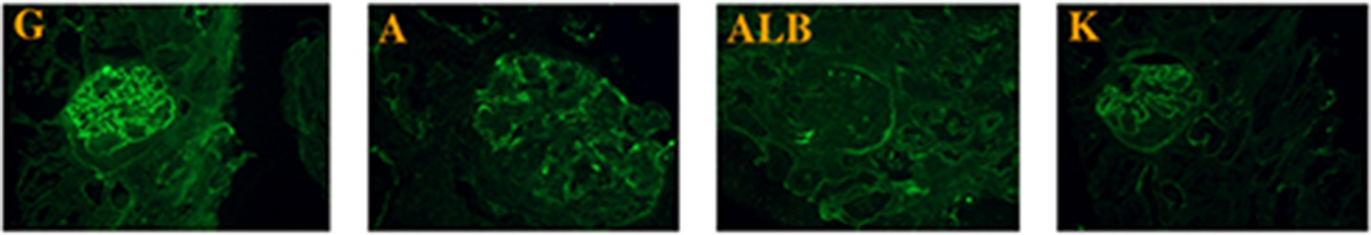
Due to further increase of urine protein, renal biopsy was conducted and showed segmental sclerosis in 3 out of 67 glomeruli in the light microscopy. The patient’s glomeruli were enlarged, and glomerular capillaries were highly dilated filled with vacuolar or laminar lipids. Endothelial cells were swollen with vacuolar degeneration, and glomerular mesangial cells and matrix exhibited moderate to severe hyperplasia with no eosinophilic deposit. Renal tubular epithelial cells showed vacuolar degeneration with some renal tubule lumen expansions, disappearance of bristle margins, and focal atrophy. The interstitium fibrosis was mild to moderate with focal inflammatory cell infiltration. The walls of renal arterioles are slightly thickened (Fig. 1). Immunofluorescence: IgM (+/-), lgG, IgA, C3, C1q, Fib, Alb (-). Electron microscopy: dilated glomerular capillary loop lumen was filled with large amounts of lipid vacuoles, podocyte foot process was segmentally fused, and no electron dense material was found (Fig. 2).
Fig. 1
The patient’s renal pathology in light microscopy. (A) laminar lipoprotein emboli in dilated glomerular capillaries shown in H&E staining. (B) vacuolar degeneration of renal tubular epithelial cells, renal tubule lumen expansions and disappearance of bristle margins shown in PAS staining. H&E staining: Hematoxylin Eosin staining, PAS staining: Periodic Acid-Schiff staining
Fig. 2
The patient’s renal pathology in electron microscopy
(A) 6000X, lipoprotein emboli in the glomerular capillary lumen, swollen endothelial cells, expanded inner loose layer, and segmentally fused foot process. (B) 20000X, lipoprotein emboli in the capillary lumen contain a large number of lipid vacuoles.
Considering the increased ApoE level and classic renal pathology, the diagnosis of LPG is of high likelihood. Thus, the patient was treated with fenofibrate in addition to irbesartan. Subsequent follow up revealed partial alleviation with Alb 45 g/L, Cr 77µmol/L, 24hUP 3.09 g, TC 4.12mmol/L and TG 2.06mmol/L, further support the LPG diagnosis.
LPG is caused by genetic mutations of APOE. But the mutations usually do not include the major APOE mutation E2 based on the amino acid residue at site 158. Accordingly, genotype E3/E3 or E3/E4 are identified in LPG as in our patient. Therefore, whole exon sequncing was applied and revealed a heterozygous mutation of the patient in APOE gene NM_000041.4:c.494G > C(p.Arg165Pro). According to the guidelines of the American College of Medical Genetics and Genomics (ACMG), this variant is classified as a suspected pathogenic variant (PS4_Supporting + PM1 + PM2 + PP4). Owing to the absence of family renal disease history, exon sequencing was also performed on the patient’s parents. The patient’s father has wild-type APOE, while the patient’s mother carries the same heterozygous mutation in NM_000041.4:c.494G > C(p.Arg165Pro), clarifying the inheritance of the pathogenic mutation.
Nevertheless, unlike the patient, his mother exhibited normal ApoE levels and preserved kidney function with no proteinuria and hematuria. According to the Online Mendelian Inheritance in Man database, some individuals with heterozygous LPG-pathogenic APOE mutation may not develop LPG, which is incomplete penetrance and in line with the genotype and clinical manifestation mismatch in the patient’s mother.
留言 (0)