
記住我


In total 142 IgAN registrations/patients from 29 different nephrology departments in Sweden were randomly selected for the validation. After contacting all primary caregivers, medical charts were received from 140 (98.6%) patients from 27 departments. One patient had the same biopsy registered twice in the SRR, and both versions had been randomly selected, hence the final number of patients undergoing review were 139.
Clinical characters in IgAN patientsThe median age at the time of the biopsy in this subset of 139 patients with a histological diagnosis of IgAN was 46 years (range: 18–85). The male:female ratio was 2.1:1. The median creatinine level at the time of the biopsy was 123 µmol/L and the median eGFR level 51 mL/min/1.73m2, with n = 80 (57.6%) of the patients having a kidney function impairment corresponding to CKD stage ≥ 3. Hypertension was seen in n = 97 (69.8%) of the patients. Proteinuria in n = 138 (99.3%) and hematuria in n = 132 (94.7%). In 12.9% (n = 18) there was a history of purpura consistent with IgAV. No patient had a documented family history of IgAN/IgAV. (Table 1)
Table 1 Clinical presentationValidationCategories i and iiOut of the 139 chart validated patients, 107 (77.0%) were categorized as Confirmed IgAN (category i) with an unambiguous diagnosis code in the biopsy chart and clear statement of IgAN in the medical records. Another 25 (18.0%) were classified as Likely IgAN (category ii). The main reason for being categorized as “Likely” and not “Confirmed”, was scarce material in the biopsy specimen and/or failed immunofluorescence staining. All these patients had a clear statement of diagnosis and clinical presentations corresponding with IgAN described in their medical charts, and in four of the 25 patients, the registration represented the patients’ second (or later) biopsy, further strengthening the IgAN diagnosis. In two patients with Likely IgAN (males, 54 and 42 years), the pathologist suggested that the histological finding was consistent with Secondary IgAN. This is a debated entity and commonly considered as having no significant differences in terms of the histological picture [8]. Nevertheless, both patients had a history of gastrointestinal symptoms and/or surgery (diarrhea from bile salt malabsorption and previous gastric bypass surgery respectively).
Categories iii and ivIn four (2.9%) patients, another clinical condition than IgAN was more likely to explain the patients´ symptoms or deteriorating kidney function, even though the histological requirements for IgAN were met. We categorized these as Secondary diagnosis (category iii). More specifically, in two patients (males, aged 54 and 64) the exact cause of the kidney failure was unclear, and even though the criteria for IgAN were met there was no proportional affection of the glomeruli in the biopsy specimen compared to the level of creatinine. The third patient, a man aged 72 with histological changes corresponding to IgAN, but additionally had changes associated with his diabetes mellitus diagnosis which also was his primary kidney diagnosis in the medical chart. Finally, the fourth patient, a woman aged 67 with granulomatosis polyangiitis (GPA) in remission, had undergone a biopsy due to a rise in her antineutrophilic cytoplasmic antibody (ANCA) level (1700 E/mL).
In three out of 139 (2.2%) patients IgAN were not considered to be the correct diagnosis, hence classified as Not IgAN (category iv): (1) man aged 81 with pulmonary fibrosis that developed proteinuria after treatment with a VEGF-inhibitor, which is a side effect previously described in case reports [16, 17], (2) woman aged 63 with a complex subset of clinical systematic symptoms not typical for IgAN (including fever, pericarditis, rashes, weight loss), inconclusive biopsy findings and where the treating physician later refuted the IgAN diagnosis, (3) woman aged 42 with relapsing nephrosis, that received treatment and clinically more resembled Minimal Change Disease or Focal Segmental Glomerulosclerosis [18]. (Table 2; Fig. 1)
Table 2 Results after medical chart reviewFig. 1 Additional clinical features
Earlier or ongoing IgA vasculitis (purpura, joint pain and/or abdominal pain), was seen in 18 of 139 (12.9%) patients (Table 2).
In three patients (males, 26, 32 and 49 years) the histological specimen showed IgAN with thrombotic microangiopathy (TMA). All of them had presented with malignant hypertension and/or severe headache. Dialysis was initiated the same year as the biopsy was performed in two of the cases.
In 16 patients, the validation biopsy did not represent the first biopsy. The main reason for having a second biopsy or more was progressively impaired kidney function and/or increased proteinuria in an already diagnosed IgAN, but also scarce material in first biopsy (1 case, category ii), highly elevated ANCA-levels in the patient with GPA in remission (1 case, category iii) and relapsing nephrosis (1 case, category iv).
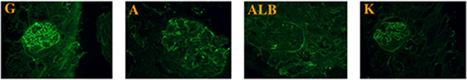
Biopsy findingsThe biopsy charts contained reports on mesangial IgA deposits in 132/132 (100%) patients with IgAN, mesangial hypercellularity or proliferation was mentioned in 102/132 (77.2%), and immunofluorescence staining for C3 was positive in 98/132 (74.2%).
In 27 (19.4%) out of the 139 biopsies the pathologist had noted a MEST score. Out of these 27 cases, 26 (96%) were classified as Confirmed or Likely IgAN (category i and ii)., even in the two cases where the score was M0 E0 S0 T0. In the last case, with the score M1 E0 S0 T0, the IgAN diagnosis was refuted by the treating clinician (Not IgAN, category iv).
C1q positivity was reported in 20.5% (27/132) of all the patients, and in 2/18 (11.1%) of the patients classified as IgAV (Table 3).
Table 3 Histology and biopsy findingsPositive predictive value (PPV) for IgAN diagnosis in the SRROur chart validation found that 132 of 139 patients belonged to category Confirmed or Likely IgAN (i or ii), i.e. had a clinical and histopathological diagnosis of IgAN/IgAV, yielding a PPV for a correct diagnosis of 95% (95% CI 90–98%). When we also included IgAN/IgAV as a Secondary diagnosis (category iii) the PPV increased to 98% (95% CI 93–99%).
ComplicationsComplications from the kidney biopsy were reported in 13/139 patients. The most common complication was flank pain that led to prolonged in-patient care and/or need for extra radiological examination. Macroscopic hematuria was identified in 3 patients but there were no reports of severe complications.
CompletenessRegistering biopsy data in the SRR was introduced in 2015 and they have increased their completeness from 37 to 58% over the first 5 years, based on numbers from the pathology departments on annually evaluated kidney biopsies compared to the total number of biopsy registrations. There has been a slight increase in the total number of kidney biopsies sent for histopathological examination in Sweden during the corresponding years, from 1255 in 2015 to 1482 in 2019, but the percentage of IgAN/IgAV diagnoses has remained stable between 15 and 18% during the whole period (Fig. 2).
Fig. 2
IgAN; IgA Nephropathy, SRR; Swedish Renal Registry
留言 (0)