Serum samples
The serum specimens were collected from 20 fasting septic AKI patients and 20 fasting healthy subjects at Shanghai Pudong New Area Gongli Hospital. These serum specimens were acquired by centrifugation and then preserved at − 80 °C prior to use. Written informed consent from all participants. The clinicopathologic characteristics of sepsis-AKI patients was listed in Table 1.
Table 1 Clinicopathologic features of Sepsis-AKI patientsStrand-specific high-throughput RNA-Seq library construction
Total RNA from serum of AKI patients were extracted with TRIzol reagent (Invitrogen, Carlsbad, CA, USA). Then 3 µg total RNA were used remove ribosomal RNA, and retain RNA classes including noncoding RNAs with VAHTS Total RNA-seq (H/M/R) Library Prep kits from Illumina (Vazyme Biotech Co., Ltd, Nanjing, China). We treated RNA through 40 U RNase R (Epicenter) at 37 °C for three hours, followed by TRIzol purification. We prepared RNA-seq libraries via KAPA Stranded RNA-Seq Library Prep kits (Roche, Basel, Switzerland) and used them for deep sequencing (Illumina HiSeq 4000 at Aksomics, Inc., Shanghai, China).
Cell culture, LPS induction and cell transfection
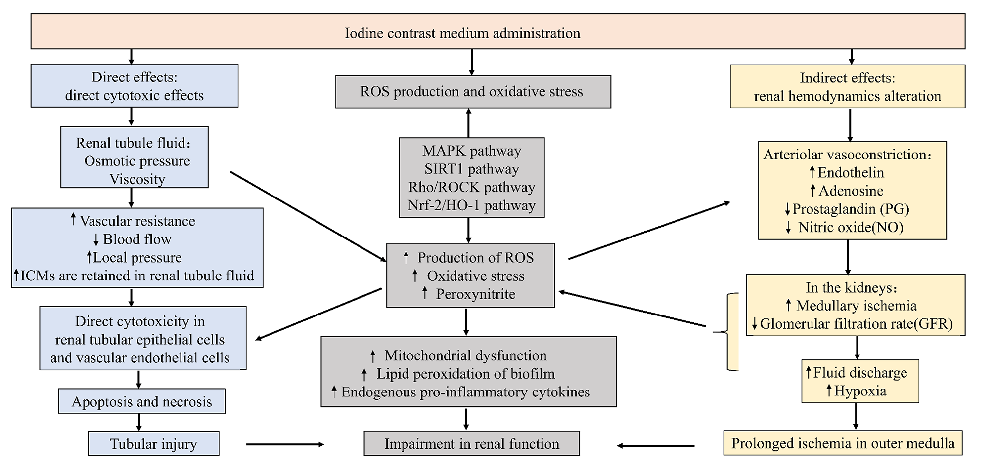
Human renal tubule epithelial cells (Human kidney-2, HK2) were purchased from Procell (Wuhan, China) and cultured in Minimum Essential Medium (MEM; Procell) plus 10% fetal bovine serum (FBS; Procell) and 1% penicillin–streptomycin (Procell) in a 37 °C, 5% CO2 humid incubator. HK2 cells were treated with LPS at different concentrations (0–15 µg/mL) for 24 h. For the establishment of sepsis-related AKI model in vitro, HK2 cells in the following experiments were exposed to 5 µg/mL LPS (Solarbio, Beijing, China) for 12 h, while the control HK2 cells were cultivated with MEM medium simultaneously.
TRPM7 overexpression vector were constructed by insert cDNA of TRPM7 into the pcDNA3.1 vector. miR-22-3p inhibitor and siRNA against circ-Gatad1 (si-circ-Gatad11) were synthesized by Genepharma (Suzhou, China). Cell transfection was then performed using Lipofectamine 2000 (Invitrogen) according to the manufacturer’s instructions.
Cell counting kit‑8 (CCK-8) assay
HK2 cells with various treatments were seeded into 96-well plates (1 × 10⁴ cells/well) and incubated for 48 h. Subsequently, 10 µL CCK-8 reagent (5 mg/mL; Beyotime, Shanghai, China) was pipetted into each well, and cells were maintained at 37 °C for another 2 h. The absorbance of each well at 450 nm measured on a microplate reader (Thermo Fisher Scientific, Waltham, MA, USA) was utilized for determining cell viability.
Enzyme-linked immunosorbent assay (ELISA)
The release of inflammatory cytokines interleukin-1β (IL-1β), IL-6 and tumor necrosis factor α (TNF-α) in culture medium was determined using the corresponding ELISA kits against TNF-α, IL-1β and IL-6 (Beyotime) according to the user’s manual. The colorimetric changes were determined at 450 nm.
RNA isolation and real-time PCR
Total RNA was extracted with TRIzol Reagent (Invitrogen), followed by cDNA synthesis using a TransScript All-in-One First-Strand cDNA Synthesis SuperMix (Transgen Biotech, Beijing, China), was performed. PCR was performed using a Bio-Rad PCR instrument (Bio-Rad, Hercules, CA, U.S.A.) with 2× Taq PCR Master Mix (Solarbio, Beijing, China) following the manufacturer’s instructions. The fold changes were calculated by means of relative quantification (2−△△Ct method).
Dual-luciferase reporter assay
The 3’UTR of HK2 gene and circ-Gatad1 containing the predicated binding sites for miR-22-3p were amplified and inserted into the multiple cloning sites of pMIR-REPORT luciferase reporter vector (Ambion, Austin, U.S.A.). Then, HK2 cells were co-transfected with 0.1 µg of luciferase reporter vectors comprising wild-type or mutant type of 3’UTR of HK2 or circ-Gatad1 and either miR-22-3p mimic or miR-control by Lipofectamine 2000 (Invitrogen, Carlsbad, U.S.A.). Relative luciferase activity was calculated by normalizing the firefly luminescence to the Renilla luminescence using the Dual-Luciferase Reporter Assay System (Promega, Madison, WI, U.S.A.) according to the manufacturer’s instructions at 48 h post-transfection.
Reactive oxygen species (ROS) activity
Intracellular generation of ROS in HK2 cells or wound skin tissue were detected using Dihydroethidium (DHE) assay (Beyotime, China). Fluorescence was measured at 488 nm excitation and 525 nm emission using a fluorescence microplate reader (PerkinElmer, USA).
Statistical analysis
Continuous variables were denoted by means ± SD (standard deviation). We used one-way variance analysis for the comparisons by GraphPad Prism (version 5.0; GraphPad, La Jolla, USA). P ≤ 0.05 indicated statistical significances.


留言 (0)