HF is a clinical syndrome with symptoms and/or signs caused by structural and/or functional cardiac abnormalities and confirmed by elevated natriuretic peptide levels and/or objective evidence of pulmonary or systemic congestion [11]. Therefore, these aspects should be taken into consideration when attempting to confirm a diagnosis of HFpEF.
Clinical suspicion
Diagnosis of HF requires the presence of symptoms with or without signs of HF. Typical symptoms include breathlessness, orthopnea, paroxysmal nocturnal dyspnea, reduced exercise tolerance, fatigue, and ankle swelling; typical signs include peripheral edema, lung rales, elevated jugular venous pressure, or third heart sound [6]. However, since symptoms and signs alone are not sufficiently accurate to confirm a diagnosis of HF, additional diagnostic tools are required to correctly diagnose HFpEF [12, 13].
Dyspnea is the cardinal symptom of HFpEF. However, patients with HFpEF are usually elderly and may have many comorbidities, some of which can mimic HF, such as coronary artery disease, lung disease, obesity, diabetes, atrial fibrillation, and anemia. Therefore, these comorbidities should be ruled out, or, at least, their contribution to symptomatology should be determined [14, 15]. Importantly, it is mandatory to investigate whether dyspnea has a respiratory or a cardiac origin. Table 1 shows key aspects that may be helpful to differentiate between them [16].
Table 1 Dyspnea: pulmonary vs cardiac originAnother key point is that no symptoms or signs by themselves can help us to determine whether a patient has HFpEF or HF with reduced EF. For example, the CHARM program included three clinical trials, two of which enrolled patients with HF with reduced EF and one with patients with HFpEF. Although some symptoms or signs could be more common in one type of HF than the other, they are frequent in both HFpEF and HF with reduced EF (Table 2) [17].
Table 2 Symptoms and signs of heart failure with preserved ejection fraction vs heart failure with reduced ejection fractionElectrocardiogram, chest X-ray, and lung ultrasound
The evaluation of patients with suspected HF should include an electrocardiogram, as a normal electrocardiogram is unusual in patients with HF. In addition, it may be helpful to consider the etiology of HF (arterial hypertension [left ventricular hypertrophy, systolic overload]; ischemic heart disease [ST-T alterations, Q waves]). Electrocardiogram abnormalities, such as atrial fibrillation/flutter, conduction disorders, left ventricular hypertrophy, pathologic Q waves, ST-T segment alterations, and left bundle branch block, are common in patients with HF [6, 18].
A chest X-ray may provide supportive evidence of HF, such as pulmonary congestion or cardiomegaly, although it can also be used to investigate other potential causes of dyspnea, particularly pulmonary diseases [6]. Moreover, the use of lung ultrasound can help in the diagnosis of HF and may have prognostic value, as the number of B-lines is associated with adverse outcomes [19].
Natriuretic peptides
European guidelines recommend determination of natriuretic peptide levels to rule out the diagnosis in patients with symptoms suggestive of HF [6]. However, natriuretic peptide levels are increased not only in HF but also in other clinical conditions (acute setting [acute coronary syndrome, atrial or ventricular arrhythmias, pulmonary embolism, acute kidney disease, sepsis]; chronic setting [increasing age, chronic kidney disease, left ventricular hypertrophy, chronic obstructive pulmonary disease, atrial fibrillation]) [6, 20]. By contrast, natriuretic peptide levels may be disproportionately low in obese patients. In fact, low NT-proBNP levels in overweight and obese patients do not rule out the diagnosis of HFpEF [21]. In this context, European guidelines recommend an upper limit of normal in the non-acute setting of 35 pg/mL for BNP and 125 pg/mL for NT-proBNP, as these values have a very high negative predictive value (from 0.94 to 0.98) and values under these levels make a diagnosis of HF very unlikely [6]. However, it should be noted that, for the same NYHA functional class, natriuretic peptide levels are higher in patients with HF with reduced EF than in patients with HFpEF and that in patients with HFpEF in NYHA functional class I or II, natriuretic peptide levels are not markedly increased [22]. In addition, many conditions that may modify natriuretic peptide levels are also common in patients with HFpEF [6]. Therefore, higher natriuretic peptide levels should be considered to rule out a diagnosis of HFpEF in this population. In fact, recent clinical trials enrolling patients with HFpEF (i.e., EMPEROR-Preserved [NT-proBNP: sinus rhythm: > 300 pg/mL; atrial fibrillation; > 900 pg/mL], DELIVER [NT-proBNP: sinus rhythm: ≥ 300 pg/mL; atrial fibrillation; ≥ 600 pg/mL], and PARAGON-HF [NT-proBNP: sinus rhythm: > 300 pg/mL; atrial fibrillation; > 900 pg/mL]) have defined higher cut-off levels of NT-proBNP as inclusion criteria (Table 3) [4, 5, 23]. As a result, we recommend as cut-off levels for NT-proBNP ≥ 300 pg/mL if sinus rhythm and ≥ 600 pg/mL if atrial fibrillation. In patients with low natriuretic peptide levels in whom HFpEF is suspected, the risk of adverse outcomes is much lower [24]. In this context, the HFA-PEFF score proposes higher levels of natriuretic peptides for a diagnosis of HFpEF (Table 4) [25,26,27,28]. On the other hand, as BNP seems a worse marker than NT-proBNP for the diagnosis of HFpEF, the latter would be better in this clinical setting [29]. Finally, other biomarkers tested in HFpEF include high-sensitivity troponins and novel biomarkers, particularly soluble suppression of tumorigenesis-2, galectin-3 (Gal-3), growth differentiation factor 15 (GDF-15), and carbohydrate antigen 125 (CA125), which have been shown to predict adverse outcomes independently from natriuretic peptide levels, as well as EF [30,31,32]. Biomarkers such as soluble glycoprotein 130 and heat shock protein 27 (hsp27) have also been proposed as biomarkers of chronic HFpEF [33].
Table 3 Cut-off levels for natriuretic peptides in the diagnosis of HFpEF in the 2021 HF ESC guidelines and in the EMPEROR-preserved, DELIVER, and PARAGON HF trialsTable 4 Scoring algorithms for diagnosis of heart failure with preserved ejection fractionEchocardiography
Echocardiography is the key diagnostic tool in HFpEF. It provides relevant information about functional and morphological aspects of the heart [6, 34]. Thus, the echocardiogram enables us to determine the left ventricular and right ventricular ejection fraction, chamber size, and valvular function, as well as the presence of regional wall motion abnormalities, eccentric and concentric left ventricular hypertrophy, pulmonary hypertension, and markers of diastolic function [6, 26].
The echocardiographic workflow should be standardized [34]. The first step in identifying HFpEF is determination of LVEF, which should be measured, rather than estimated, ideally from biplane or three-dimensional images. Left ventricular diameters and volumes should then be recorded, with a special focus on assessing the presence of concentric remodeling or left ventricular hypertrophy and non-dilated left ventricle and left atrial enlargement [26, 34]. Although the presence of concentric left ventricular remodeling or hypertrophy renders a diagnosis of HFpEF more likely, its absence does not necessarily exclude the diagnosis of HFpEF. On the other hand, after excluding valvular heart disease, left atrial enlargement reflects chronically elevated left ventricular filling pressure (with or without atrial fibrillation) [6, 26].
The next stage should involve estimation of left ventricular filling pressure or pulmonary capillary wedge pressure using transthoracic echocardiography. These parameters include early (E) and late diastolic mitral inflow velocity (mitral E/A ratio), septal and lateral mitral annular early diastolic velocity (e′), ratio of early diastolic mitral inflow and annular velocity (E/e′ ratio), maximal left atrial volume index, and tricuspid regurgitation peak velocity, which enables measurement of pulmonary artery systolic pressure [35, 36]. The E/e′ ratio is usually considered the first step when assessing diastolic function. A mean E/e′ index ≥ 15 at rest identifies patients with high mean pulmonary capillary wedge pressure, thus making a diagnosis of HFpEF more likely. However, a value of 9–14 is less sensitive and should be considered a minor criterion. As E/e′ is subject to limitations [37, 38], this parameter should not be considered alone and should be included within a comprehensive echocardiographic approach for the diagnosis of HFpEF. A recent study showed that a multivariable-based approach including different parameters assessed using echocardiography increases accuracy in the diagnosis of HFpEF. In other words, the greater the number of echocardiographic abnormalities, the higher the likelihood of HFpEF [39]. The structural and functional alterations for the diagnosis of HFpEF using echocardiography are summarized in Table 5 [6, 26, 40, 41].
Table 5 Structural and functional alterations for the diagnosis of HFpEF by echocardiographyIt should be noted that, in some cases, access to a rapid echocardiography examination is difficult. Better coordination between healthcare levels is mandatory if we are to improve the diagnostic approach to patients with suspicion of HFpEF [26, 35]. In this context, the development of artificial intelligence–assisted echocardiography of HFpEF has been shown to be an accurate prescreening method capable of automatically generating quantitative metrics that could prove very valuable for clinicians [42].
Additional imaging techniques, such as cardiac magnetic resonance, can prove useful in cases of a doubtful diagnosis of HFpEF or when a particular etiology is suspected. In fact, cardiac magnetic resonance imaging provides relevant measurements for cardiac structure and function, enables tissue characterization, and could facilitate the early diagnosis of HFpEF. The main problem is its availability in daily clinical practice [43, 44].
Scores
In this context, two scoring systems have been proposed to simplify the diagnostic approach to patients with HFpEF, namely H2FPEF and HFA-PEFF. While the H2FPEF score relies mostly on comorbidities, the HFA-PEFF scoring system is based on echocardiographic structural and functional parameters and natriuretic peptide levels (Table 4) [25, 26]. Different studies have analyzed the validity of these scores for the diagnosis of HFpEF in real-world practice. Although most have shown that they are reliable diagnostic tools in HFpEF, with high diagnostic accuracy, and are associated with diastolic dysfunction, lower cardiac output, and exercise intolerance, they are barely used in clinical practice and their results may be discordant in patients affected by unexplained dyspnea, with relevant differences in sensitivity and specificity according to the clinical setting [45,46,47,48,49,50].
Additional diagnostic tools
Although the diagnosis of HFpEF can be reasonably performed in most patients after a clinical history, physical examination, measurement of biological parameters, and echocardiography, additional confirmatory tests may be needed when the diagnosis is not clear. In these cases, further investigation is required.
Diastolic stress test
Diastolic stress tests, mainly exercise echocardiography, can unmask left ventricular diastolic and systolic dysfunction and should be the next step when attempting to confirm a diagnosis of HFpEF. Of note, this is a mainly submaximal exercise stress test, whereas the maximal exercise stress test is generally used to exclude ischemia [6, 26, 51,52,53,54].
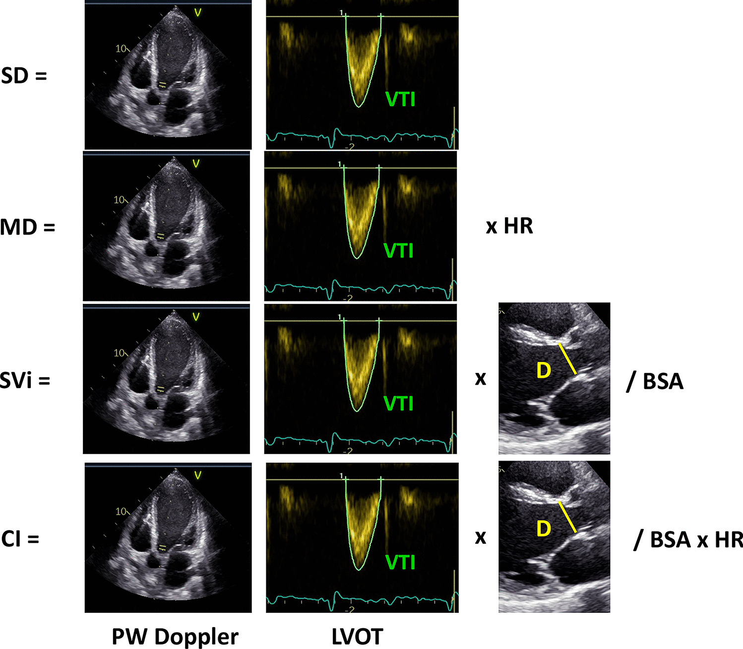
The parameters most commonly analyzed to rule out HFpEF are mitral E/e′ ratio and tricuspid regurgitation peak velocity, which are closely associated with mean pulmonary capillary wedge pressure and pulmonary artery systolic pressure, respectively. These parameters should be measured during standardized exercise. In addition, stroke volume and its change during exercise should also be determined. An average E/e′ ratio at peak stress ≥ 15 and a tricuspid regurgitation velocity > 3.4 m/s increase the probability of a diagnosis of HFpEF. In fact, an average E/e′ ratio at peak stress ≥ 15 adds two points to the HFA–PEFF score and three points when the two conditions are present. Additionally, the absence of increased cardiac output during exercise also favors HFpEF as the etiology of dyspnea [6, 26, 51,52,53,54].
Right heart catheterization
If exercise echocardiography cannot be performed or data are inconclusive, an invasive hemodynamic test is recommended. If the patient has an invasively measured pulmonary capillary wedge pressure of ≥ 15 mmHg or left ventricular end-diastolic pressure ≥ 16 mmHg at rest, then a diagnosis of HFpEF can be considered. If not, an invasive hemodynamic measurement of pulmonary capillary wedge pressure should be taken during exercise. In the case of pulmonary capillary wedge pressure ≥ 25 mmHg, the patient has HFpEF; if not, HFpEF can be ruled out [6, 26, 55]. It is important to note that this diagnostic procedure is subject to risks and may not always be available. In addition, invasive exercise hemodynamics is limited for the diagnosis of HFpEF, for example, it is subject to respiratory pressure swings that may impact on the results in up to 30% of patients [55]. Therefore, it should be limited to specific cases, particularly when therapy depends on the results [6, 26, 56].
Additionally, although further studies are required, the use of specific microRNA panels could add value to current biomarkers in the diagnosis of HFpEF [57].


留言 (0)