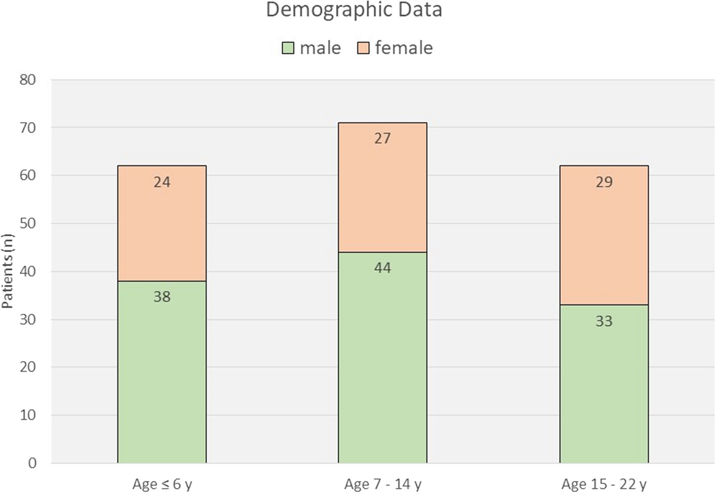
This study describes an analysis of a clinical cohort of 39 patients with FIL. To our knowledge, this is the largest number of cases reported by a single center to date. In these cases, left-sided disease was less common than right. Most patients exhibited facial asymmetry at birth, while a minority showed more noticeable enlargement after one year of age. Female patients outnumbered males, and all cases were nonhereditary. PIK3CA, a widely expressed lipid kinase, regulates signaling pathways associated with cell proliferation, movement, survival, and metabolism [17]. Somatic PIK3CA missense mutations were detected in the subcutaneous tissue of FIL patients [7], and subsequent studies identified PIK3CA hotspot mutations in various types of FIL tissues (skin, mucosa, adipose, bone) [8], confirming postzygotic PIK3CA mutations as the underlying cause of FIL. Genetic testing was performed on lesion tissues of 21 patients in our center, contingent upon patient preferences and the timing of the introduction of NGS. Nineteen specimens were found to harbor somatic PIK3CA mutations. Furthermore, our previous analysis [13], revealed that hotspot mutations were associated with more severe FIL phenotypes, further enriching our understanding of FIL's molecular pathogenesis.
EN is common in our center's patients. We found that approximately 40% of FIL patients had EN on the affected side, distributed across the dermatomes innervated by the three branches of the trigeminal nerve. The highest frequency was observed in the mandibular branch (V3) dermatome. Three patients underwent genetic sequencing using EN specimen, with two patients (patients 4 and 6) found to harbor the PIK3CA p.Cys420Arg mutation. We recommend using EN specimens or subcutaneous adipose tissue for genetic testing in patients who are not prepared for open surgery. The reasons are as follows: (1) both epidermal nevi and subcutaneous adipose tissue are directly involved in the disease process. Changes in these tissues are often visible or palpable, making them readily accessible for sampling, (2) these tissues are expected to carry disease-associated genetic alterations. Sampling from these tissues, therefore, provides us with a good chance of detecting disease-related genetic mutations. (3) DNA extraction from these tissues is relatively straightforward and yields high-quality DNA suitable for NGS.
CT bone reconstruction revealed varying degrees of hyperplasia and deformity in the affected maxillofacial bones, with pronounced abnormalities in the zygomatic bones. Our observations mirrored Padwa's findings [2], where hyperplasia of adipose tissue could be discerned at an early stage, and potential skeletal deformities gradually became apparent over time. Notably, in two patients, the affected side of the maxillofacial bones exhibited a pronounced anterior displacement compared to the contralateral side. This resulted in facial asymmetry, not only in the coronal plane but also in an anterior–posterior orientation within the sagittal plane.
MR can accurately identify and diagnose the adipose nature of FIL, thus excluding potential confounding vascular malformations and obviating the need for histological examination [18]. In our cohort, MR imaging revealed that the masticatory and parotid glands, situated closest to the subcutaneous fat layer, were the soft tissues most prone to involvement, followed by the medial pterygoid, lateral pterygoid, and submandibular glands. The utility of MR in identifying central nervous system lesions is also well established. Even in the absence of neurological features, MR should be employed for further screening for possible HMEG presence when FIL is suspected or diagnosed. The occurrence of HMEG is associated with various cortical developmental disorders, encompassing neuronal differentiation, proliferation, and apoptosis [19]. To date, no simultaneous genetic testing of adipose and brain tissues has been performed in patients with FIL and concomitant HMEG. However, considering the association of both conditions with PIK3CA mutation [20], it is plausible to hypothesize a shared etiological origin. Notably, all previously reported cases of FIL with concurrent HMEG had lesions on the same side. In contrast, we observed a patient with HMEG and FIL located on opposite sides. While this patient has not yet undergone genetic testing, subsequent molecular examination of the bilateral facial lesions may provide insights into the underlying causes of this unique phenotype.
The co-occurrence of diverse tissue abnormalities, ranging from EN distribution to overgrowth of maxillofacial soft and hard tissues, coupled with potential hemimegalencephaly, has stimulated our exploration into the underlying etiology of FIL. Prasad proposed that FIL may result from neurogenic dysregulation and compared the similarities between lipomatosis of nerve (LN) and FIL [21]. LN commonly affects the median nerve in the wrist and palm, resulting in excessive growth within the nerve territory, such as macrodactyly and hemihypertrophy, and is also associated with PIK3CA mutations [22]. In FIL, commonly affected sites such as the masseter muscle, parotid gland, and the most frequent distribution area of EN, are innervated by the maxillary or mandibular branches of the trigeminal nerve. However, histological examination of the nerves in FIL did not reveal fusiform expansion of the nerve with fibroadipose tissue infiltration between nerve bundles seen in LN [22], suggesting that the role of neurogenic abnormalities in FIL remains to be explored. Maclellan hypothesized that FIL may potentially originate from a mutation occurring in the first pharyngeal arch, given its subsequent derivation into the maxilla, mandible, masticatory muscles, and anterior two-thirds of the tongue [7]. Another theory is that FIL may originate from aberrant development and migration of neural crest cells. The affected tissues in FIL predominantly arise from the mesoderm (bone, muscle, fat) and neuroectoderm (brain, epidermal nevi) [12]. The neural crest is responsible for much craniofacial development. After neural tube formation, neural crest cells migrate along established pathways to give rise to structures of mesodermal origin (such as blood vessels, melanocytes, adipose tissue, membranous bone, and connective tissue) throughout the embryo. Additionally, the neural crest appears in a segmental manner in all three primary brain vesicles: rhombencephalon, mesencephalon, and prosencephalon. The prosencephalic neural crest migrates rostrally to the head as a series of vertically oriented cell columns [23]. It has been suggested that adipose tumors are terminal overgrowth arising from dysregulation of multipotent neural crest cells [24]. We speculate that if a small proportion of neural crest cells randomly carry PIK3CA mutations at an early stage, it may lead to overproliferation of maxillofacial tissue from which they differentiate, generating the clinical phenotype of FIL. The fourth potential reason is that cells carrying PIK3CA mutations may exert indirect growth-promoting effects on adjacent or distant cells, which may involve direct cell–cell contact, paracrine signaling molecules, exosomes, changes in extracellular matrix components, and other unknown cell–cell interactions [12]. Further experimental studies are needed to substantiate this hypothesis.
Differential diagnosis of FIL has been extensively discussed in previous literature [3, 25]. Surgery remains the mainstay of treatment for FIL, but there is currently no standardized guideline regarding the timing and approach of surgical intervention. Due to the nonmalignant nature of FIL, complete excision is generally not recommended by most scholars. The optimal timing for operation is still a subject of debate. Slavin advocated for early excision to control excessive growth [1], while Wingerden recommended delayed excision to achieve facial nerve preservation and better symmetry [5]. Padwa advocated for delayed surgery and temporary measures in young patients [2]. We believe that the timing of surgery should be personalized based on the patient's phenotype and severity. For patients with mild facial enlargement, a conservative approach such as liposuction and adjustments to the lips and tongue can be considered. However, for rapidly progressing cases, delaying surgery is impractical as additional facial overgrowth can lead to loss of normal contour of the cheeks, nose, and lips, and may impair functions such as chewing, swallowing, and speech, and even compromise the airway [26]. Furthermore, parents of pediatric patients often request early surgery for aesthetic reasons to prevent their children from experiencing self-esteem issues related to their appearance during their school years. The postoperative recurrence rate in our center was 24.2%, which is lower than the reported data in previous literature, where the highest reported rate reaching 79% [27]. Among all the recurrent cases, 87.5% were children under the age of 14. Padwa suggested that growth hormone may play a role in recurrence, as surgeries performed before the end of adolescence were more likely to experience recurrence [2]. We also agree with this viewpoint, as there have been few complaints of recurrence among adult patients who underwent surgery in our center. Early removal of the lesion may help normalize maxillofacial structures. A previous report found that open bite deformity due to large maxillofacial venous malformations with macroglossia spontaneously improved significantly after sclerotherapy and laser therapy [28]. We found one patient's underbite was naturally corrected after two liposuction procedures, suggesting that relieving the pressure of adipose tissue on the bones may potentially correct occlusal disorders. However, further evidence is required to support this finding.
Assessment of treatment outcomes primarily hinges on clinical manifestations and patient's subjective feedback. This evaluation includes symptom relief and improvements in functional aspects. Additionally, improvements in appearance are assessed by comparing preoperative and postoperative photographs and imaging data to evaluate changes in facial symmetry and contour. With regard to potential complications, monitoring is also in place for any postoperative issues such as infection, pain, and facial nerve damage. Regular follow-ups are conducted to assess whether there is recurrence of the lesion. Currently, there isn't a comprehensive standardized criterion for evaluating the effectiveness of interventions in FIL. Future research may require a larger patient cohort and a more comprehensive evaluation method, such as pain scoring, functional assessment scales, quality of life questionnaires, etc. to accurately assess treatment effects. With the advancement of genetic testing technology, the detection and analysis of genetic mutations may also aid in assessing disease prognosis and treatment response.
Recently, targeted therapies focusing on PIK3CA mutations and downstream signaling pathways have shown promising progress. The PI3K inhibitor alpelisib has been found to reduce the adipose volume of FIL and improve functionality [4]. Additionally, the AKT inhibitor miransertib has been reported to improve the quality of life and seizures in patients with FIL and HMEG [29]. Although the disease cannot be cured, targeted inhibitors may prevent progression or recurrence, but more clinical data are needed to support this perspective.
Limitations of this study include the fact that all data were derived from a single center, which may introduce biases in phenotype assessment and treatment strategies. Additionally, genetic testing was only performed on a subset of newly enrolled patients. Linking genetic mutation analysis with disease classification, diagnosis, and prognosis may provide insights for future treatment of FIL and other overgrowth disorders. Another disease associated with PIK3CA mutations, CLOVES syndrome, has been reported to have an increased risk of developing Wilms tumor [30]. We did not perform systemic examinations, such as hematological and visceral evaluations on our patients. To establish the correlation between mutations and clinical phenotypes and prognosis, a more comprehensive analysis is required, involving a larger number of cases and more advanced methodologies.


留言 (0)