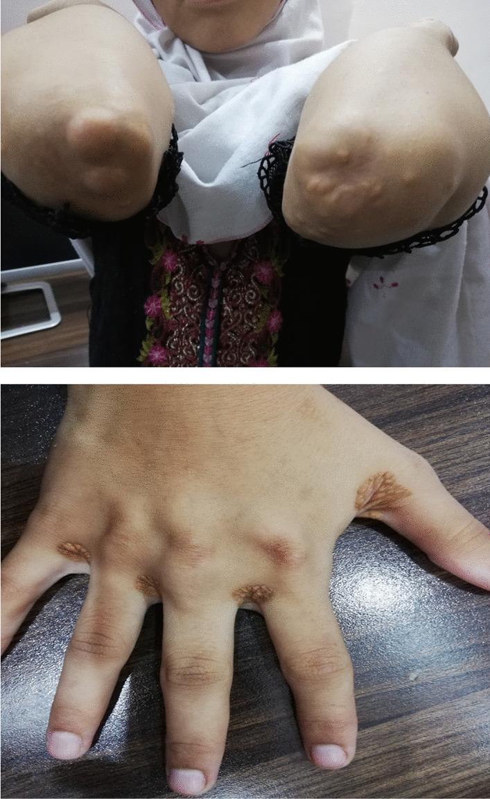
The complications of infectious endocarditis are a significant source of morbidity and mortality [2]. Despite advancements in IE management [3], the number of patients experiencing at least one complication requiring surgical intervention has remained constant over time. In fact, primarily bacteria induce irreparable valvular damage. Secondly, during the acute stage of IE, vegetation particles enter the bloodstream and cause localized vascular inflammation and vascular embolism. While the majority of pulmonary embolisms are caused by particles from the right side of the heart, the bacteria-carrying particles mostly affect individuals with left-sided IE [4]. According to studies [5], there are several characteristics that increase the likelihood of embolization in infective endocarditis, including the vegetation’s size (> 10mm), motility, location on the mitral valve as opposed to the aortic valve, and a CRP level of > 40mg/l. Aerococcus Viridans (AV), as isolated in our patient, is not the most frequently isolated germ in simple or complicated endocarditis; only a few cases have been reported [6]. It is a coccus that is microaerophilic, Gram-positive, catalase-negative, and has a propensity to form tetrads. Its growth properties are comparable to those of enterococci and streptococci. Aerococci are environmental isolates that are regularly found in dust, raw vegetables, animals and their products, as well as human skin, as well as the air of residential buildings (hospitals, schools, industries, and offices) [7]. Meningitis, vertebral osteomyelitis, endocarditis, para-aortic abscess, urinary tract infections [8], bacteremia, and septic arthritis are all brought on by AV. Although the risk factors for AV systemic infections are not fully understood, granulocytopenia, oral mucositis, prolonged hospitalization, prior antibiotic therapy, invasive procedures, and implantation of foreign bodies have all been linked to severe infections with AV [9]. In all reported cases [6], vegetations were identified on the mitral or aortic valves. As in our patient, it often had a long latency period (subacute 73%); in every case recorded, blood cultures and echocardiography were used to make the diagnosis. However, there has only ever been one documented case of splenic embolization. Currently, the imaging methods used to assess embolic endocarditis include ultrasound, MRI, CT, and PET‐CT. The brain is the most common site of embolization, followed by solid organs, including the spleen, kidney, and lung. Less common sites of embolization include peripheral arteries, coronary circulation [10], and the eyes [5]. The true incidence of embolic events is unknown, with estimates ranging from 10 to 50% of IE [11]. Cerebral embolisms are sometimes inaugural and associated with the worst prognosis, with a mortality rate of 21–81% [12]. In several European studies, ischemic cerebrovascular accident constitutes 20–60% of the neurological complications of infective endocarditis, especially in the territory of the middle cerebral artery [13]. A brain abscess is more frequently a feature of acute endocarditis than subacute endocarditis. The abscesses may be single or multiple, and their clinical presentation may be that of a space-occupying lesion, toxic encephalopathy, or meningitis [14]. Involvement of the spinal cord or peripheral nerves is exceptional. Signs and symptoms related to spine involvement can be nonspecific. Patients can simply present with low back pain, a common complaint in the elderly population with degenerative joint disease [15]. Splenic and renal embolisms and certain cerebral embolisms are frequently completely asymptomatic and discovered by systematic paraclinical examinations when looking for remote complications [16]. Appropriate antimicrobial therapy remains the favorite treatment to prevent embolic endocarditis. However, there is no evidence to suggest that prolonged antimicrobial treatment can effectively reduce the incidence of embolic endocarditis. Guidelines recommend that the selection of antibiotics be based on the sensitivity of the newly isolated bacteria, and the duration of antibacterial treatment is usually two to six weeks [5]. According to the 2015 European Society of Cardiology guidelines [17], the primary indications for the use of surgery to prevent embolic endocarditis are the presence of persistent vegetations > 10 mm and one or more previous episodes of embolic endocarditis despite appropriate antibiotic therapy (Additional files 1 and 2).


留言 (0)