
記住我

Liver, biliary tree, and ventral pancreas share a common origin from an endodermal bud of the foregut [1–3]. Only recently, the precise lineage hierarchy and succession of events leading to the segregation of an endoderm progenitor compartment into hepatic, biliary and pancreatic structures have been depicted which demonstrated that a multipotent progenitor subpopulation persists in the pancreato-biliary organ rudiment, contributing cells not only to the pancreas and gall bladder but also to the liver [4]. These results are crucial to understand trajectories taken by the biliary progenitor cells along ongoing organogenesis and in cholangiopathies. On the same line, another significant and recent investigation demonstrated that intrahepatic cholangiocyte regenerate upon resumption of Jagged/Notch signaling, from multipotent progenitors originating from an Fgf-dependent extrahepatic stem cell niche in a zebrafish model of Alagille Syndrome [5]. In normal human livers, epithelial cell adhesion molecule (EpCAM) positive stem/progenitors cells are mostly restricted in two distinct niches, which are the bile ductules or canals of Hering and the mucous glands present inside the wall of large intrahepatic bile ducts [the so-called peribiliary glands (PBGs)] [1,2]. The elegant single-cell transcriptomic atlas compiled by Aizarani et al. confirmed the presence of transcriptomic heterogeneity in the EpCAM+ population in human and, within this population, identified the cell fraction with the highest potential to form liver organoids and to putatively serve as a stem cell compartment: a Mucin 6 high EpCAM+ population residing in intrahepatic large bile ducts furnished of PBGs [6]. The organoid technique was precious instrument to analyze the phenotype and functional heterogeneity of the biliary tree [intrahepatic bile ducts (IHBD) vs. extrahepatic bile ducts (EHBD)]. Taken together, results demonstrated that differences exist between IHBD and EHBD organoids [7–14]. Although, the existence of small and large cholangiocytes is a historical discovery made by Alpini and Coll. almost 30 years ago [15–17], only recently, thanks to a wise experimental approach, it has been clarified that IHBD and EHBD have distinct lineage fate and that EHBD-derived organoids provide a competent model to study bile duct diseases like cystic fibrosis [11]. As a result of an international long lasting collaboration started in 2009 [1,2,18–21], we have participated to the discovery of the presence of cells equipped with a constellation of markers suggestive of the primitive endodermal phenotype in the PBGs [1,2,18–21], the bile duct glands residing within the duct wall or even in the peribiliary tissue outside the wall, of the IHBD and EHBD. These cells are able to be isolated and cultured easily, which demonstrates the persistence of a stable phenotype during in vitro expansion [1,2,18–21]. These cells showed the ability to self-renew in vitro, a fundamental property of stem cells. They differentiated between hepatocyte and biliary and pancreatic islet fates in defined mediums. Notably, transplantation into the liver of immune-deficient (SCID) mice or into the epididymal fat of diabetic SCID mice resulted in differentiation towards the hepatic or pancreatic islet lineage and detectable levels of human c-peptide, respectively [1,2,18–21]. For this reason, we conclude that biliary glands contain cells with a differentiating capacity for mature endodermal fates. We have called them human biliary tree stem/progenitor cells (hBTSCs) [1,2,18–21]. At anatomical level in human, a longitudinal axis exists in the biliary tree: from the hepatopancreatic ampulla, where the most primitive stem cells reside, to intrahepatic bile ducts [2,18,22]. hBTSCs (adult and fetal liver) constitute a physiologic source of hepatocytes and β-cells and a possible target for therapeutic strategies [23,24]. New specific markers for study and isolation are under investigation. hBTSCs from fetal liver have already been used in cell therapy of cirrhotic patients and are among the few identified sources ready for liver regenerative medicine [25▪]. hBTSCs are a tool for in vitro disease modeling in 2D and 3D [26]. Once discovered a new tissue progenitor niche, a sort of cognitive knowledge could be identified, in which the areas of interest range from the role of the stem/progenitor in development/embryogenesis, up to pathophysiology and carcinogenesis. In this review we will follow this theoretical streamline by presenting experimental and translational evidence regarding the study of progenitor populations of the biliary tree: the tissue regeneration and homeostasis, the pathobiology of diseases and malignancies, the application into the regenerative medicine.
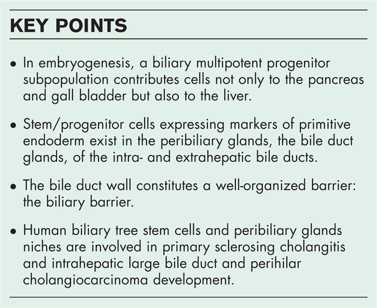
 Box 1:
 no caption available
RADIAL (TRANSVERSAL) AXIS, AND THE BILIARY BARRIERAs largely demonstrated for the intestine, similarly, the bile duct wall constitutes a well-organized barrier capable of counteracting the toxic environment of bile through active energy expenditure [27]. The histo-morphological, molecular and functional features of the biliary barrier have not largely investigated [27]. The biliary barrier and defense systems include mechanisms shared with the gut, for example, immunoglobulins A (IgA), defensins, Toll-like receptor (TLR)-mediated immune activation, etc. Microorganisms must possess tolerance mechanisms in order to resist bile action [26,27]. Experiments in germ-free mdr 2 knockout mice show exacerbated biochemical and histological features of sclerosing cholangitis [28]. These results demonstrate the physiological role of the commensal microbiota [28]. In human, primary sclerosing cholangitis (PSC) is characterized by an altered microbiome of the upper alimentary tract and bile ducts [29]. The biliary tree possesses a discrete vasculature: the peribiliary vascular plexus (PBVP) [30]. PBVP represents the substrate for a recirculation of biliary components in the liver. Thus, maintaining the analogy with gut-liver axis, it could be conceivable to define such a communication as bilio-hepatic axis.
PERIBILIARY GLAND NICHE IN HOMEOSTASIS OF BILE DUCTSThe existence of multiple cell lines within PBGs and robust proliferation of PBG cells following duct injury were confirmed in mice [31]. Moreover, using an ex vivo model based on precision-cut slices it was demonstrated that human biliary progenitor cells within PBGs are able to respond to bile duct epithelial loss with proliferation, differentiation, and maturation to restore epithelia [32]. In the colony formation assay, PBG cells showed significantly higher colony formation capacity than cholangiocytes lining the biliary epithelia [33]. Notably, different epithelial tissues showed a similar functional organization as demonstrated in experimental models of lineage tracing [34–39]. The ablation of lining epithelia cells reactivates the multipotency of basal cells from multiple epithelia both in vivo in mice and in vitro in organoids [34]. On this line, investigations in experimental model of large bile duct/extrahepatic bile duct injury demonstrated clearly the contribution of PBG cells in the regeneration of mature biliary epithelium in experimental models of sclerosing cholangitis, being governed in concert by Wnt and Notch pathways [40]. The differentiation of PBG cells into mature cholangiocytes implied the switch from a glycolytic to an oxidative metabolism [41,42].
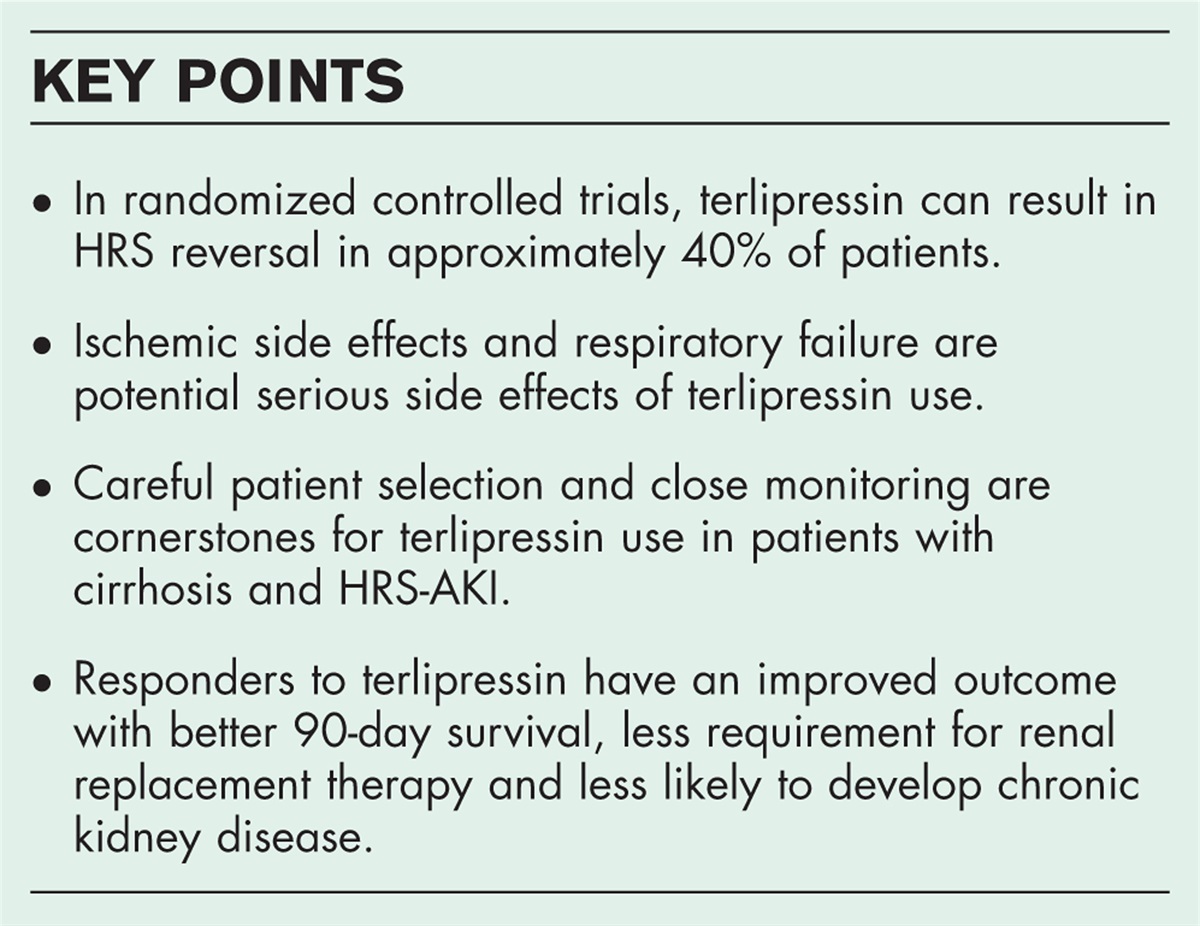
HUMAN BILIARY TREE STEM/PROGENITOR CELLS AND PERIBILIARY GLAND NICHE INVOLVEMENT IN PATHOLOGYFollowing the conceptual framework of the cell/tissue of origin within defined pathological process (benign or malignant), in the last decade in a collaborative international effort the role of the two defined stem/progenitor niche of the liver (hepatic progenitors within the bile ductules/canals of Hering and BTSCs in PBGs) have been investigated to shed light into the patho-biology of cholangiopaties and cholangiocarcinoma [2,43]. Historically PSC and primary biliary cholangitis (PBC) have been coupled within the spectrum of cholangiopathies [2,43]. Both diseases are characterizing by the appearance of ductular reaction (DR) as the results of hepatic progenitor cell activation [43]. Nevertheless, progenitor cell activation differs between PSC and PBC and is characterized by a divergent fate commitment and different signaling pathway predominance [43]. In other words, it appeared that PBC is sustained by a liver parenchymal injury directed vs. interlobular cholangiocytes with a consequential biliary fate committed DR, while on the contrary, PSC-induced ascending cholestasis damages hepatocytes with the appearance of intermediate hepatocytes in the portal space. This evidence highlights the relevance of phenotyping DR to define undetermined cholestasis. In PSC samples, progressive hyperplasia, and mucinous metaplasia of PBGs characterize fibrotic large bile ducts [44]. PBGs undergo massive hyperplasia in relation to the worsening of histology and clinical stage in PSC [44]. Hyperplasia of PBGs is determined by the expansion of BTSCs, which also contributes to biliary fibrosis through epithelial-to-mesenchymal transition and is sustained by the signaling pathway mediated by hedgehog (Hh) ligands [44]. Histologically, dysplastic lesions and CCA in PSC patients mainly arise in diseased large IHBD and EHBD [45]. Pretumoral and tumor lesions are associated with high inflammation and PBG area and further determine the increase of bile duct wall thickness [45]. These observations indicate a ‘field cancerization’ in PSC, in analogy with inflammatory bowel diseases’ carcinogenesis occurring in ulcerative colitis, in which the epithelium is preconditioned to the future development of neoplastic lesions, for example, trough hyper-vascularization [45]. So, it is rational nowadays to insert PBG at the center of PSC pathogenesis [2,44,46,47]. In a condition of genetic predisposition background, gut and biliary microbial dysbiosis and an increased intestinal permeability (‘leaky gut’) characterizing inflammatory bowel disease and/or unhealthy diet/and or metabolic disorders, culminate in bile composition alteration and toxicity, which in turn induce a stem/progenitor niche microenvironment alteration with several consequences such as, stem cell activation, differentiation imbalance, mucinous metaplasia, Hh pathway mediated EMT and fibrosis, antigens exposures, vascular suffering, cell metabolism sufferance, etc. This bile duct wall ongoing chronic injury may predispose to gut-primed lymphocyte homing, which in turn perpetuate the inflammatory injury of the bile duct in a virtually irreversible vicious circle leading the sclerosing duct fibrosis and CCA development (Fig. 1). Clearly, cholangiopaties, both benign and malignant, are lineage-dependent diseases, leading to a crucial question whether we should consider an advancement into the classification and nomenclature for cholangiopathies, as done recently for CCA. Biliary barriers and biliary tree stem cell niche could be a therapeutic target in cholangiopathies or constitute a conceptual framework to conceive innovative early diagnostic tools [48]. The depiction of the molecular anatomy of PBG could lead to innovative biomarkers [49,50]. Large bile duct involvement in PSC evaluated by MRI and radiomic is becoming more and more relevant for risk stratification [51–53,54▪]. The field cancerization appears a relevant translation finding that in our opinion should inform the surgical approach in PSC liver transplantation in order to prevent CCA recurrence [54▪,55]. PBGs are closely associated with several diseases, e.g. IgG4 associated SC, PSC, hepatolithiasis, liver flukes, CCA, etc. [56,57]. PBGs, since the discovery of hBTSCs became a very relevant pathogenic tool to understand post transplantation biliary complications, the nonanastomotic biliary strictures, largely understudied before [32,41,58–61]. Human PBGs contain biliary progenitor cells and are able to respond to bile duct epithelial loss with proliferation, differentiation, and maturation to restore epithelial integrity [41]. PBGs have a key role in the pathophysiology of ischemia-mediated cholangiopathies after liver transplantation, implying alterations in the peribiliary vascular plexus and in nutrient and oxygen inflow [41]. Clinically, these recent data have important implications, indicating the rationale for the use of normothermic machine perfusion (NMP) in orthotopic liver transplantation (OLT) procedures [61]. Favorable bile chemistry during NMP correlates well with better-preserved biliary microvasculature and PBGs, with a preserved capacity for biliary regeneration [61]. PBGs and BTSCs are currently under investigation to depict biliary atresia pathogenesis both in experimental models [62,63], and in children undergoing Kasay procedure [64,65]. Interestingly, even in genetic determined intrahepatic cholestasis, such as ABCB4-related LPAC syndrome associated with an ABCB4 gene variant, large and extrahepatic bile duct injury mimicking a sclerosing cholangitis has been demonstrates [66]. Thus, also the raising knowledge determined by the advent of NGS to define cholestasis and cholangiopathies suggest to re-consider the classification and nomenclature for cholestasis and cholangiopathies based on cell/tissue of origin and patho-biological determinant over the clinical appearance.
 FIGURE 1: Peribiliary glands at the center of PSC pathogenesis: a working hypothesis [2,19,20,40,43,45,46]. In the situation of genetic predisposition background, gut and biliary microbial disbiosis, intestinal permeability, or ‘leaky gut,’ which characterizes inflammatory bowel disease and/or unhealthy diet and metabolic disorders, result in altered and toxic bile composition, which in turn induces an alteration in the stem/progenitor niche microenvironment, including activation of stem cells, differentiation imbalance, mucinous metaplasia, fibrosis mediated by the Hh pathway, exposure to antigens, vascular suffering, impaired cell metabolism, etc. This bile duct wall ongoing chronic injury may predispose to gut-primed lymphocyte homing, which in turn perpetuate the inflammatory injury of the bile duct in a virtually irreversible vicious circle leading the sclerosing duct fibrosis and CCA development. APC, adenomatosus polyposis coli; BMP, bone morphogenetic protein; CK1, Casein kinase 1; EMT, epithelial-mesenchymal transition; FZ, frizzled; GLI, glioma-associated oncogene; GS, gamma secretase; GSK3β, glycogen synthase kinase-3-beta; IBD, inflammatory bowel disease; LRP, low-density lipoprotein receptor-related proteins; NF-κB, nuclear factor-kappa-light-chain-enhancer of activated B cells; NICD, notch intracellular domain; PTCH, protein patched homolog; SHH, sonic hedgehog; SMO, smoothened; TLRs, toll-like receptors; WNT, wingless-related integration site.LINEAGE-BASED CARCINOGENESIS
To further demonstrate the importance of biliary tree progenitor niche, its role in neoplastic development must be considered [67]. It has been suggested that intraductal papillary neoplasm of bile duct (IPNB) derived from PBGs are preneoplastic lesions of mucin-producing CCAs that morphologically resemble PBGs [68]. Interestingly, interleukin (IL)-33-mediated biliary epithelial injury-induced regenerative response accelerates the development of extrahepatic CCA from peribiliary glands, as demonstrated by lineage tracing models [69,70]. The role of PBGs in the genesis of ampullary tumors has been demonstrated in lineage tracing models [71]. Based on the existence of multiple cells of origin, the histology of CCA has been recently reclassified in the ICD-O 3.2 (5th edition of the WHO classification) [72–74]. This classification is based on the anatomical organization of the intrahepatic biliary tree and recapitulates the level or size of the displayed bile duct. Different intrahepatic cholangiocarcinoma (iCCA) histological subtypes can be identified, including large bile duct type [which histologically resembles perihilar cholangiocarcinoma (pCCA) and distal cholangiocarcinoma (dCCA)] and small bile duct type, which comprises also the so called cholangiolocarcinoma [72–75]. The molecular alterations of CCA ranges from SNVs, insertions and deletions, to copy number alterations and chromosome rearrangements/functions [76–82,83▪,84,85]. This leads to a very complex pathway through analysis in routine clinical practice [76–82,83▪,84,85]. There is a wide heterogeneity of the molecular alterations based on anatomical classification [76–82,83▪,84,85]. From the point of view of markers for target therapies, intrahepatic CCA is among the tumors with the greatest actionable mutations, in particular IDH1/2, FGFR2, whereas p/dCCA present a very low percentage of cases with actionable mutations, mainly in ERBB2 [75–82,83▪,84,85]. Notably, etiology of chronic biliary or hepatic inflammation impact strongly on the molecular pathogenesis [79]. In other words, defined etiologies co-segregate with defined molecular alterations, for example, liver flukes and TP53 and/or ARID1A, PSC and KRAS [81,82]. Notably, a recent meta-analysis of n = 1481 iCCA (52.1% surgical specimens), where pair-wise co-occurrences or mutual exclusivities of seven recurred genetic driver mutations have been evaluated, showed an interesting clinico-pathological and molecular clustering [83▪]. Namely, cluster 1 [KRAS (17%), TP53 (22%), and/or SMAD4 (6%)] resulted constituted by large bile duct iCCAs, and cluster 2 [FGFR2-fus (7%), IDH (15%), or BAP1 (12%)] by small bile duct iCCAs. Cluster 1 demonstrated the worst outcome in term of overall survival (OS) or recurrence-free survival (RFS) [81]. Importantly, there is a significant difference when it comes to treatment choice; the small duct type is known to harbor isocitrate dehydrogenase (IDH)-1 and -2 mutations and fibroblast growth factor receptor (FGFR) fusions which are treatable with currently available targeted therapies. On the other hand, the large duct type often presents with KRAS, and SMAD4 mutations, also observed in the perihilar and distal CCA [76–82,83▪,84,85]. These data indicate the utility of subtyping iCCA in terms of clinical outcome and treatment choice [83▪]. The inter-tumor heterogeneity of CCA might be due to the interplay of distinct tissues/cells of origin, the underlying disease, and the associated molecular clustering based on driver mutations which shape the pathobiological features of the different CCA subtypes [84]. Multiparametric and holistic approach to the patho-biological characterization of the CCA patients, is useful in a prognostic-therapeutic sense and also for defining patients at risk for screening/early diagnosis purposes. Studies that depict the correlates of homogenous clusters of subjects based on a tissue-based patho-biological classification could lead to have effective noninvasive tools (liquid biopsy) for precision medicine in CCA.
CONCLUSIONA systemic rational approach based on the concept of keeping the fidelity through the multiple domains (clinical and high throughput-based) depicting overall each patient with her/his disease, as per the network medicine precepts, will bring us into future tools for an effective precision medicine. The existence of a network of stem/progenitor cell niches within the liver and along the entire biliary tree should informs a patho-biological-based translational approach to biliary diseases and CCA [2,86].
AcknowledgementsFinancial support and sponsorship: This review was created with the co-financing of the Next Generation Europe Grant PE 6 FONDAZIONE HEAL ITALIA ‘Health Extended Alliance for Innovative Therapies, Advanced Lab-research and Integrated Approaches of Precision Medicine’; SPOKE 8: ‘Molecular, mutational, radiomic and histo- morphologic profile of HBP (hepati-biliary-Pancreatic) cancers: assigning the right treatment to the right patient at the right time’; Next Generation Europe Grant National Center 3 - Spoke 2. Rna based therapeutics in cancer: from discovery to preclinical studies. Numero Protocollo CN 312184522E9D9A: ‘Nanosystems for the delivery of antitumor compounds of natural origin and miRNA inhibitors for cancer molecular target treatment’; Next Generation Europe Grant Piano Nazionale Complementare Salute - PNC1221852F49EDDD: Clinical use cases for the generation of Digital Twins: Cancer, T1 Diabetes and Multiple Sclerosis.
Financial support and sponsorshipNone.
Conflicts of interestThere are no conflicts of interest.
REFERENCES AND RECOMMENDED READINGPapers of particular interest, published within the annual period of review, have been highlighted as:
▪ of special interest
▪▪ of outstanding interest
REFERENCES 1. Cardinale V, Wang Y, Carpino G, et al. The biliary tree—a reservoir of multipotent stem cells. Nat Rev Gastroenterol Hepatol 2012; 9:231–240. 2. Lanzoni G, Cardinale V, Carpino G. The hepatic, biliary, and pancreatic network of stem/progenitor cell niches in humans: a new reference frame for disease and regeneration. Hepatology 2016; 64:277–286. 3. Tan CEL, Moscoso GJ. The developing human biliary system at the porta hepatis level between 29 days and 8 weeks of gestation: a way to understanding biliary atresia. Part 1. Pathol Int 1994; 44:587–599. 4. Willnow D, Benary U, Margineanu A, et al. Quantitative lineage analysis identifies a hepato-pancreato-biliary progenitor niche. Nature 2021; 597:87–91. 5. Zhao C, Lancman JJ, Yang Y, et al. Intrahepatic cholangiocyte regeneration from an Fgf-dependent extrahepatic progenitor niche in a zebrafish model of Alagille syndrome. Hepatology 2022; 75:567–583. 6. Aizarani N, Saviano A, Sagar, et al. A human liver cell atlas reveals heterogeneity and epithelial progenitors. Nature 2019; 572:199–204. 7. Rimland CA, Tilson SG, Morell CM, et al. Regional differences in human biliary tissues and corresponding in vitro-derived organoids. Hepatology 2021; 73:247–267. 8. Sampaziotis F, Justin AW, Tysoe OC, et al. Reconstruction of the mouse extrahepatic biliary tree using primary human extrahepatic cholangiocyte organoids. Nat Med 2017; 23:954–963. 9. Sampaziotis F, Muraro D, Tysoe OC, et al. Cholangiocyte organoids can repair bile ducts after transplantation in the human liver. Science 2021; 371:839–846. 10. Sampaziotis F, De Brito MC, Geti I, et al. Directed differentiation of human induced pluripotent stem cells into functional cholangiocyte-like cells. Nat Protoc 2017; 12:814–827. 11. Verstegen MMA, Roos FJM, Burka K, et al. Human extrahepatic and intrahepatic cholangiocyte organoids show region-specific differentiation potential and model cystic fibrosis-related bile duct disease. Sci Rep 2020; 10:21900. 12. Tysoe OC, Justin AW, Brevini T, et al. Isolation and propagation of primary human cholangiocyte organoids for the generation of bioengineered biliary tissue. Nat Protoc 2019; 14:1884–1925. 13. Marsee A, Roos FJM, Verstegen MMA, et al. Building consensus on definition and nomenclature of hepatic, pancreatic, and biliary organoids. Cell Stem Cell 2021; 28:816–832. 14. Huch M, Gehart H, Van Boxtel R, et al. Long-term culture of genome-stable bipotent stem cells from adult human liver. Cell 2015; 160:299–312. 15. Alpini G, Roberts S, Kuntz SM, et al. Morphological, molecular, and functional heterogeneity of cholangiocytes from normal rat liver. Gastroenterology 1996; 110:1636–1643. 16. Marzioni MM, Glaser SS, Francis H, et al. Functional heterogeneity of cholangiocytes. Semin Liver Dis 2002; 22:227–240. 17. Zhou T, Meadows V, Kundu D, et al. Mast cells selectively target large cholangiocytes during biliary injury via H2HR-mediated cAMP/pERK1/2 signaling. Hepatol Commun 2022; 6:2715–2731. 18. Carpino G, Cardinale V, Onori P, et al. Biliary tree stem/progenitor cells in glands of extrahepatic and intraheptic bile ducts: an anatomical in situ study yielding evidence of maturational lineages. J Anat 2012; 220:186–199. 19. Cardinale V, Wang Y, Carpino G, et al. Multipotent stem/progenitor cells in human biliary tree give rise to hepatocytes, cholangiocytes, and pancreatic islets. Hepatology 2011; 54:2159–2172. 20. Carpino G, Cardinale V, Gentile R, et al. Evidence for multipotent endodermal stem/progenitor cell populations in human gallbladder. J Hepatol 2014; 60:1194–1202. 21. Wang Y, Lanzoni G, Carpino G, et al. Biliary tree stem cells, precursors to pancreatic committed progenitors: evidence for possible life-long pancreatic organogenesis. Stem Cells 2013; 31:1966–1979. 22. Carpino G, Renzi A, Franchitto A, et al. Stem/progenitor cell niches involved in hepatic and biliary regeneration. Stem Cells Int 2016; 2016:3658013. 23. Semeraro R, Carpino G, Cardinale V, et al. Multipotent stem/progenitor cells in the human foetal biliary tree. J Hepatol 2012; 57:987–994. 24. Riccio M, Carnevale G, Cardinale V, et al. The Fas/Fas ligand apoptosis pathway underlies immunomodulatory properties of human biliary tree stem/progenitor cells. J Hepatol 2014; 61:1097–1105. 25▪. Cardinale V, Lanthier N, Baptista PM, et al. Cell transplantation-based regenerative medicine in liver diseases. Stem Cell Rep 2023; 18:1555–1572. 26. Nicoletti A, Ponziani FR, Nardella E, et al. Biliary tract microbiota: a new kid on the block of liver diseases? Eur Rev Med Pharmacol Sci 2020; 24:2750–2775. 27. Bragazzi MC, Venere R, Vignone A, et al. Role of the gut-liver axis in the pathobiology of cholangiopathies: basic and clinical evidence. Int J Mol Sci 2023; 24:6660. 28. Tabibian JH, O’Hara SP, Trussoni CE, et al. Absence of the intestinal microbiota exacerbates hepatobiliary disease in a murine model of primary sclerosing cholangitis. Hepatology 2016; 63:185–196. 29. Liwinski T, Zenouzi R, John C, et al. Alterations of the bile microbiome in primary sclerosing cholangitis. Gut 2020; 69:665–672. 30. Gaudio’ E, Onori P, Pannarale L, Marinozzi G. Microcirculation of the extrahepatic biliary tree: a scanning electron microscopy study of corrosion casts. J Anat 1993; 182:37–44. 31. Dipaola F, Shivakumar P, Pfister J, et al. Identification of intramural epithelial networks linked to peribiliary glands that express progenitor cell markers and proliferate after injury in mice. Hepatology 2013; 58:1486–1496. 32. de Jong IEM, Matton APM, van Praagh JB, et al. Peribiliary glands are key in regeneration of the human biliary epithelium after severe bile duct injury. Hepatology 2019; 69:1719–1734. 33. Matsui S, Harada K, Miyata N, et al. Characterization of peribiliary gland-constituting cells based on differential expression of trophoblast cell surface protein 2 in biliary tract. Am J Pathol 2018; 188:2059–2073. 34. Centonze A, Lin S, Tika E, et al. Heterotypic cell-cell communication regulates glandular stem cell multipotency. Nature 2020; 584:608–613. 35. Wang Y, Qin J, Wang S, et al. Conversion of human gastric epithelial cells to multipotent endodermal progenitors using defined small molecules. Cell Stem Cell 2016; 19:449–461. 36. Lynch TJ, Anderson PJ, Rotti PG, et al. Submucosal gland myoepithelial cells are reserve stem cells that can regenerate mouse tracheal epithelium. Cell Stem Cell 2018; 22:653–667. e5. 37. Tata A, Kobayashi Y, Chow RD, et al. Myoepithelial cells of submucosal glands can function as reserve stem cells to regenerate airways after injury. Cell Stem Cell 2018; 22:668–683. e6. 38. Van Keymeulen A, Lee MY, Ousset M, et al. Reactivation of multipotency by oncogenic PIK3CA induces breast tumour heterogeneity. Nature 2015; 525:119–123. 39. Koren S, Reavie L, Couto JP, et al. PIK3CA(H1047R) induces multipotency and multilineage mammary tumours. Nature 2015; 525:114–118. 40. Carpino G, Nevi L, Overi D, et al. Peribiliary gland niche participates in biliary tree regeneration in mouse and in human primary sclerosing cholangitis. Hepatology 2020; 71:972–989. 41. de Jong IEM, Overi D, Carpino G, et al. Persistent biliary hypoxia and lack of regeneration are key mechanisms in the pathogenesis of posttransplant nonanastomotic strictures. Hepatology 2022; 75:814–830. 42. Yanes O, Clark J, Wong DM, et al. Metabolic oxidation regulates embryonic stem cell differentiation. Nat Chem Biol 2010; 6:411–417. 43. Carpino G, Cardinale V, Folseraas T, et al. Hepatic stem/progenitor cell activation differs between primary sclerosing and primary biliary cholangitis. Am J Pathol 2018; 188:627–639. 44. Carpino G, Cardinale V, Renzi A, et al. Activation of biliary tree stem cells within peribiliary glands in primary sclerosing cholangitis. J Hepatol 2015; 63:1220–1228. 45. Carpino G, Cardinale V, Folseraas T, et al. Neoplastic transformation of the peribiliary stem cell niche in cholangiocarcinoma arisen in primary sclerosing cholangitis. Hepatology 2019; 69:622–638. 46. Overi D, Carpino G, Cardinale V, et al. Contribution of resident stem cells to liver and biliary tree regeneration in human diseases. Int J Mol Sci 2018; 19: https://doi.org/10.3390/IJMS19102917 47. Graham JJ, Mukherjee S, Yuksel M, et al. Aberrant hepatic trafficking of gut-derived T cells is not specific to primary sclerosing cholangitis. Hepatology 2022; 75:518–530. 48. Lattanzi B, Covotta F, Cardinale V. The potential role of endoscopic ultrasound in primary sclerosing cholangitis. J Hepatol 2023; 79:e199–e200. 49. MacIas RIR, Cardinale V, Kendall TJ, et al. Clinical relevance of biomarkers in cholangiocarcinoma: critical revision and future directions. Gut 2022; 71:1669–1683. 50. Lapitz A, Azkargorta M, Milkiewicz P, et al. Liquid biopsy-based protein biomarkers for risk prediction, early diagnosis, and prognostication of cholangiocarcinoma. J Hepatol 2023; 79:93–108. 51. Trivedi PJ. Risk stratification in primary sclerosing cholangitis: It's time to move on from replicating imperfection and break the glass ceiling. J Hepatol 2019; 71:867–870. 52. Cristoferi L, Porta M, Bernasconi DP, et al. A quantitative MRCP-derived score for medium-term outcome prediction in primary sclerosing cholangitis. Dig Liver Dis 2023; 55:373–380. 53. Goldfinger MH, Ridgway GR, Ferreira C, et al. Quantitative MRCP imaging: accuracy, repeatability, reproducibility, and cohort-derived normative ranges. J Magn Reson Imaging 2020; 52:807–820. 54▪. Chazouilleres O, Beuers U, Bergquist A, et al. EASL Clinical Practice Guidelines on sclerosing cholangitis. J Hepatol 2022; 77:761–806. 55. Morelli MC, Gambato M, Martini S, et al. Nationwide survey of liver transplantation for Primary Sclerosing Cholangitis in Italy. Dig Liver Dis 2023; 55:S1. 56. Matsubara T, Kozaka K, Matsui O, et al. Peribiliary glands: development, dysfunction, related conditions and imaging findings. Abdom Radiol (NY) 2020; 45:416–436. 57. Zaccari P, Cardinale V, Severi C, et al. Common features between neoplastic and preneoplastic lesions of the biliary tract and the pancreas. World J Gastroenterol 2019; 25:4343–4359. 58. Sutton ME, Op den Dries S, Koster MH, et al. Regeneration of human extrahepatic biliary epithelium: the peribiliary glands as progenitor cell compartment. Liver Int 2012; 32:554–559. 59. Op Den Dries S, Westerkamp AC, Karimian N, et al. Injury to peribiliary glands and vascular plexus before liver transplantation predicts formation of nonanastomotic biliary strictures. J Hepatol 2014; 60:1172–1179. 60. de Vries Y, von Meijenfeldt FA, Porte RJ. Posttransplant cholangiopathy: classification, pathogenesis, and preventive strategies. Biochim Biophys Acta Mol Basis Dis 2018; 1864:1507–1515. 61. De Jong IEM, Bodewes SB, Van Leeuwen OB, et al. Restoration of bile duct injury of donor livers during ex situ normothermic machine perfusion. Transplantation 2023; 107:e161–e172. 62. PROM1+ biliary progenitor cells reside in peribiliary glands and proliferate into cholangiocytes in response to cholestatic injury in murine biliary atresia models. Pediatrics. American Academy of Pediatrics n.d. Available at: https://publications.aap.org/pediatrics/article/149/1. Meeting Abstracts February 2022/902/186609/PROM1-Biliary-Progenitor-Cells-Reside-in?autologincheck=redirected (Accessed December 9, 2023). 63. Uemura M, Ozawa A, Nagata T, et al. Sox17 haploinsufficiency results in perinatal biliary atresia and hepatitis in C57BL/6 background mice. Development 2013; 140:639–648. 64. de Jong I, Hunt M, Gupta K, et al. Hyaluronan in the prenatal extrahepatic bile duct increases in response to injury. J Hepatol 2022; 77:S740. 65. Bove KE, Finegold MJ, Harpavat S. Biliary atresia remnants revisited: myogenesis, hepatic duct-like structures, and fate of peribiliary glands. Pediatr Dev Pathol 2023; 26:259–272. 66. Biyoukar M, Corpechot C, El Mouhadi S, et al. A BCB4 variant is associated with hepatobiliary MR abnormalities in people with low-phospholipid-associated cholelithiasis syndrome. JHEP Reports Innov Hepatol 2022; 4: https://doi.org/10.1016/J.JHEPR.2022.100590 67. Carpino G, Cardinale V, Reid L, et al. Cells of origin and cancer stem cells in cholangiocarcinoma. Transl Gastrointest Cancer 2012; 1:33–43. 68. Nakanuma Y, Sato Y. Cystic and papillary neoplasm involving peribiliary glands: a biliary counterpart of branch-type intraductal papillary mucinous [corrected] neoplasm? Hepatology 2012; 55:2040–2041. 69. Nakagawa H, Suzuki N, Hirata Y, et al. Biliary epithelial injury-induced regenerative response by IL-33 promotes cholangiocarcinogenesis from peribiliary glands. Proc Natl Acad Sci USA 2017; 114:E3806–E3815. 70. Tomita H, Hara A. Development of extrahepatic bile ducts and mechanisms of tumorigenesis: lessons from mouse models. Pathol Int 2022; 72:589–605. 71. Hayata Y, Nakagawa H, Kurosaki S, et al. Axin2+ peribiliary glands in the periampullary region generate biliary epithelial stem cells that give rise to ampullary carcinoma. Gastroenterology 2021; 160:2133–2148. e6. 72. Cardinale V. Classifications and misclassification in cholangiocarcinoma. Liver Int 2019; 39:260–262. 73. Beaufrère A, Calderaro J, Paradis V. Combined hepatocellular-cholangiocarcinoma: an update. J Hepatol 2021; 74:1212–1224. 74. Washington MK, Goldberg RM, Chang GJ, et al. Diagnosis of digestive system tumours. Int J Cancer 2021; 148:1040–1050. 75. Cho MT, Gholami S, Gui D, et al. Optimizing the diagnosis and biomarker testing for patients with intrahepatic cholangiocarcinoma: a multidisciplinary approach. Cancers 2022; 14:392. 76. Lamarca A, Barriuso J, McNamara MG, Valle JW. Molecular targeted therapies: ready for ‘prime time’ in biliary tract cancer. J Hepatol 2020; 73:170–185. 77. Valle JW, Kelley RK, Nervi B, et al. Biliary tract cancer. Lancet 2021; 397:428–444. 78. Bekaii-Saab TS, Bridgewater J, Normanno N. Practical considerations in screening for genetic alterations in cholangiocarcinoma. Ann Oncol 2021; 32:1111–1126. 79. Jusakul A, Cutcutache I, Yong CH, et al. Whole-genome and epigenomic landscapes of etiologically distinct subtypes of cholangiocarcinoma. Cancer Discov 2017; 7:1116–1135. 80. Goeppert B, Folseraas T, Roessler S, et al. Genomic characterization of cholangiocarcinoma in primary sclerosing cholangitis reveals therapeutic opportunities. Hepatology 2020; 72:1253–1266. 81. Wang XY, Zhu WW, Wang Z, et al. Driver mutations of intrahepatic cholangiocarcinoma shape clinically relevant genomic clusters with distinct molecular features and therapeutic vulnerabilities. Theranostics 2022; 12:260–276. 82. Banales JM, Marin JJG, Lamarca A, et al. Cholangiocarcinoma 2020: the next horizon in mechanisms and management. Nat Rev Gastroenterol Hepatol 2020; 17:557–588. 83▪. Alvaro D, Gores GJ, Walicki J, et al. European Association for the Study of the Liver. EASL-ILCA Clinical Practice Guidelines on the management of intrahepatic cholangiocarcinoma q 2023; https://doi.org/10.1016/j.jhep.2023.03.010 84. Vicent S, Lieshout R, Saborowski A, et al. Experimental models to unravel the molecular pathogenesis, cell of origin and stem cell properties of cholangiocarcinoma. Liver Int 2019; 39: (Suppl 1): 79–97. 85. Casadio M, Biancaniello F, Overi D, et al. Molecular landscape and therapeutic strategies in cholangiocarcinoma: an integrated translational approach towards precision medicine. Int J Mol Sci 2021; 22:5613. 86. Alvaro D, Gaudio E. Liver capsule: biliary tree stem cell subpopulations. Hepatology 2016; 64:644.
留言 (0)