Experimental groups and surgical procedures
Animal experiments were approved by the Animal Experimentation Committee of Nihon University School of Dentistry (AP21D009). All applicable international, national, and/or institutional guidelines for the care and use of animals were followed.
The sample size for the study was determined using a rigorous power analysis conducted using G*Power software v. 3.1 (University of Dusseldorf, Dusseldorf, Germany). The analysis utilized an alpha level of 0.05 and a statistical power of 95%. Data from a previous study using the same animal model [26] were collected to determine the appropriate sample size. This previous study compared the percentages of newly formed tissues using two different barriers and demonstrated a statistically significant difference. The results indicated percentages of 40 ± 4.5% and 31.5 ± 4.7% for the respective barriers. Minimizing the use of animals was prioritized to a significant extent from an animal welfare perspective. Forty surgical sites were included in the study, involving 9-week-old male Fisher rats weighing 250–300 g.
The animals were acclimatized for 1 week and housed in pairs in standard cages in a controlled environment in terms of temperature, humidity, light/dark cycle, and diet. Surgery was performed under general anesthesia, initially using 4% isoflurane inhalation for 2 min, followed by an intraperitoneal injection of a mixture of dexmedetomidine hydrochloride (0.15 mg/kg), midazolam (2.0 mg/kg), and butorphanol tartrate (2.5 mg/kg). Local anesthesia was achieved with a 0.5 ml solution of 2% lidocaine with 1:80,000 epinephrine dilution to control pain and bleeding.
All the surgical procedures were performed by an experienced surgeon (N.W.). The surgical procedure involved shaving and disinfecting the area between the eyes and the posterior end of the skull with 70% ethanol. A 6.0-cm midline incision was made, and a mucoperiosteal flap was elevated to expose the cranial vertex. Bilateral circular grooves, 5 mm in diameter, were created at the center of each parietal bone using a trephine bur. Within the experimental site, five small holes (diameter: 0.5 mm) were drilled inside the circular grooves without penetrating the inner dura of the cranial bone to induce bleeding from the marrow space, thereby allowing the infusion of the bone graft materials with blood. Plastic cylinders measuring 5.0 and 3 mm in diameter and height, respectively were tightly fixed into the circular grooves on the denuded bone.
Surgical sites were randomly categorized into four distinct groups based on the type of bone graft and mechanical barriers employed. To establish a robust basis for comparison with PLACL, a widely acknowledged natural collagen membrane (COL, Bio-Gide®, Geistlich-Pharma, Wolhusen, Switzerland) was selected as the control membrane owing to its well-established reputation as the preferred absorbable membrane in clinical practice for over two decades. Considering the pivotal role of bone grafts in providing a structural scaffold for clot development, maturation, and remodeling, which is essential for supporting bone formation in osseous defects, our study incorporated two types of bone graft materials: carbonate apatite (CO3AP, Cytrans®, GC, Tokyo, Japan) and deproteinized bovine bone mineral (DBBM, Bio-Oss®, Geistlich-Pharma, Wolhusen, Switzerland), both utilized in conjunction with membranes. The four treatment combinations are as follows: (1) CO3AP + PLACL; (2) CO3AP + COL; (3) DBBM + PLACL; and (4) DBBM + COL.
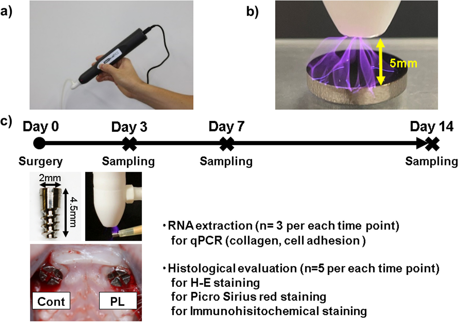
Each plastic cylinder was filled with the assigned bone graft and aligned with the circular grooves. The cylinders were then covered with the designated mechanical barriers (Fig. 1B). Two surgical sites were identified in the calvaria of each animal (Fig. 1C). Finally, the mucoperiosteal flaps were repositioned using resorbable interrupted sutures (VSORB 4-0, Washiesu Medical, Tokyo, Japan). The day of surgery was designated as Day 0.
Outcome assessmentsMicro-CT analysis
Mineral volume at the surgical sites was assessed using an in vivo micro-CT system (R_mCT2 system; Rigaku, Tokyo, Japan) without euthanizing the rats. The rats were anesthetized with an oxygen–isoflurane mixture administered via a facemask and positioned on an imaging stage. The exposure parameter was set to 90 kV. The region of interest for micro-CT assessment was the circular grooves (diameter: 5.0 mm, height: 3.0 mm) on the calvaria. Images were reconstructed using the i-View software (i-View Image Center, Tokyo, Japan) on a personal computer. The bone volume within the plastic caps in the three-dimensional voxel images was analyzed using the same software. The enhanced volume was calculated by subtracting the volume on Day 0 from that at each follow-up. Micro-CT assessments were performed by a single, blinded examiner (T.W.) under the supervision of an experienced examiner (Y.A.).
Histomorphometric analysis
After 24 weeks postoperatively, all the rats were euthanized by administering excess CO2 gas after the last micro-CT scan. The calvarial bone defects were resected following skin dissection. Bone segments containing the cylinders were surgically removed and fixed in 10% neutral-buffered formalin. After fixation, the bone specimens were decalcified in 5% formic acid for 14 days and embedded in 2-hydroxyethyl methacrylate using routine methods. Each embedded bone specimen was cut into 5-μm sections (HistoCore AUTOCUT; Leica, Wetzlar German) and air-dried at 50 ℃. The samples were stained with hematoxylin and eosin (HE).
Histomorphometric analyses were performed using histological sections obtained from within the plastic cylinders under a light microscope and an image analyzer computer system using FIJI (ImageJ 1.50b or 1.52i; National Institutes of Health, Bethesda, MD, USA). We assessed osteogenesis (bone formation) at the experimental GBR sites.
Quantitative measurements were performed on each HE-stained histological section. These measurements included the percentage of newly generated bone area; the percentage of remaining bone substitute particles; and the heights of the total, tissue, and non-calcified tissues (Fig. 1D). Each height measurement was calculated as the average of three different locations: the horizontal center of the plastic cylinder and both parts located 1 mm away from the center. The measurements for histomorphometric assessment were performed by an experienced and blinded examiner (S.W.).
Statistical analysis
The means and standard deviations (SD) of each variable were calculated and analyzed using EZR (Saitama Medical Center, Jichi Medical University, Saitama, Japan), a graphical user interface for R 2.13.0 (R Foundation for Statistical Computing, Vienna, Austria). To assess the statistical significance of the bone volumes obtained from the micro-CT images and the percentages of residual bone substitutes in HE stain, a Kruskal–Wallis test with a Steel post-hoc test was performed. Statistical significance was set at p < 0.05.


留言 (0)