
記住我


Maxillary transverse deficiency can lead to several orthodontic problems, including dental crowding, unilateral or bilateral posterior crossbite, functional shift, mandible deviation, and even nasal airway obstruction.[1-3] The prevalence of maxillary transverse deficiency is approximately 23.3% in the primary dentition and approximately 30% in the permanent dentition.[4]
Rapid maxillary expansion (RME) has long been used to correct maxillary transverse deficiency. The maxilla in the permanent dentition can be expanded using three methods: Conventional tooth-borne, micro-implant-assisted, and surgically assisted RME. Although conventional RME does not require surgery, it uses the teeth for anchorage, involves less skeletal expansion, and can lead to unwanted side effects as interdigitation of the midpalatal suture (MPS) occurs. Surgically assisted RME ensures skeletal expansion but involves invasive surgery and general anesthesia. Mini-screw-assisted RME or micro-implant-assisted rapid palatal expansion (MARPE) was recently introduced and has been shown to expand the MPS in adult patients. However, the prognosis for MARPE remains unpredictable. Moreover, clear guidelines regarding the type of RME to be selected by the clinician are lacking. Chang et al. conducted a finite element study and concluded that MPS ossification plays a key role in determining the reaction force of bone-anchored RME.[5] Furthermore, according to the results of Persson’s study, orthopedic treatment should be effective in patients with an MPS obliteration index of <0.5.[6] Therefore, assessment of MPS ossification or obliteration is essential for decision-making. Chronological age has traditionally guided the clinical decision between conventional RME and surgically assisted RME. However, several histological studies have found no signs of suture fusion in patients aged 27, 32, 54, or even 71 years,[7,8] and the youngest patient with obliteration in the posterior part of the MPS was a 15-year-old girl.[6] The results of studies related to the correlation between chronological age and the MPS maturation stage remain controversial.[6-9]
Angelieri et al. proposed a classification method for identifying an individual’s MPS maturation stage through cone-beam computed tomography (CBCT). This method was proposed to predict the prognosis of conventional RME, and avoid unnecessary surgically assisted RME for adolescents and young adults.[10] According to the results of their research, RME can successfully expand the MPS without surgical intervention when the patient is in maturation stages A–C.[11] However, performing a CBCT scan in orthodontic patients involves exposure to radiation, which hinders its routine usage, especially in adolescents or young adults. A lateral cephalometric radiograph has long been a routine procedure for orthodontic diagnosis and treatment planning. It is not only used for skeletal and dental analyses but also provides information about cervical vertebral morphology on the same film. Baccetti et al. presented six stages of cervical vertebral maturation (CVM) that have been proven to predict the patient’s growth status.[12] To reduce radiation exposure during CBCT, some studies have attempted to correlate CVM stage with Angelieri’s MPS maturation stage. However, this correlation remains controversial.[13-17] In this study, therefore, we evaluated whether a simple diagnostic index – age or CVM stage – could predict MPS maturation stages by investigating the correlation between the patient’s age, CVM stage, and MPS maturation stage. We also assessed the relationship between the prognosis for RME and these three factors.
MATERIAL AND METHODSThis retrospective study was conducted after approval from the Institutional Review Board of our institution had been received (IRB No. B-ER-103-290). In total, 109 participants (age: 7–43 years; 54 males and 55 females) were included in the study. To be included, participants had to have undergone both CBCT and lateral cephalometric X-ray examination. Patients who had undergone previous orthodontic treatments, such as RME and orthognathic surgery were excluded, as were those with any developmental or genetic anomaly related to the craniofacial region, such as cleft lip and palate. Patients with poor-quality images were also excluded from the study.
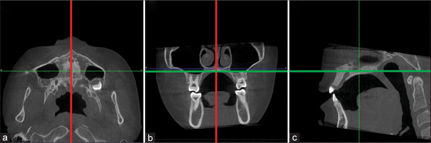
According to a previous study, the average age of peak velocity of growth in body height is approximately 14 years for males and 12 years for females.[18,19] Surgically assisted RME has, thus, been recommended for patients older than 16 years[20] or 25 years.[21] We, therefore, divided the participants into three age groups: under 15 years, between 15 and 25 years, and older than 25 years. All CBCT images were taken using a DCT 100 (Taiwan CareTech, Kaohsiung, Taiwan), and the settings were as follows: 120 kV, 5.0 mA, field of view of 15 × 9 cm, voxel size 0.25 mm, and exposure time 11 s. The lateral cephalometric X-ray examination was conducted with the patient holding their head in its natural position using an ASAHI Hyper-X CM (Asahi Roentgen, Kyoto, Japan). Image reading was performed using Planmeca Romexis image software (Planmeca, Helsinki, Finland). The CBCT image slices were obtained according to the following protocols: The median sagittal plane was aligned with the nasal septum, and the horizontal plane was aligned with the hard palate [Figure 1]. For those patients in whom the palate had a pronounced curvature or was very thick, two distinct axial image slices were obtained.[10] The MPS maturation stage was classified (from A to E) using the Angelieri et al. method,[10] and the CVM was classified (from cervical stage -CS1 to CS6) according to the method of Baccetti et al.[12]
Export to PPT
All images were analyzed by a senior orthodontic resident. One month later, the same examiner reclassified all samples in random order. The weighted kappa coefficient was used to evaluate the intra-examiner agreement. Spearman’s correlation coefficient was used to evaluate the correlation of MPS maturation stage with age and CVM stage. Statistical Package for the Social Sciences (SPSS) software version 24 (SPSS, Chicago, IL, USA) was used for statistical analysis, and P < 5% was considered significant.
RESULTSThe weighted kappa coefficients were 0.849 and 0.923 for the classification of MPS maturation stage and CVM stage, respectively. This shows that there was substantial agreement between the two sets of classifications.
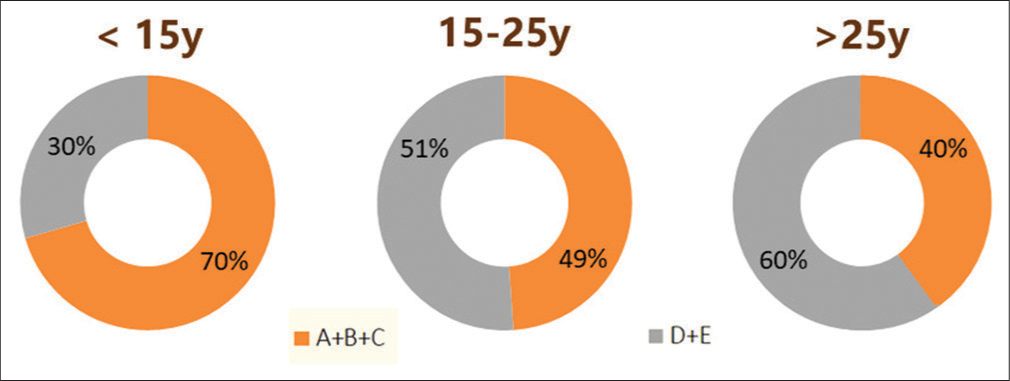
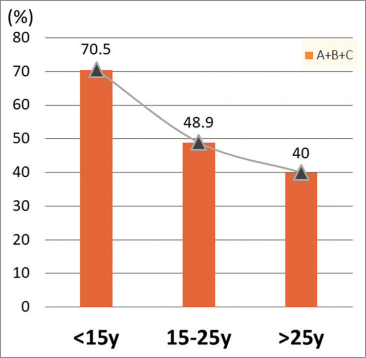
MPS maturation stage versus ageThe r value for the correlation between MPS stage and age was 0.313, and P < 0.05, which indicates a significant but weak correlation. [Table 1] shows the distribution of MPS maturation stage for the three age groups. Only one participant was in stage A and four participants in the under-15 age group were in stage B. One participant in stage B was in the over-25 age group. [Figure 2] shows that stage C was the most prevalent stage across all age groups. We divided the five MPS maturation stages into two groups: Non-fused (stages A, B, and C) and fused (stage D [partially fused] and stage E [completely fused]). The pie and bar charts [Figures 3 and 4] show the percentage of non-fused and fused sutures in each age group. Both the charts reveal a clear tendency for the percentage of non-fused sutures to decrease with age.
Table 1: Distribution of MPS maturation stages according to chronological age groups.
Chronological age group (years) MPS maturation stage Correlation A B C D E Total r P n % n % n % n % n % n % < 15 1 2.3 4 9.1 26 59.1 5 11.3 8 18.2 44 100 0.313 0.001* 15–25 0 0 0 0 22 48.9 16 35.6 7 15.5 45 100 >25 0 0 1 5 7 35 6 30 6 30 20 100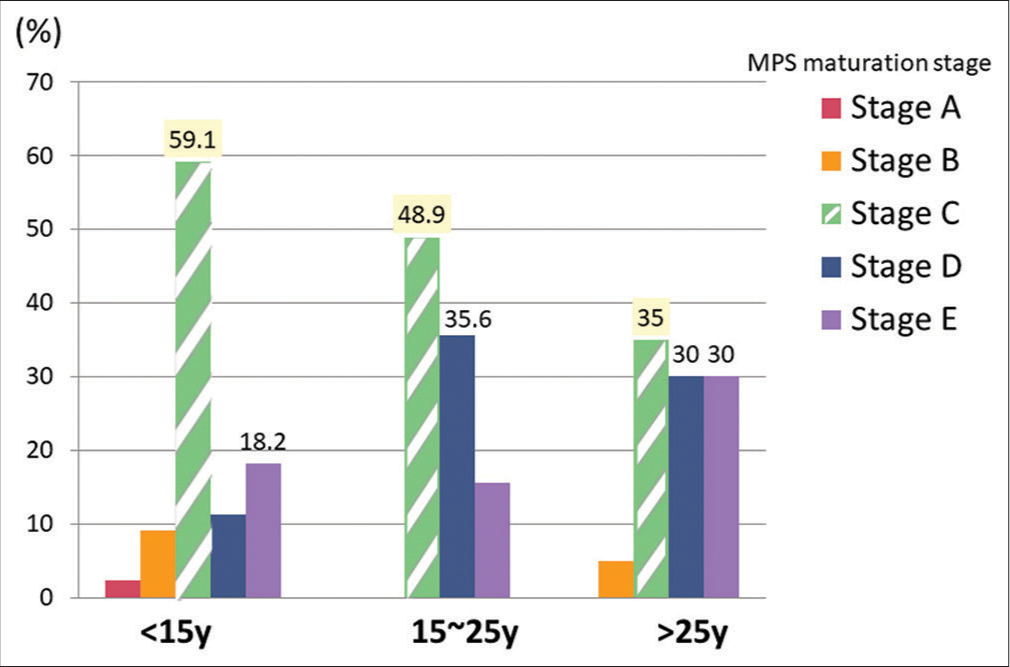
Export to PPT
Export to PPT
Export to PPT
MPS maturation stage versus CVM stageThe distribution of MPS maturation stages according to CVM stages is shown in [Table 2]. The correlation coefficient was 0.287 and P < 0.05, which indicates a significant but weak correlation. [Figure 5] shows the following patterns: CVM stages 1–3 generally corresponded to MPS stages A–C, and CVM stages 4–6 generally corresponded to MPS stages C–E. MPS maturation stages A and B were mainly distributed in CS1–CS3. However, one 29-year-old female participant was at CVM stage 5 but exhibited MPS maturation stage B.
Table 2: Distribution of MPS maturation stages according to CVM stages.
CVM stage MPS maturation stage Correlation Stage A Stage B Stage C Stage D Stage E Total r P CS1 0 2 7 0 0 9 0.287 0.002* CS2 1 1 1 0 1 4 CS3 0 1 9 2 1 13 CS4 0 0 6 4 4 14 CS5 0 1 14 7 7 29 CS6 0 0 18 14 8 40
Export to PPT
Age, CVM stage, and MPS maturation stage versus RME treatment outcomeOf the 109 participants, 14 received RME treatment. All successful cases for whom conventional RME was used were in stage C. One participant in stage E was successfully treated with MARPE, and MARPE failed in one participant who was in stage D [Table 3].
Table 3: Subjects’ distribution according to treatment outcomes, chronological age, CVM stages, MPS maturation stages, and the types of RME.
No. of subjects Chronological age CVM stage MPS maturation stage Treatment outcome Type of expansion <15 15–25 >25 1 2 3 4 5 6 A B C D E Success Conventional 6 0 0 3 0 0 2 1 0 0 0 6 0 0 MARPE 2 5 0 0 0 0 0 4 3 0 0 2 4 1 Failure Conventional MARPE 0 0 1 0 0 0 0 0 1 0 0 0 1 0 DISCUSSIONThe first aim of this study was to identify a simple diagnostic index – age or CVM stage – to help orthodontists assessing MPS maturation stages before selecting maxillary expansion appliances. However, both correlations were significant, they were weak, especially for the oldest age group. Angelieri et al. found great variability in the distribution of MPS stages in patients older than 11 years.[11] The previous studies, which included younger patients, reported a moderate to strong correlation,[14,16,17] while studies including older patients[15,22,23] found no significant correlation or only weak correlations. In our study, 88% of the participants were older than 11 years. The age distribution of our sample may explain the weak correlation between MPS maturation stage and age. Further, Angelieri et al.[10] and Ram and Kannan[24] found that it was mostly patients younger than 13 years who were in MPS maturation stages A and B. We obtained a similar result, in that most of the stages A and B patients were under 12 years, except the one 29-year-old female.
In our study, stage C was the most prevalent stage in all age groups. However, the percentage of non-fused sutures decreased with increasing age. This finding is similar to those of the previous studies involving individuals aged <15 years.[14,25] However, the results of some studies were contradictory to ours; these studies reported stage E as the most prevalent stage in patients older than 15 years.[22,23] Those findings remind us that the correlation between MPS maturation stage and chronological age becomes weaker with increasing age.
Angelieri et al.[13] and Jang et al.[14] revealed a strong positive correlation between CVM and MPS maturation stage, while Lee and Mah[16] and Mahdian et al.[17] found only moderate correlations and Gorucu-Coskuner et al.[15] reported no significant correlation. However, our study produced a contradicting result: A significant but weak correlation. In the studies by Jang et al.[14] and Lee and Mah,[16] CVM stages 1–3 corresponded to MPS stages A–C, except in one participant who was in stage D. In our study, CS1– CS3 also corresponded to MPS stages A–C, except in two patients who were in stage D and two patients who were in stage E. However, other studies have found considerable percentages of non-fused sutures (27–61.9%) in CS5 and CS6 patients.[14-16] We found 51.7% and 45.0% of non-fused sutures in CS5 and CS6 patients, respectively. These findings suggest that even when the patients were in CVM stage 5 or 6, some percentage of non-fused sutures still existed, and suture opening without surgical intervention was still possible.
In this study, all the cases in which conventional RME was successful were at MPS maturation stage C. Similar to the study conducted by Angelieri et al., expanding the maxilla with conventional RME is possible until stage C.[11] In the study of Jesus et al.,[26] which included 25 patients, all patients classified as MPS maturation stage B or C were successfully treated with micro-implant-assisted RME. In contrast, in stages D and E, both failed and successful cases were observed. These findings were similar to those of our study, which was that one patient in stage E was successfully treated with mini-implant-assisted RME while the same treatment failed in one patient in stage D.
CONCLUSIONAfter a comprehensive investigation, the following conclusions can be drawn:
Significant but weak correlations were observed between MPS maturation stage and age and CVM stage. Therefore, neither patient age nor CVM stage appears to be an accurate index for predicting MPS maturation stage
MPS stage C was the most prevalent stage in all age groups
45–51% of non-fused sutures were found in patients in CVM stage 5 or 6
The prognosis for micro-implant-assisted RME may not be related to MPS maturation stage or CVM stage
Using CBCT X-rays for individual MPS assessment before selecting the maxillary expansion treatment is still recommended and, therefore, cannot be deemed as unnecessary radiation exposure.
Ethical approvalThe author(s) declare that they have taken the ethical approval from IRB.
留言 (0)