Plant material
OM fresh leaves were collected from El-Orman Botanical Garden, Giza, Egypt. Engineer Therese Labib, the consultant at El-Orman Botanical Garden and the National Gene Bank at the Ministry of Agriculture, Egypt, kindly authenticated the plant. A voucher specimen of OM numbered (PHBL-00324) was deposited at the Pharmaceutical Biology Department at the German University in Cairo (GUC). No permission for plant collection was required, as OM is not an endangered species or at risk of extinction. Air-dried powdered OM leaves (2 kg) were extracted for 2 h using 5 L of distilled water at 60 to 70 °C to extract all polar compounds. The obtained extract was subjected to filtration and evaporation under reduced pressure in vacuo (BUCHI, Rotavapor, R-210; Switzerland) until complete dryness. The obtained dried residue (150 g) was extracted with purified 90% ethanol (Sigma-Aldrich, Darmstadt, Germany). Afterward, the ethanolic extract was subjected to filtration and evaporation under reduced pressure in vacuo. The residue was then washed with acetone (Sigma-Aldrich, Darmstadt, Germany), and lyophilization was done to produce the crude phenolic content (28 g) [32]. Isolated and purified rosmarinic acid (RA) was obtained from another study conducted by our research group [20, 33].
Determination of total phenolic and flavonoid content
Folin-Ciocalteu method was conducted to calculate the phenolic content of OM extract. It was measured as mg gallic acid equivalent (GAE)/mg extract [34]. Briefly, OM extract of 10 mg/ml in methanol (Sigma-Aldrich, Germany) and serial dilutions of Gallic acid standard (0 to 200) µg/ml were prepared. Afterward, 200 µl of each prepared sample was transferred to a 2 ml tube, followed by the addition of 1 ml Folin-Ciocalteu reagent and left to stand for 5 min at room temperature. Next, 800 µl sodium carbonate solution (7.5%) (El-Nasr Pharmaceuticals Co., Egypt) were added and left for 2 h at room temperature in the dark, and then the absorbance was measured at 750 nm (Perkin Elmer, Waltham, MA, USA). Preparation of the blank was done using a similar protocol. Serial dilutions of Gallic acid standard (Sigma-Aldrich, Germany) were used for the calibration curve. The flavonoid content was estimated according to the method of Schofield, Mbugua, and Pell as mg quercetin equivalent QE/mg extract [35]. OM extract of 0.4 mg/ml in methanol was prepared. In a 96-well plate, 150 µL of OM extract was added to 100 µL of 2% (w/w) AlCl3 (El-Nasr Pharmaceuticals Co., Egypt). Same experimental steps were performed for the standard quercetin (Sigma-Aldrich, Germany) (0–80 µg/ml). All samples were then left for 30 min, including the blank, at room temperature. A microplate reader (Perkin Elmer, Waltham, MA, USA) was used to measure the absorbance at 430 nm.
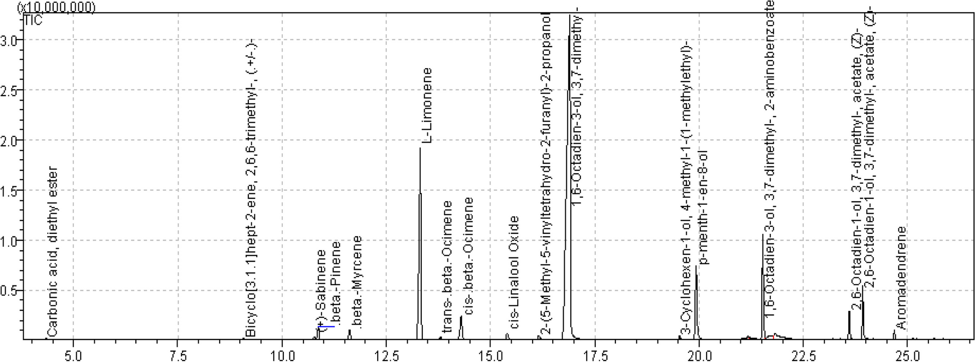
Phytochemical characterization
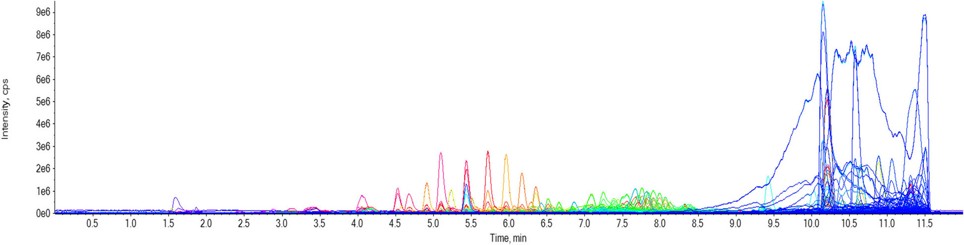
In order to profile the active metabolites in OM active extract, high-performance liquid chromatography HPLC/PDA/ESI-MSn was applied using HPLC Agilent 1200 series instrument provided with high-performance autosampler, binary pump, and PDA G 1290 C (SL; Agilent Technologies, Waldbronn, Germany). For analytes separation, acquity BEH C18 100 mm × 2.1 mm column (particle size, 1.7 μm) was used with RP C18 100 A° guard column with dimensions (5 × 3 mm, i.d.; 5 µm). A mobile phase gradient of 2% acetic acid in purified water (A) and 90% methanol in purified water (B) was implemented. The flow rate operation of the mobile phase was adjusted at 50 µL/min. OM extract was solubilized in 5% methanol and 2% acetic acid at 1 mg/mL concentration and filtered with a 0.2 µm syringe filter. The gradient was as follows: 0 to 60 min, 5% B; 60 to 70 min, 50% B; 70 to 80 min, 90% B; and 80 to 90 min, 5% B. The volume of the injected sample was 10 µL. A Fourier transform ion cyclotron resonance mass analyzer was supported with an Electrospray ionization (ESI) system. The mass was evaluated in Fourier Transform Ion Cyclotron Resonance (FT-ICR) in the full scan and in trap in Mass/Mass (MS/MS) mode (fragmentation). X-calibur® software was used for system control and data acquisition. MS scan and detection was applied in the negative ion mode with the followings: capillary voltage of 36 V, temperature of 275 °C, API source voltage of 5 kV, desolvation temperature of 275 °C and the nitrogen was used as desolvation and cone gas at a flow rate of 15 L/min. The entire run duration was 89 min. The mass range covered was from 150 to 2,000 m/z and resolving power to 100,000 [20].
Antioxidant effect of OM hydroalcoholic extract and rosmarinic acid
BV2 mouse microglial cell lines were cultured in 96-well plates, for 24 h, at a density of 10,000/well in Roswell Park Memorial Institute medium (RPMI; Lonza, Belgium) containing 10% fetal bovine serum (Lonza, Belgium) and 1% penicillin–streptomycin (Lonza, Belgium). The safety of OM extract and RA was tested by adding increasing concentrations of OM (25, 50, 100, 200, 400 µg/ml) or RA (5, 10, 20 µg/ml) to the cells for 72 h. WST-1 cell viability assay kit (Takara, Canada) was used to evaluate the cell viability, and absorbance was measured at 450 nm by a microplate reader (Perkin Elmer, Waltham, MA, USA). In another experiment, oxidative stress was induced by incubating BV2 cells with hydrogen peroxide (H2O2) (Sigma Aldrich) for 24 h at increasing concentrations (50, 100, 150, 200, 250, 300, 350 µM) to determine its IC50. The antioxidant effect on BV2 cells was assessed by pre-treating the cells with different concentrations of OM or RA for 24 h prior to the stimulation with H2O2 at IC50, followed by the measurement of cell viability.
Animals
Healthy adult male Swiss albino mice aged 2 to 3 months weighing 20 to 30 g were purchased from the National Institute of Research animal facilities, Cairo, Egypt. The weight of the mice was recorded daily for an accurate measurement of the treatment dose. Mice were acclimatized for one week at the GUC animal house at room temperature under a 12-h light/12-h dark cycle, with access to food and water ad libitum. Mice were first subjected to behavioral tests; then brains were harvested for further research and analysis. In order to avoid any interference of other chemical compounds with the scientific outcomes of the study, the use of anesthetics was avoided based on their previously reported effects on memory and cognition [36]. The cervical dislocation was chosen as a physical method for rapid sacrifice [37] that was shown to be completely safe on the brain [38]. Cervical dislocation was applied by a trained operator to achieve the highest level of animal welfare. Animal care and treatments were conducted according to the National Research Council's Guide for the Care and Use of Laboratory Animals and in accordance with ARRIVE guidelines. The study was approved by the Research Ethics Committee at the German University in Cairo (project ID 2015–04-HH). This study was randomized and blinded whenever possible. Due to the color of the given plant extract, the experimenter could not be blinded to the drug allocation. Randomization was carried out using Graphpad online random number generator.
LPS mouse model
The neuroinflammatory mouse model used in the current study was induced by daily i.p. injection of 250 µg/kg/day LPS for 7 successive days. This model was previously shown to induce oxidative stress, neuroinflammation, neurodegeneration and memory impairment [17, 20, 39]. The experimental unit used was a single animal, where mice were randomly divided into four experimental groups of eight mice each (without exclusion) [17, 20]. The total number of mice (32) in the study was decided based on previous studies using the same neuroinflammatory model [17]. LPS (Sigma Aldrich, Strain: Escherichia coli, 055:B5) was dissolved in saline and injected i.p at 250 µg/kg/day. OM extract was dissolved in saline and injected i.p at 100 mg/kg/day, based on previous work from our group [20, 40]. Mice were treated for 12 consecutive days: the control group received saline only, the LPS group received LPS starting day 6, the LPS + OM group received OM extract starting day one and LPS starting day 6, and the OM group received OM extract for 12 consecutive days [41]. Any two subsequent injections on the same day were separated by 2 h.
Behavioral tests
In this study, memory and cognitive functions were considered the primary outcomes and were assessed on day 12. All mice were subjected to both Y-maze and novel object recognition tests, and blinding was applied during testing and analysis of the results. The testing followed a randomized order of the tested mice.
Novel object recognition
This test was carried out as previously described [42]. The test apparatus is an empty wooden box attached to an elevated camera to afford a full image of the arena. Transportation of the mice to the experimental room was performed 2 days before the test day for acclimatization (60 min). Mice were permitted to habituate for 10 min in the empty arena a day prior to the test day. In the training phase, each mouse was given 10 min to explore the box with two identical objects, either cubes or pyramids. After 2 h, one of the familiar objects was switched with a novel object in the testing phase and mice were left to freely explore both objects for 5 min. The videos were recorded and analyzed to calculate the discrimination ratio as the time spent by the mouse exploring the novel object divided by the exploration time of both objects. Mice with intact memory are expected to explore the novel object longer than the familiar one. The pyramids and the cubes were randomly used as ‘the novel objects’ to avoid bias.
Y-maze spontaneous alternation test
To evaluate the spatial working memory, Y-maze test was conducted as described previously [43]. The test apparatus has 3 arms that are equally distanced, extending from a central platform at 120°. Transportation of the mice to the experimental room was performed 2 days before the test day for acclimatization (60 min). During the test, each mouse was positioned in the center of the Y-maze to explore it for 8 min. The sequence of arm entries was manually recorded, the entry of three consecutive arms represented one alternation, and alternations were only considered when the mouse entered three different arms consecutively without repetitions. Mice with intact memory tend to explore the three arms equally with no repetitions. The spontaneous alternation percentage was calculated using this formula: [number of actual alternations/total number of entries-2] × 100. A higher spontaneous alternation percentage indicates a better memory.
Histopathology
Brains were fixed and processed using 10% formalin within 24 h. Subsequently, fixed brains were washed by water and were dehydrated using serial dilutions of alcohol. Paraffin-embedded samples were sectioned at 4–5 µm thickness using sledge microtome. Tissues were later deparaffinized to be stained with hematoxylin and eosin (H&E) (VMR, Darmstadt, Germany).
Immunohistochemistry
Brain sections of 5 µm thickness were moved onto poly-lysine-coated slides and were deparaffinized using xylene. Next, they were boiled in 10 mM citrate buffer (pH 6.0) and cooled to room temperature. The endogenous peroxidase was blocked by hydrogen peroxide for 10 to 15 min. The sections were incubated with primary antibodies against cyclooxygenase 2 (COX-2; 1:50) (Invitrogen, Karlsruhe, Germany) or glial fibrillary acidic protein (GFAP; 1:50) (Invitrogen, Karlsruhe, Germany) for 60 min; then rinsed with phosphate-buffered saline (Lonza, Belgium) four times. The sections were treated with biotinylated secondary antibodies for 10 min followed by the addition of streptavidin peroxidase (Carl Roth, Karlsruhe, Germany) for another 10 min, and then visualized with peroxidase-compatible chromogen (3, 3'-diaminobenzidine) DAB mixture (Sigma Aldrich, Munich, Germany).
Statistical analysis
Statistical analysis was conducted using the one-way analysis of variance (ANOVA) followed by Tukey's multiple range tests and expressed as mean ± SEM (standard error of mean). GraphPad Prism (version 7.00) was used for analysis. A probability of P value < 0.05 was considered statistically significant (***P < 0.001, **P < 0.01, *P < 0.05). The immunohistochemistry (IHC) results were analyzed using the open-source plugin IHC profiler for fully automated digital image processing [44]. After proper separation of the DAB color spectra by the spectral deconvolution method, pixels were counted, and their percentage contribution was calculated according to pixel intensity. A semi-quantitative IHC profiler score (high positive, positive, low positive or negative) was then automatically generated. To calculate the exact number of “IHC optical density score” (from 1 to 4), the following algebraic formula was used based on IHC profiler percentage contribution (Seyed Jafari & Hunger, 2017). IHC optical density score = [(Percentage contribution of high positive × 4) + (Percentage contribution of positive × 3) + (Percentage contribution of low positive × 2) + (Percentage contribution of negative × 1)] ÷ 100.

留言 (0)