
記住我


The ECMO circuit consists exclusively of commercially available components. By default, a Thoratec Centrimag centrifugal magnetic levitation pumps (Abbott) (Fig. 1) and ECMOLIFE centrifugal magnetic levitation pumps (Eurosets SPA, Medolla, Italy) (Fig. 2) were used, in synergy with Landing system for pressure monitoring (Blood Flow, Pressure Drop); Gas Transfer (O2 uptake and CO2 removal), (Eurosets SPA, Medolla, Italy). As a standard, the Eurosets A.L.ONE ECMO Adult oxygenator was used (Fig. 3). The tubing and the oxygenator were treated with phosphorylcholine-coated surface (Eurosets SPA, Medolla, Italy). The system has a priming volume of 500 ml and features connectors for other emergency extracorporeal devices, such as renal replacement devices or rapid infusion systems for advanced in-center intensive care treatment during further courses of therapy. The main determinants of cannula sizing in peripheral VA ECMO are anatomical considerations and the targeted flow rate. Generally, cannulas are chosen to support a flow equivalent to a cardiac index of 2.2–2.5 L/m2/min, which is considered full flow. Femoral Arterial cannulas that we used were 17–25 Fr and Femoral venous cannulas were usually 19–25 Fr Biomedicus (Medtronic, Minneapolis, USA). For Central VA ECMO Aortic Arterial cannulas were 20–24 Fr EOPA (Medtronic, Minneapolis, USA) and Atrial venous cannulas were 32/40–36/46 Fr (Medtronic, Minneapolis, USA). For Peripheral VV ECMO for Femoral venous cannulas were 19–25 Fr for reinfusion in jugular vein cannulas were 17–21 Fr Biomedicus (Medtronic, Minneapolis, USA) (Fig. 4).
Fig. 1
Adult A.L.ONE ECMO Oxygenator (Eurosets SPA, Medolla, Italy) configuration with Thoratec Centrimag, centrifugal magnetic levitation pumps (Abbott) during VA ECMO
Fig. 2
Adult A.L.ONE ECMO Oxygenator configuration with ECMOLIFE, centrifugal magnetic levitation pump (Eurosets SPA, Medolla, Italy) during VA ECMO
Fig. 3
Adult A.L.ONE ECMO Oxygenator (Eurosets SPA, Medolla, Italy)
Fig. 4
Trend of PO2 and PCO2 out Oxygenator with and without continuous Heparin
Anticoagulation and blood product managementThe ECMO patients are i our center are anticoagulated with a heparin infusion, with a goal activated partial thromboplastic time (aPTT) of 50–65 s unless the clinical setting (e.g., active bleeding) dictates otherwise. The heparin infusion is titrated with a nurse-managed nomogram, whereby the initial infusion dose is based on the patient’s weight. Six hours after the infusion begins, an aPTT is again drawn, and the rate of infusion is increased if subtherapeutic (< 50 s), decreased if supratherapeutic (> 65 s), or kept constant if within goal (50–65 s). Another aPTT is drawn in 6 h until the second consecutive aPTT is within target range, at which point the aPTT is checked daily. In our institution the usual practice is to transfuse platelets when counts fall below 80,000/μL, although several experienced centers use a more conservative approach and transfuse platelets only when they fall below 40,000–50,000/μL, or even as low as 20,000 in non- bleeding patients. The strategies in RBC transfusion depending on Hb level—restrictive when transfusion is performed at a Hb level of 7–9 g/dL, and liberal with a Hb level between 10 and 12 g/dl, in relation to Blood Flow (BF), Cardiac Output (CO) and Oxygen Delivery (DO2) [6].
Veno-arterial (VA) ECMO indicationECMO was initiated for circulatory instability during or immediately after weaning from the cardiopulmonary bypass (CPB) in the primary cardiac procedure or for hemodynamic support for high risk interventional cardiology procedures. The clinical criteria for hemodynamic support included the following: left atrial pressure > 15 mmHg; central venous pressure > 12 mmHg; metabolic acidosis (i.e. pH < 7.3 with serum lactate > 3.0 mmol/L); end-organ hypoperfusion (urine output < 30 mL/h); cardiac index < 2.2 L/min/m2; and systolic blood pressure < 80 mmHg despite adequate filling volumes, use of multiple adrenergic agents (epinephrine > 0.1 µg/kg/min or dobutamine > 10 µg/kg/min, norepinephrine > 0.1 µg/kg/min), or an intra-aortic balloon pump (IABP). VA-ECMO support was initiated via peripheral cannulation through the femoral route with the semi-open method, and an additional 6 Fr catheter was systematically inserted distally into the femoral artery to prevent leg ischemia ECMO blood flow was adjusted on based on clinical assessments (e.g. pre-oxygenator venous oxygen saturation, evidence of hypoperfusion, resolution of hyperlactatemia, normalization of mean arterial pressure). ECMO-related complications were carefully monitored. ECMO weaning was performed in patients who fulfilled our published institutional weaning criteria and passed an ECMO weaning trial consisting in decreasing and clamping ECMO flow. In general, the patient should have a pulsatile arterial waveform for at least 24 h; be hemodynamically stable, with baseline mean arterial pressure greater than 60 mmHg with no or low doses of catecholamines; should have left ventricular ejection fraction (LVEF) of 35%, and an aortic velocity time integral (VTI) of ≥ 12 cm; and have recovered from major metabolic disturbances. Weaning was considered unsuccessful if ECMO re-cannulation was required within 2 days of decannulation [2,3,4,5,6,7].
Veno-venous (VV) ECMO indicationThe indication for VV-ECMO are typically severely hypoxemic and/or hypercapnic and unresponsive to optimal medical management, including protective ventilation with low-tidal volumes and plateau pressure less than 28–30 cmH2O, high levels of PEEP, prone positioning, neuromuscular blockers and/or other adjunctive therapies, including nitrous oxide or almitrine. The recent literature suggests that a PaO2/FIO2 ratio of 70–80 mmHg, Murray score > 3, and pH < 7.2 provide a reasonable threshold for considering VV-ECMO in adults with ARDS. It is crucial to determine the acute nature of the pulmonary failure, exclude cardiac and/or other organ failure and verify that the respiratory failure cannot be improved with optimal ventilator management [8].
Indication and cut-off parameters used for oxygenator or circuit replacementThe polymethylpentene fiber oxygenator is responsible for oxygen uptake and carbon dioxide removal. The non-biologic surface of the oxygenator activates inflammatory and coagulation pathways with thrombus formation, fibrinolysis, and leukocyte activation leading to fiber dysfunction. Activation of coagulation and fibrinolysis can precipitate systemic coagulopathy or hemolysis, while clot deposition can obstruct blood flow. Additionally, moisture buildup in the gas phase and protein and cellular debris accumulation in the blood phase may contribute to shunt and dead-space physiology, respectively, impairing gas exchange. These three categories—hematologic abnormalities, mechanical obstruction, and inadequate gas exchange—prompt the majority of oxygenator exchanges. Principal Cut-off parameters for replacement the oxygenator or the circuit, Gas Transfer: Arterial oxygen partial pressure (PO2) post oxygenator (< 200 mmHg), Carbon dioxide partial pressure PCO2 (> 40 mmHg) post oxygenator, the oxygen transfer across the oxygenator membrane V′O2 (< 100–150 ml/min/m2),
$$V^ O_ = BFR\left( - CPreO_ } \right)$$
where V′O2 = O2 transfer across the oxygenator (mL/min), BFR = blood flow rate (L/min), Cx O2 = O2 content of(pre-/post-oxygenator) blood (mL/L) for
$$CxO_ = 13.4 \cdot Hb \cdot SxO_ + 0.03 \cdot PxO_$$
where Hb = hemoglobin (g/dL), Sx O2 = O2 saturation of (pre-/post-ML) blood, Px O2 = O2 partial pressure of (pre-/post-oxygenator) blood (mmHg).
Measurement of V′O2 provides an objective measure of oxygen transfer and can confirm oxygenator dysfunction, when clinically indicated. Differential CO2 across the oxygenator “pre oxygenator PCO2–post oxygenator PCO2” (< 10 mmHg); Pressure monitoring: pressure Drop across the oxygenator “Pressure Pre oxygenator–Pressure Post oxygenator” (> 80 mmHg) in relation to Blood flow rate (BFR) (ΔP); Hematologic profiles: Fibrinogen (< 200 mg/dl), Platelets (< 80,000 109/L), aPTT (> 65 s), D-Dimer (> 25–30 ng/ml), LDH (> 250 mg/dl) [1].
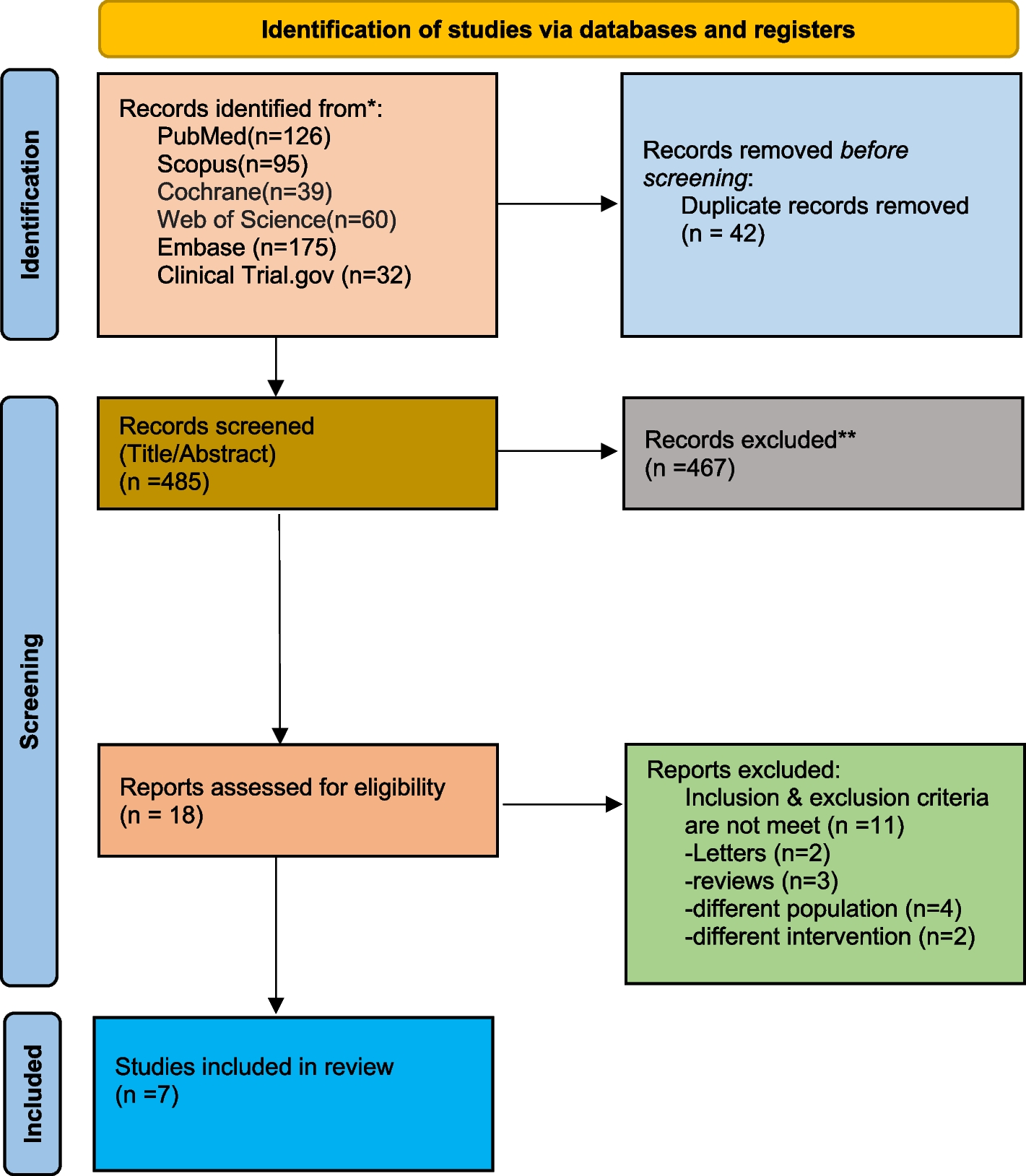
Patients and data collectionWe recruited retrospectively from January 2014 to May 2022 at Institution of Anthea Hospital GVM Care & Research, Bari, Italy, long-term ECMO procedures (exceeding 14 days) that use the Eurosets A.L.ONE ECMO Adult oxygenator. The procedures analyzed including: Veno-arterial (VA) ECMO post-cardiotomy or not, veno-venous (VV) ECMO. ECMO characteristics are described and presented as means with sd or medians with interquartile range. The primary end point was the substitution of oxygenator incidence in relation to the oxygenator performance were Gas Transfer: O2 uptake and CO2 removal were collected in relation to the blood flow rate (BFR), maximum rate per minute of pump (RPM), hemoglobin value (Hb), ventilation indices FiO2 (%)/ Air (L/min), PO2 post oxygenator (mmHg), PCO2 post oxygenator, the transfer across the oxygenator membrane V′O2 (ml/min/m2), Indexed Oxygen Delivery (iDO2) (ml/min/m2) only for VA ECMO patient, the partial pressure of carbon dioxide from the gas exhaust of oxygenator (PECO2) (mmHg), differential CO2 across the oxygenator (mmHg); Hematologic profiles: Fibrinogen (mg/dl), Platelets (109/L), aPTT (sec), D-Dimer (ng/ml), LDH (mg/dl), and incidence of Heparin‐induced thrombocytopenia (HIT) I, II, Temperature in arterial and venous line (°C) and Pressure monitoring: pressure Drop (ΔP) (mmHg).
Statistical analysisContinuous data were expressed as mean ± standard deviation or a median with the interquartile range and categorical data as percentages. Cumulative survival was evaluated with the Kaplan–Meier method. All reported p-values were two-sided, and p-values of < 0.05 were considered to indicate statistical significance. All statistical analyses were performed with SPSS 22.0 (SPSS, Inc., Chicago, IL, USA).
留言 (0)