
記住我

This prospective, observational study was approved by the Institutional Ethics Committee of Fujian Maternity and Child Health Hospital, Fuzhou, China on September 24th, 2021 (2021KLR09068). It is registered in the Chinese Clinical Trial Registry (ChiCTR2100051796). A total of 110 patients who received elective gynecological laparoscopic surgery with general anesthesia in our hospital were recruited from October 2021 to January 2022. All patients had physical status at I ~ II according to the American Society of Anesthesiologists (ASA). The informed consent has been obtained from all patients. They were awake and spontaneously breathed, and voluntarily took measurements. Emergency surgery, peripheral vascular disease, cardiovascular disease, cardiopulmonary, liver and kidney complications, or circulatory instability, were excluded. The patients with unclear ultrasound images were also excluded.
All patients who were conscious, breathing spontaneously, supine, and had routine ECG monitoring, were monitored for electrocardiogram (ECG), blood pressure (BP), and oxygen saturation (SPO2). If the patients in the operating room had MBP greater than 30% during the preoperative visit, 1 mg/ml midazolam would be given by intravenous injection, and the case would be excluded if high MBP persisted.
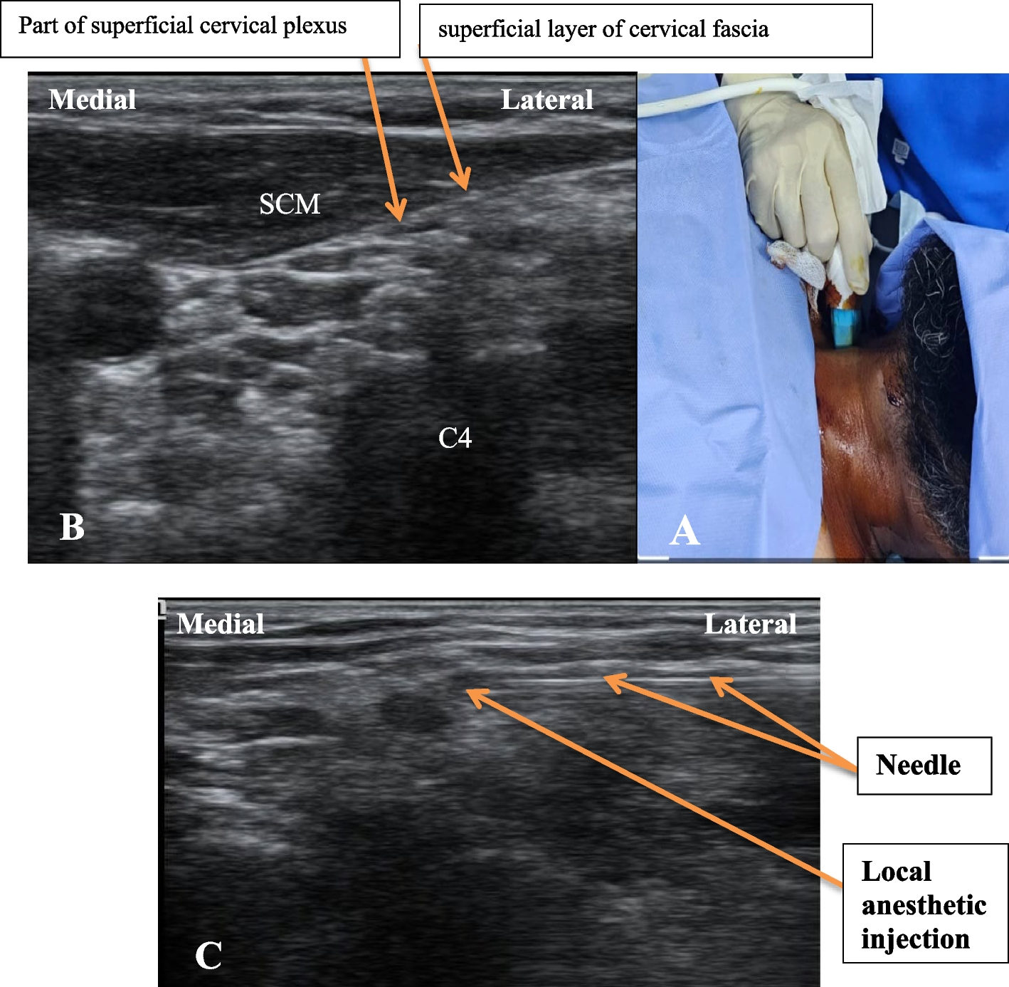
Sonographic measurements of the SCV were performed for all patients by ultrasound diagnostic machine (SonoSite M-Turbo, Bothell, Washington, USA) with a high-frequency linear array probe (6-12 MHz). The linear array probe was initially located on the middle of the clavicle, with the mark point facing the head, and then scanned along the lateral third of the clavicle for optimal SCV short-axis view (Fig. 1). All patients were instructed to breathe, used a linear array probe and M-mode, and take three breathing cycles for the average of three measurements. The maximum value of DSVC (DSCVmax) and the minimum value of DSVC (DSCVmin) were obtained as well (Fig. 2) and recorded as DSCVmax1 and DSCVmin1 in the supine position. The Collapsibility Index of DSCV(DSCV-CI) in the supine position was calculated by the formula [9] below and recorded as DSCV-CI1:
Fig. 1
The sonographic location of subclavian vein using the linear array probe
Fig. 2
M-mode assessment of the subclavian vein
$$DSCV-CI1\:=\:(DSCVmax1\;-\;DSCVmin1)/DSCVmax1\:\times\:100\%$$
Patients then changed their body positions for the PLR test (trunk in supine position and legs elevated at 45°), and the DSCVmax and DSCVmin were measured again and recorded as DSCVmax2 and DSCVmin2, respectively. DSCV-CI2 calculation was the same as DSCV-CI1:
$$DSCV-CI2\:=\:(DSCVmax2\;-\;DSCVmin2)/DSCVmax2\:\times\:100\%$$
ΔDSCV expresses the change rate of DSCV by the PLR test, and was calculated by the following formula [13]:
$$\triangle\mathrm=\left(\mathrm2-\mathrm1\right)/\mathrm2\times100\%$$
Ultrasound measurements during the experiment were performed by the same anesthesiologist with ultrasound training experience.
The MBP and Heart Rate (HR) before induction was collected as baseline. After all data had been collected, all patients received 2 mg/kg propofol and 0.4 μg/kg sufentanil for anesthesia induction. Point 6 mg/kg rocuronium was administered after the patient lost consciousness. The volume-controlled ventilation mode was adopted, tidal volume was 8–10 ml/kg of ideal body weight, respiratory rate was at 12–16 times/min, the fraction of inspired oxygen (FiO2) was at 40%, and Positive end-expiratory pressure (PEEP) was at 0 cmH2O. Anesthesia was maintained with 4–6 mg/kg·h propofol and 0.5–1 mg/kg·h remifentanil. HR and MBP were measured every 1 min after anesthesia induction, and the lowest Mean Blood Pressure (MBPmin) during the 10 min period after anesthesia induction was recorded. Crystal fluid infusion rate was at 10 ml/kg·h. Monitoring ended as surgery began.
Hypotension was defined as Mean Blood Pressure (MBP) less than 60mmhg or more than 30% below the baseline [6]. If the MBP was less than 60 mmhg, the crystalloid infusion rate would be accelerated, and if there is still no improvement, 6 mg/ml ephedrine would be administered by intravenous injection.
Statistical analysesThe sample size was estimated using PASS 15.0 software, the minimum area under receiver operating characteristic (ROC) curve was 0.75, the test level α was 0.05, the test efficiency 1-β was 0.9, and the ratio of the positive group to negative group was 1:1. The calculated sample size is N1 = N2 = 46, and the minimum sample size is N = 92. Due to the inevitable loss of follow-up and non-compliance of the study subjects, the estimated sample size increased by 10% ~ 20% to 110 cases.
SPSS 24.0 software was used to analyze the data, including age, ASA, NPO period, BMI, Propofol, HR, MBP, DSCVmax1, DSCVmax2, DSCV-CI1, DSCV-CI2 and ΔDSCV parameters. Continuous variables were expressed by mean ± standard deviation (x ± s), and t test was used for normal distribution, while for parameters not following a normal distribution, data was expressed by median (interquartile range), and Mann–Whitney U test was used.
ROC curves were drawn to determine the diagnostic capability of DSCV, DSCV-CI, ΔDSCV. The cut off values were defined as the value (sensitivity + specificity – 1) [14]. Binary logistic regression was applied to evaluate the associations of related indicators with hypotension, and P < 0.05 was considered as statistically significant.
留言 (0)