1. IntroductionThe use of closed-circuit rebreathers (CCRs) has become increasingly common in the recreational scuba diving community over the past two decades. Their use allows longer and deeper dives than classical open-circuit (OC) scuba equipment. CCRs bring major advantages in terms of gas consumption, an optimal oxygen mix, and warm humidified breathing gas [
1]. Conversely, since the breathing system is much more complicated to use, it exposes the diver to technical failures or specific emergencies [
2].During a dive, the cardio-pulmonary system is challenged by various combinations of stressors and adaptive mechanisms such as blood shift, thermal strain, exercise, gas density, hypercapnia, narcosis and hyperoxia [
3,
4,
5]. In addition, the breathing apparatus by itself may add to the respiratory workload (work of breathing) that could be increased by the negative transpulmonary pressure gradient in the prone position with back-mounted counterlungs on the CCR used [
6]. A number of studies have investigated pulmonary function following OC diving under varying conditions, but the results remain contradictory [
4]. Many did not show any spirometric alteration after a single OC dive for a maximum depth of 65 m [
7,
8]. However, changes in pulmonary function have been found to be associated with depth, cold temperatures, oxidative or decompression stress and duration. These post-dive obstructive pattern changes appeared to be limited and transient [
9,
10]. Exposure to pure oxygen, even at shallow depths (5 msw), leads to a lung diffusing capacity alteration [
11]. CCR diving exposes to high, and potentially prolonged, PpO2 and specific mechanical constraints [
1,
6]. There is a lack of data about cardio-pulmonary effects during CCR diving. A CCR deep diving study has shown an almost 30% decrease in forced vital capacity (FVC) after bounce dives at 100 msw [
12]. Conversely, CCR use did not affect the spirometry despite the long duration at a maximum depth of 20 msw [
13,
14]. The impact of CCR repeated dives has not been evaluated in cold conditions (
15,
16]. Most studies have shown an accumulation of B-lines with incomplete resolution between each in repetitive deep OC dives to 60–80 msw, which is not observed at a 33 msw depth [
3,
8,
17]. With CCR, a lung aeration loss was detected, even in shallow water, between 1 and 10 msw, and was substantially amplified by exercise and negative-pressure breathing. The right-to-left heart imbalance and increase in pulmonary vasoconstriction seem to be related to these impairments [
5,
6]. This phenomenon was already described during breath-hold diving and was related to diaphragmatic spasms with a closed glottis adding to the negative pressure gradient explanation [
18].All immersion constraints, such as blood centralization, pulmonary workload, or hyperoxia, also modulate the autonomic nervous system (ANS). Heart rate variability (HRV) reflects the constant fluctuation of the interaction between pulmonary ventilation, blood pressure, and cardiac output to maintain homeostasis [
19]. It can be used to indirectly study changes in parasympathetic (PNS) and sympathetic (SNS) nervous system activity, which express the level of intensity in physiological adaptation. There are marked changes in autonomic cardiac activity during and after scuba diving with a predominance of PNS activity [
20,
21]. There are complex and sometimes conflicting additional ANS modulations, and CCR diving seems to provoke a different HRV response in divers as compared to OC diving [
22].
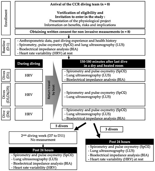
We hypothesize that in-water breathing constraints may have a negative impact on the lung after CCR dives, especially in case of repetitive exposures. Better knowledge of the physiological impacts of CCR appears essential given the growing diving community and technical developments. Data regarding the cardio-pulmonary effects of repetitive CCR diving are still needed for different depths and environments. The aim of this study was to examine the impact of CCR diving on lung function together with autonomous cardiac activity in asymptomatic healthy volunteers after a recurrent diving exposure in cold water.
5. Discussion
The present study depicts that lung aeration disorders are observed during repeated CCR dives. However, these abnormalities, associated with a slight but significant SpO2 decrease, may be transient and not associated with lung spirometry modifications.
A long-term FVC increase has already been reported in the experienced diving community, in relation to the chest muscular workload at depth. This change suggests a distension of the alveoli wall that may cause narrowing of small airways [
4]. It could explain the moderate but not significant basal FEF 25–75 alteration observed in our study. However, the absence of any significant spirometric alteration after such shallow dives is similar to previous studies on CCR diving. There is a general consensus in pulmonary medicine and anesthesiology that breathing oxygen at an oxygen partial pressure (PpO2) higher than 50 kPa causes acute pulmonary injury, which can result in atelectasis, interstitial oedema, and inflammation [
38]. In such diving conditions, there was no significant clinically relevant impairment of clinical airway physiology. After breathing PpO2 at 140 kPa for 20 min at 15 msw, an increase in oxidative stress urinary markers has been described but was not considered sufficient to affect the spirometry [
14]. The pulmonary function also remained unchanged either after a prolonged 3- and 12 h exposure at 5 msw and 20 msw, respectively, with a PpO2 of 150 kPa or after repeated dives (20 dives within 11 days) at an average depth of 69 msw during 112 min with a PpO2 set at 130–140 kPa. It should be noted that the recommended maximal repetition excursion oxygen exposure (REPEX) threshold was approached in these studies [
11,
13,
39]. In contrast, for deeper dives (90–120 msw) with a duration of 2 or 3 h, we previously found a gradual FVC decrease from 109 to 73% of the predicted value after a second dive, without returning to baseline between dives. No alteration of pulmonary resistance was observed, which might suggest other physiological mechanisms than hyperoxia. Considering all these arguments, one might consider that the alteration of spirometry data seems more likely to result from the effects of prolonged and deeper immersion at depth than from oxygen toxicity by itself [
12].A loss of lung aeration was observed after dives, as shown by the accumulation of B-lines without the alteration of spirometry. B-lines are an index of extravascular congestive lung fluid, which has been previously validated with high sensitivity and intra-patient reliability, allowing good interrater consistency of pulmonary fluid assessment using radiographic imaging [
16,
28]. Some authors have reported up to 75% of divers showing extravascular lung water detected as B-line accumulation. Many factors seem to be associated with asymptomatic changes in cardiovascular and pulmonary physiology in diving, therefore linked to the development of extravascular lung water [
40]. However, B-lines are not specific, and their occurrence may also reflect any interstitial disorder or ventilation impairment. Some studies indicate a good correlation between their number and the intensity of damages [
41]. An aeronautic study has shown that hyperoxia and hypergravity are independent risk factors of pulmonary atelectasis formation in healthy humans after a long arm centrifuge session. The increase in B-lines has been reported to reflect the onset of hyperoxic atelectasis [
42]. Our study does not enable us to distinguish extravascular lung water or atelectasis contribution. It is interesting to note, while not significant, that a higher LUS number was observed during the first two days. Dives were shallower but the total immersion time and oxygen exposure were longer. Moreover, helium mixed gases were used for the deepest following dives, thus inducing a lesser gas density and a decrease in breathing workload. Two or three hours after surfacing from a deep Trimix dive, the B-lines were already largely resolved, similar to what has been reported by a previous Croatian study with similar dives [
8]. Our results suggested that most of the pulmonary changes including loss of aeration lasted only for a short time after dives with a return to baseline 24 h post-dive.A reduction in pulmonary diffusing capacity was shown only after a wet shallow oxygen dive as compared to a dry similar dive that suggested the implication of cardiopulmonary changes in immersion [
11]. This impairment was inconsistent and was not correlated with the presence of B-lines [
8]. In our study, SpO2 slightly decreased after dives but remained within physiological values considered to be normal [
43]. This oxygenation decrease may be related to the lung aeration loss and a potential alteration of alveolo-capillary gas exchange, even though it persists while LUS values have decreased and/or returned to baseline values. Atelectasis could lead to a pulmonary ventilation/perfusion mismatch and shunt opening [
42]. Although non-pathological, this may interfere with gas elimination and decompression. Similar results were found after CCR dives at 10 msw [
5] but not after deep dives despite spirometric alteration [
12]. This SpO2 decrease might be compensated by a prolonged high PpO2 up to 150 kPa during long decompression stop after deep dives. Several artefacts can interfere with SpO2 monitoring. However, data were always recorded after rewarming, in dry conditions, and after hydration in order to reduce these methodological artefacts [
43].It is well known that immersion induces hyperdiuresis, which in turn alters the hydration status with a loss of body weight of up to 3% and a potential impact on the cardio-pulmonary system [
12,
44]. Conversely, our results showed an increase in body water after diving. Considering that dehydration plays a role in decompression stress and that water intake could provide a decrease in the risk of decompression sickness (DCS), no specific instruction was given to fluid management for the team [
45,
46]. Technical divers were aware of this problem, and they probably rehydrated themself effectively during hours prior to measurements. There is no direct evidence within the literature that immersion pulmonary oedema is related to hydration in healthy divers [
47,
48]. In our study, there is no clear evidence that the observed state of hyperhydration could have contributed to lung ultrasound abnormalities.HRV monitoring during scuba diving is only available from a limited number of studies, and CCR diving seems to induce a different HRV response than OC diving [
22,
49]. Immersion stimulates both the PNS and SNS branches. A predominant PNS is usually observed during descent and bottom stay, in accordance with human diving responses [
49]. After emersion and continuation of atmospheric air breathing, the SNS takes over the PNS. A PNS tone increase in dive, related to dive length and depth, has been demonstrated [
50]. Nitrox diving seems to induce a higher PNS activation [
51] but also to be the principal dynamic component of SNS [
50]. Short-term HRV is in fact influenced by many other factors, including PNS/SNS balance, as well as respiration via the respiratory sinus arrhythmia, heart and vascular tone via baroreceptor and cardiac stretch receptor activity, the central nervous system (CNS), the endocrine system, and chemoreceptors [
21]. The PNS activity is a major contributor to the HF component (which reflects the power of vagally modulated respiratory sinus arrhythmia) [
19]. Non-linear analysis revealed complexity in heart-rate patterns, which could not be perceived from time–domain [
52]. Compared to OC diving, no variation in HF power or SDNN at depth was found in CCR diving, while the non-linear analysis increased. This suggests a lower PNS dynamic and variability in CCR dives [
22]. In CCR cold diving (2 to 4 °C), the PNS index significatively decreased at submersion and increased gradually throughout the dive. At the same time, the SNS index sharply increased during immersion and then slowly declined back to the pre-dive rest levels [
21]. Our results demonstrate a similar PNS dynamic. However, the SNS index appeared mostly predominant while global HRV activity and PNS decreased, which is contradictory to previous observations during OC diving. The SNS activity can be stimulated by many factors including physical activity or psychologically stressful situations [
53]. The divers wore heavy equipment and were not necessarily previously familiar with the diving conditions that they experienced in this dynamic. Our HRV analysis has some limitations as we were only able to record 23 measurements due to interference between the heating system and the sensor. Moreover, the dive profiles varied over time, and we are not able to compare the responses at these different depths.


留言 (0)