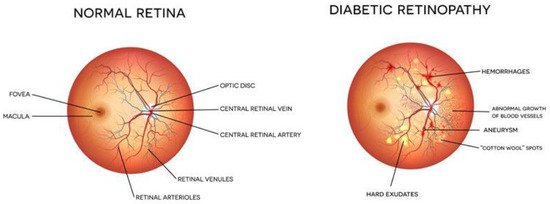
Detection of diabetic retinopathy is critical in the treatment of patients. Presently, a trained ophthalmologist performs manual analysis of the fundus images to detect the presence of DR. This method requires the availability of trained individual and is prone to human errors. Thus, there is a need for automated methods that can analyse fundus images. The automated methods are fast and accurate. In the literature, many methods based on image processing and machine learning are available for DR detection and classification. Many researchers have used segmentation techniques for the identification of exudates and discs. Maximum principal curvature is used for the segmentation of exudates or any other abnormality in the eye [
1]. Another author presented a blood vessel segmentation technique for analysis of the retinal image vessels. Image morphological operators along with K-means clustering are utilized for the segmentation of blood vessels [
2]. Many other authors have proposed different image segmentation-based techniques for detection of DR [
3,
4,
5]. The literature review reveals that mostly Gaussian methods [
6], mathematical morphology [
7] and multi-scale analysis [
8] are being widely used for detection of DR. All these methods based on image processing do not show very high accuracy for the detection of DR. Thus, more efficient methods are required that can identify and classify the retinal images more accurately. Nowadays, deep learning is being widely used for image classification problems. Many researchers have made use of various deep learning models for detection and classification of DR [
8]. Convolutional neural networks have been widely used for DR detection and classification [
9,
10,
11]. The performance of these networks was further improved using transfer learning along with CNN [
12]. With transfer learning, the pretrained models can be tuned as per the medical image database consisting of retinal images. These models demonstrated improved accuracy as compared to the traditional CNN [
13,
14]. Many researchers proposed ensemble methods that combined the advantages from different classifiers and produced highly accurate results. The ensemble models have a higher information gain, as the information from standalone models is combined [
15]. There are many ensembling techniques that can be used for the combining of complementary information amongst models. Many ensemble classifiers are reported in the literature that are presented for the DR problem [
15,
16,
17,
18,
19]. The literature review reveals that there are certain limitations in the existing methods. Due to a limited database of medical images, the accuracy of machine learning models is not high. In addition, the traditional methods have the efficiency of a single model. However, the proposed method performs augmentation; thus, the model is immune to orientation variations.
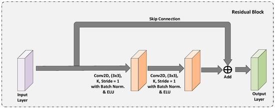
In this paper, to further improve the accuracy and speed of DR detection and classification DRRest is proposed. DRRest is a deep learning ensemble model that uses ResNext architecture. The ResNeXt architecture comprises a shortcut from the previous block to the next block, stacking layers and adapting split–transform–merge strategy. The model has a cardinality parameter that specifies the number of transformations. The model is trained on the third largest dataset APTOS19 which includes more than 5000 retinal images. The images are pre-processed using CLAHE method for histogram equalization. The dataset has a high-class imbalance, and the images of the non-proliferative type are very low, therefore, GAN-based augmentation technique is used for data augmentation. The results obtained are accurate and outperform the other existing methods.

留言 (0)