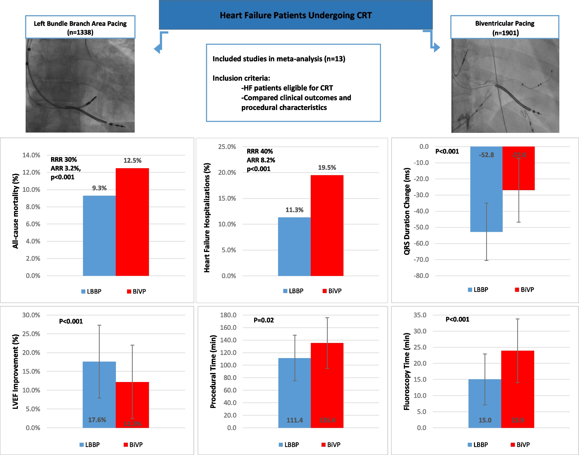
The major methods for mapping PVCs include activation mapping and pace mapping. PVCs originate from the cardiac conductive system, which consists of bundles and fascicles, and the earliest presystolic Purkinje potential was a valid target for catheter ablation. The strategies of mapping PVCs originating from the HPS include the following: (1) mapping the earliest presystolic FP of PVCs, (2) predicting the FP-V interval at the target site. The earliest FP-V interval at the successful ablation site was similar to half of the additive value of the His-ventricular (HV) interval during SR and PVC [7]; (3) comparing the FP-V interval during SR and PVCs. The FP-V interval in sinus rhythm is equal to the FP-V interval of PVCs at the earliest FP activation. If the FP-V interval in sinus rhythm is greater than the FP-V interval in PVCs, this suggests that the location of origin of the PVCs is at the distal end of the mapping electrode [3].
In our cases, the presystolic FP was testified to be safe and effective for RFCA of PVCs originating from the HPS. In cases 2 and 3, the earliest activation of presystolic FP was recorded in PVCs and showed very tiny and sharp morphology features. The morphology of these low-amplitude and high-frequency potentials may be of importance in targeting the earliest activation sites of PVCs originating from the DET and the distal HPS. In addition, comparing predicted FP-V intervals with SR and PVC intervals is not of significant utility in our patients. Ventricular depolarization begins with action potentials that are propagated down the left and right bundle branches on either side of the ventricular septum. The left side of the septum is the first to depolarize owing to quicker conduction velocities of the left posterior branch, which results in the earliest onset of the QRS deflection [8]. As for PVCs originating from the normal proximal to middle left posterior fascicle, it is reasonable that the HV interval in sinus rhythm and HV interval in PVCs are used to infer the FP-V interval. Similarly, the FP-V value in sinus rhythm on the target electrogram being equal to the FP-V value in PVCs applies to these PVCs. During mapping, these methods are subject to errors for PVCs originating from diseased fascicles, DET, and distal fascicles.
Jinglin Z et al. reported that PVCs arose from the left fascicular system, and the earliest FP potential could be recorded in both sinus rhythm and PVC at the site of origin, which provides a definitive diagnosis of PVCs originating from the HPS [3]. In our study, FP could be recorded in PVC but not in sinus rhythm in cases 1 and 3. In case 1, the PVCs originated from the diseased fascicle, so it was impossible that the FP was recorded in sinus rhythm for the local ventricular activation preceding the diseased fascicle activation. In case 3, the PVCs originated from the distal fascicles, so it was sometimes difficult to use an ablation catheter to map these tiny potentials in some regions without FP distribution in sinus rhythm. According to these findings, it is not clear whether the PVCs originate from the fascicles, leading to misinterpretation of the PVCs as non-fascicle origin and activation mapping by the earliest ventricular activation. The simple adoption of mapping the earliest ventricular myocardial activation could lead to failure of catheter ablation or prolonged operation time.
The DET is part of the HPS and is a stump of the extended branch of the atrioventricular conduction loop, which links proximally to the distal LBB or the proximal LAF, with a structure that is insulated from the surrounding myocardium [9]. The PVC activation from here could only be conducted retrogradely to the insertion of the proximal segment of the conduction system [10]. In case 2, the morphological features of the PVCs are easily misinterpreted as having proximal LAF and distal LBB origin, and ablation at these regions may potentially increase the risk of atrioventricular block or left bundle branch block. The true origin for these so called “proximal LAF and proximal LBB” PVCs might be the DET. A precise grasp of the DET anatomy and electrophysiological characteristics and the detailed mapping of an earliest low-amplitude FP of the DET in the right coronary cusp or in the left ventricle beneath the valve can help prevent incorrect, high-risk ablation at the proximal LAF and distal LBB.
Some studies report pace mapping might be ineffective in identifying the target site of PVCs originating from bundles and fascicles. Since the pacing threshold of fascicles was usually higher than the ventricular myocardium, it is difficult to selectively capture local Purkinje fibers only, without activating the surrounding ventricular myocardium [3]. In case 3, the PVC origin was located at the distal fascicle, which connected electrically with the neighboring ventricular myocardium. The ectopic activation can not only directly excite the neighboring ventricular myocardium but also simultaneously conduct retrogradely to the proximal fascicle from where it activated the entire HPS. It is worth thinking that pacing will simultaneously activate local Purkinje fibers and the neighboring ventricular myocardium, resulting in complex pacing QRS morphology that might be totally similar to the clinical PVC itself. Pace mapping could be utilized to guide ablation of the idiopathic origin of PVCs from the terminal of the HPS.
In conclusion, our study analyzed the different intracardiac electrophysiological characteristics of PVCs originating from diseased fascicles, the distal His-Purkinje system, and the DET. Ablation of these PVCs can be safely and effectively performed by identifying the earliest FP.


留言 (0)