
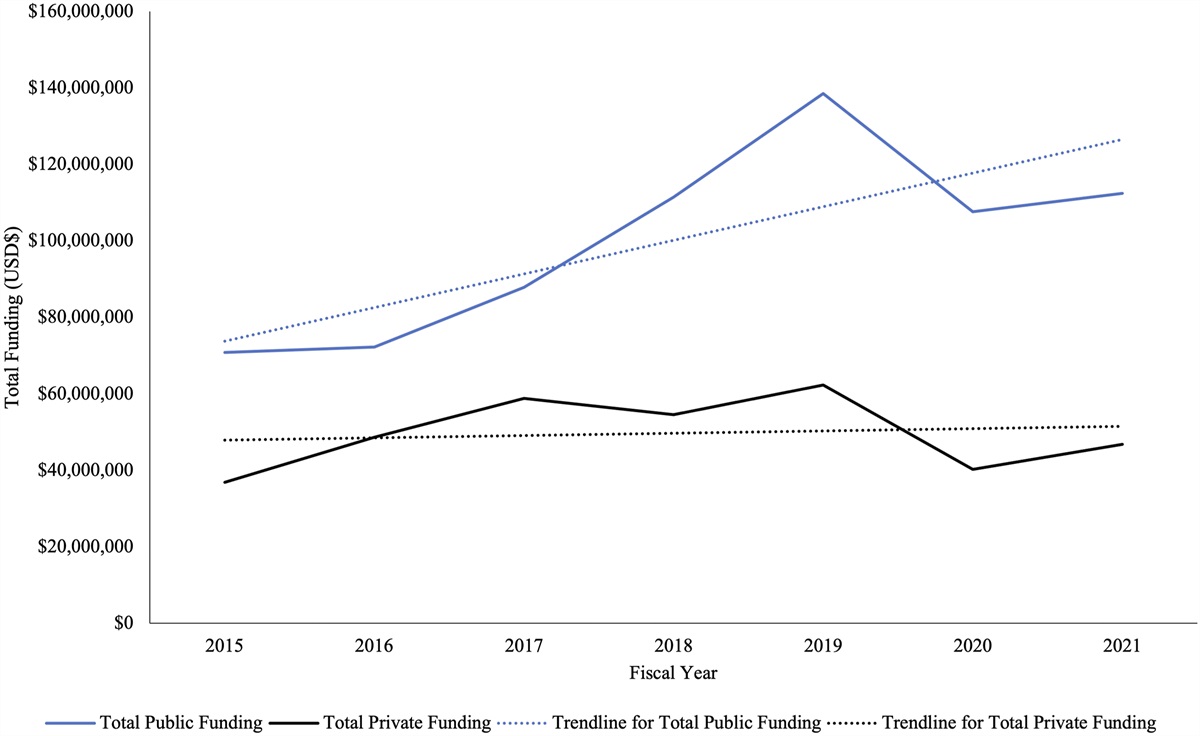
記住我


Complex elbow instability, resulting in damage to the articular surface and ligamentous structures that stabilize the elbow joint, is a challenging condition to manage1. Posterolateral rotatory instability2 of the elbow is the most common type of instability and pattern of injury in complex elbow instability. Posterolateral rotatory instability has been mainly thought to occur as a result of laxity and detachment of the lateral ulnar collateral ligament (LUCL), which is included in the lateral collateral ligament (LCL), due to conditions such as dislocation3-5. Ligament reconstruction has been reported for chronic injuries; however, some studies have demonstrated poor results consisting of persistent or recurrent instability6-8. Precise understanding of the LUCL anatomy is essential to improve the clinical results of LUCL reconstruction.
The LUCL has been identified as the posterior fibers of the LCL connecting the lateral epicondyle and supinator crest9. However, the anatomical understanding of the LUCL remains inconclusive, as its existence and morphology have varied among specimens10-14. On the medial side of the elbow joint, the anterior bundle of the ulnar collateral ligament has recently been interpreted as part of the aponeuroses and tendinous septum of the flexor digitorum superficialis and pronator teres muscles15. On the lateral side of the elbow joint, the LUCL has been described as related to the surrounding ligaments but the relationship with the surrounding aponeuroses and joint capsule has rarely been discussed16,17.
In this anatomical study, we reconsidered the LUCL in terms of the link between the joint capsule and surrounding aponeuroses rather than as a specific ligament. We hypothesized that the deep aponeuroses of the superficial extensors and supinator formed a relevant portion of the joint capsule that was previously defined as the LUCL.
Materials and Methods Preparation of CadaversThe original study sample consisted of 25 elbows (3 bilateral pairs and 19 unilateral; 13 right) from 22 cadavers (12 female; average age when the person died, 78.0 years [range, 54 to 99 years]), which were donated to the anatomical laboratory of Tokyo Medical and Dental University. All of the cadaveric specimens were fixed in 8% formalin and stored in 30% ethanol. We obtained the arm by sectioning the humerus in the middle. The skin and subcutaneous tissues of the arm were removed. Three-dimensional (3D) osseous images of the elbow were obtained using micro-computed tomography (micro-CT; inspeXio SMX-100CT; Shimadzu) with a resolution of 200 µm and ImageJ software (version 1.52; National Institutes of Health). One elbow (right, unilateral) treated with arthroplasty was excluded from the study. No specimens were affected by osteoarthritis with a grade of >3 according to the modified Kellgren and Lawrence classification18. Of the remaining 24 elbows, 20 and 4 were chosen at random for macroscopic and histological analyses, respectively.
Macroscopic AnalysisFor the macroscopic observation of the 20 elbows, we carefully dissected the extensor muscles and joint capsule in a repeated specific sequence to observe their attachments. Initially, each extensor was reflected to its proximal end and removed to expose the supinator and joint capsule. The deep aponeurosis was also analyzed to assess the connection with the supinator tendon and joint capsule. The muscular parts of the supinator were removed with forceps to evaluate the fibrous link between the supinator tendon and joint capsule. Moreover, the supinator aponeurosis, which was separated from the outer surface of the joint capsule, was removed. The joint capsule was separated from the humerus, ulna, and radius. The width of the capsular attachment to the humeral lateral condyle was measured, using calipers (accuracy, 0.1 mm), anterior to the extensor carpi radialis brevis (ECRB) origin, distal to the extensor digitorum communis and extensor digiti minimi (EDC/EDM) origin, distal to the extensor carpi ulnaris (ECU) origin, and posterior to the anconeus origin. To validate accuracy, 40 measurements made on 2 different days by the same assessor were compared with each other, and the intraclass correlation coefficient (ICC) was calculated.
Thickness of the Lateral Elbow Joint Capsule on Micro-CTThe detached whole-capsule specimens were flattened and were analyzed using micro-CT with a resolution of 200 μm as previously described19,20. The images of the joint capsule were reoriented to be level with the flattened joint capsule. By specifying the CT intensity value, we extracted binarized images for all sequential frames from the input micro-CT images. An image stack was projected along the axis perpendicular to the image plane. A real image, which was the sum of the binary slices in the stack, was created by the projection. The thickness of the joint capsule was determined using the slice unit number and length. Based on the calculated thickness, the mean thicknesses in 1 × 1-cm regions (value based on the width of the humeral attachments of the extensors21) corresponding to the anterior, middle, and posterior parts of the humeroradial joint were measured. We created a color look-up table to demonstrate the distribution of capsular thickness. To validate the capsular thickness measurements, we measured wooden-block thicknesses with both micro-CT and calipers and compared these measurements using regression analysis. They were confirmed to be highly correlated (r2 = 0.9996).
Histological AnalysisTo reveal the relationship between the deep aponeuroses of the extensor and supinator muscles and the joint capsule, the coronal and axial sections of the humeroradial joint were analyzed in 2 elbows each. To observe the capsular attachment to the humerus, the coronal section was analyzed at the level of the capitellar ossification center. To demonstrate the connection between the joint capsule and the aponeuroses, we analyzed the axial section at the most distal level of the radial head. The lateral part of the elbow, including the lateral half of the humerus and radius, was harvested using a diamond pathology saw (EXAKT 312; EXAKT Advanced Technologies). The en bloc specimen was decalcified for a week with Plank-Rychlo solution (AlCl3·6H2O [126.7 g/L], HCl [85.0 mL/L], and HCOOH [50.0 mL/L]; Wako Pure Chemical Industries) and then dehydrated. We embedded the specimens in paraffin. Then the blocks were serially sectioned at a thickness of 5 µm and stained using the Masson trichrome staining protocol.
Statistical AnalysisR for Windows (version 4.0.3; R Development Core Team) was used for statistical analyses. Comparison of the capsular attachment width and thickness among regions was performed using the Friedman test, and the significance level was set at p < 0.01. If the Friedman test gave a significant result, the values for the regions were compared using the Wilcoxon signed-rank test with a Bonferroni correction. As a result of the correction, the significance levels of the comparisons of the capsular attachment width and thickness among regions were <0.0017 and <0.0033, respectively. We calculated the ICCs using a measurement process analysis. A score of >0.75 was considered to indicate excellent agreement. All ICCs were ≥0.75 (range, 0.87 to 0.94). Data are presented as the average with the standard deviation (SD).
Source of FundingThis study received financial support from the JA Kyosai Research Institute (Agricultural Cooperative Insurance Research Institute).
Results Relationships Between the Extensor Muscles and Elbow Joint CapsuleThe ECRB deep aponeurosis and anterior part of the EDC/EDM were loosely connected with the supinator aponeurosis and joint capsule (Fig. 1). Compared with the anterior part, the deep aponeuroses of the posterior part of the EDC/EDM and ECU were relatively tightly connected with the supinator aponeurosis. Proximal to the radial head, the supinator aponeurosis and joint capsule intermingled to form a thick membrane, which we termed the “capsulo-aponeurotic membrane” (Fig. 2). Distal to the radial head, the superficial aponeurosis of the supinator and the joint capsule were separated by the interposing muscular part of the supinator.
 Fig. 1:
 Figs. 1-A through 1-F Humeral origins of the superficial extensors. ECRB = extensor carpi radialis brevis, EDC = extensor digitorum communis, ECU = extensor carpi ulnaris, ANC = anconeus, Dist = distal, and Prox = proximal. The asterisks and daggers indicate the posterior deep aponeuroses of the EDC and ECU, respectively, connected to the aponeurosis of the supinator. Fig. 1-A The brachioradialis and extensor carpi radialis longus were detached from the humerus, and the ECRB was reflected proximally. Fig. 1-B The ECRB was detached from the humerus, and the EDC was reflected proximally. Fig. 1-C The EDC was detached from the humerus, and the ECU was reflected proximally. Fig. 1-D The ECU was detached from the humerus. The anterior tendinous part of the anconeus is shown. Fig. 1-E The anconeus was reflected proximally. Fig. 1-F The anconeus was detached from the humerus. Each positional relationship of the extensor origins is seen.
 Fig. 2:
 Figs. 2-A through 2-F Spatial geometry of the aponeurosis of the supinator and joint capsule. ECRB = extensor carpi radialis brevis, EDC = extensor digitorum communis, ECU = extensor carpi ulnaris, ANC = anconeus, Prox = proximal, and Dist = distal. The black-dotted areas indicate the insertion of the brachialis and the origins of the extensors. The black arrows indicate the distal border of the area where the joint capsule and supinator aponeurosis are connected. The blue arrows indicate the supinator crest. Figs. 2-A and 2-B The lateral and anterior appearances of the supinator aponeurosis, respectively. Fig. 2-C Muscular parts of the supinator were removed, and the aponeurosis was proximally reflected. Figs. 2-D, 2-E, and 2-F The membrane that consists of the supinator aponeurosis and joint capsule is shown anteriorly (Fig. 2-D), laterally (Fig. 2-E), and posterolaterally (Fig. 2-F).
Capsular Attachment on the Humerus and UlnaOn the coronoid process, the capsulo-aponeurotic membrane had a wide attachment proximal and lateral to the brachialis tendon insertion (Fig. 3). On the lateral epicondyle of the humerus, the capsulo-aponeurotic membrane had a significantly (p < 0.001) wider attachment on the distal edge of the EDC/EDM origin (L2 in Fig. 4) compared with the anterior edge of the ECRB origin (L1 in Fig. 4), distal edge of the ECU origin (L3 in Fig. 4), and posterior edge of the anconeus tendon origin (L4 in Fig. 4). The results of subgroup analyses in terms of age and sex are shown in Supplementary Table 1 (see Appendix). The capsulo-aponeurotic membrane had substantial attachments posterior to the radial notch of the proximal part of the ulna (Fig. 3).
 Fig. 3:
 Figs. 3-A, 3-B, and 3-C Capsular attachment on the humerus, radius, and ulna in the elbow joint. ECRB = extensor carpi radialis brevis, EDC = extensor digitorum communis, ECU = extensor carpi ulnaris, ANC = anconeus, Prox = proximal, and Med = medial. The black-dotted areas show the insertion of the brachialis and the origins of the extensors. The white-dotted areas show the osseous attachment of the capsulo-aponeurotic membrane. The white triangles indicate the attachment on the lateral aspect of the coronoid process. The white stars indicate the attachment on the distal part of the lateral epicondyle of the humerus. The white circles indicate the attachment on the posteroproximal part of the radial notch. The white diamonds indicate the attachment on the posterodistal part of the radial notch. The same symbols indicate the corresponding joint capsule. The blue arrows indicate the supinator crest. The capsulo-aponeurotic membrane was detached from the humerus and anterior aspect of the ulna (Fig. 3-A), radius (Fig. 3-B), and around the radial notch (Fig. 3-C).
 Fig. 4:
 Figs. 4-A and 4-B Measurements of the attachment of the joint capsule on the lateral aspect of the humerus. ECRB = extensor carpi radialis brevis, EDC = extensor digitorum communis, EDM = extensor digiti minimi, ECU = extensor carpi ulnaris, ANC = anconeus, Dist = distal, and Post = posterior. Fig. 4-A The black-dotted areas indicate the origins of the superficial extensors. The white-dotted area indicates the capsular attachment of the elbow joint. The attachment width was measured at the anterior edge of the origin of the ECRB (L1), distal edge of the origin of the EDC/EDM (L2), distal edge of the origin of the ECU (L3), and posterior edge of the origin of the ANC (L4). Fig. 4-B Scatterplot showing the attachment widths (mean and standard deviation) at the locations indicated in Fig. 4-A. The attachment at L2 (red asterisk) was wider than other areas (Friedman test and Wilcoxon signed-rank test with Bonferroni correction, p < 0.001).
Gross Appearance of the Capsulo-Aponeurotic Membrane and Thickness DistributionViewed from the outer aspect of the joint capsule, the supinator aponeurosis originated from the distal part of the EDC origin and extended anteriorly and posteriorly to form the thick capsulo-aponeurotic membrane (Figs. 5-A, 5-B, and 5-C). From the inner aspect, the radiocapitellar plica was identified proximal to the radial head (Fig. 5-D). The thickness distribution analysis using micro-CT demonstrated that the capsulo-aponeurotic membrane distal to the EDC origin and the radiocapitellar plica proximal to the radial head were brighter in color than the anterior and posterior parts (Fig. 6-A). The capsulo-aponeurotic membrane (R2 in Fig. 6) was significantly thicker (p < 0.001) than the anterior part (R1 in Fig. 6), which corresponded to the capitellum and trochlea, and posterior part (R3 in Fig. 6), which underlies the anconeus muscle. The results of subgroup analyses in terms of age and sex are shown in Supplementary Table 2 (see Appendix).
 Fig. 5:
 Figs. 5-A through 5-D Appearance of the lateral elbow joint capsule. The white triangles indicate the capsular attachment on the coronoid process of the ulna. The white stars indicate the widest capsular attachment on the lateral condyle of the humerus. The white circles and white diamonds indicate the proximal and distal capsular attachments posterior to the radial notch, respectively. Dist = distal, Prox = proximal, Ant = anterior, Post = posterior, and Lat = lateral. The white-dotted areas on the capsulo-aponeurotic membrane correspond with the osseous attachment. The black arrows indicate the distal edge of the supinator aponeurosis. The red arrows indicate the radiocapitellar plica. The black open square corresponds to the radial head. Figs. 5-A and 5-B The 3D bone models of the anterior (Fig. 5-A) and posterolateral (Fig. 5-B) aspects of the right elbow show the locations of representative attachments of the joint capsule. Figs. 5-C and 5-D Gross appearances of the outer (Fig. 5-C) and inner (Fig. 5-D) aspects of flattened joint capsules.
 Fig. 6: Figs. 6-A and 6-B Distribution of the local thicknesses of the joint capsule. Fig. 6-A The same specimen as shown in Fig. 5 is seen after data processing. Boxed regions R1, R2, and R3 correspond to the anterior, middle, and posterior parts of the humeroradial joint, respectively. The approximate thicknesses (in millimeters) are shown as the different colors in the color bar. The white triangle indicates the capsular attachment on the coronoid process of the ulna. The white star indicates the widest capsular attachment on the lateral condyle of the humerus. The white circle and white diamond indicate the proximal and distal capsular attachments posterior to the radial notch, respectively. Ant = anterior and Prox = proximal. Fig. 6-B Scatterplot showing the thicknesses (mean and standard deviation) of the regions indicated in Fig. 6-A. R2 (red asterisk) was thicker than the other regions (Friedman test and Wilcoxon signed-rank test with Bonferroni correction, p Histological Analysis
In the coronal section at the level of the anteroposterior center of the capitulum, the capsulo-aponeurotic membrane was attached to the lateral aspect of the capitellum via a fibrocartilaginous structure (Figs. 7-A, 7-B, and 7-C). The capsulo-aponeurotic membrane was confirmed to transition into the superficial aponeurosis of the supinator (Fig. 7-D). In the axial section at the most distal portion of the radial head, the deep aponeurosis of the ECRB was separated from the capsulo-aponeurotic membrane by interposing loose connective tissue (Figs. 7-E and 7-F). The deep aponeuroses of the EDC/EDM and ECU were connected to the capsulo-aponeurotic membrane through dense connective tissue (Figs. 7-E and 7-G).
 Fig. 7: Figs. 7-A through 7-G Histological images of the joint capsule (Masson trichrome stain). Scale: Figs. 7-B and 7-E = 5 mm; Figs. 7-C, 7-D, and 7-F = 1.5 mm; and Fig. 7-G = 2 mm. ECRL = extensor carpi radialis longus, ECRB = extensor carpi radialis brevis, EDC = extensor digitorum communis, EDM = extensor digiti minimi, ECU = extensor carpi ulnaris, ANC = anconeus, SUP = supinator, Dist = distal, Post = posterior, Lat = lateral, and Med = medial. Fig. 7-A The locations of the sections as labeled in the images to the right. Fig. 7-B Coronal section at the level of the most projecting part of the lateral epicondyle. Fig. 7-C Enlarged image of the box labeled C in Fig. 7-B. The capsulo-aponeurotic membrane attaches to the lateral part of the humerus through fibrous tissue. Fig. 7-D Enlarged image of the box labeled D in Fig. 7-B. The supinator aponeurosis is connected with the joint capsule more proximally than the radial head. Fig. 7-E Axial section at the level of the most distal portion of the radial head. Fig. 7-F Enlarged image of the box labeled F in Fig. 6-E. Loose connective tissue intervenes between the deep aponeurosis of the ECRB and the capsulo-aponeurotic membrane. Fig. 7-G Magnified image of the boxed region labeled G in Fig. 7-E. The deep aponeuroses of the EDM and ECU were connected tightly to the capsulo-aponeurotic membrane through dense connective tissue.Discussion
This study revealed that the supinator aponeurosis and joint capsule intermingled to form the thick capsulo-aponeurotic membrane. The capsulo-aponeurotic membrane had a wide attachment on the distal part of the EDC/EDM origin of the humerus, lateral part of the coronoid process, and posterior part of the radial notch of the ulna. Histologically, the humeral attachment was shown as a fibrocartilaginous structure. Comprehensive and quantitative analysis of the thickness revealed that the capsulo-aponeurotic membrane and radiocapitellar plica proximal to the radial head were thicker than the anterior and posterior parts of the joint capsule. The EDC/EDM and ECU aponeuroses were connected to the capsulo-aponeurotic membrane through dense connective tissues.
Given that the LCL has been described to include the LUCL posteriorly, the LCL needs to be considered before discussing the LUCL. The LCL has a fan-like shape, originates from the lateral epicondyle of the humerus, inserts anterior and posterior to the radial notch of the proximal part of the ulna, and connects to the anular ligament22,23. The LCL has been previously described to have proximity to and an ambiguous boundary with the supinator muscle and the joint capsule9,10,14,17,24,25. Histologically, ligaments are defined as fibrous connective tissues, which are inferior to tendons and superior to aponeuroses in terms of fibrous orientation26. However, because the differences among tendons, ligaments, aponeuroses, and joint capsules are unclear, previously defined ligamentous structures have been reconsidered as parts of the surrounding fibrous structures in several joints15,27,28. In this study, we demonstrated that the supinator aponeurosis is connected with the joint capsule to form the thick capsulo-aponeurotic membrane, which is widely attached to the humerus distal to the EDC/EDM origin, and the ulna both anterior and posterior to the radial notch. Taking cues from these previous studies, we could interpret the LCL as a capsulo-aponeurotic membrane from the perspective of periarticular fibrous structures. Because the fibrocartilaginous distribution implies adaptation to loaded mechanical stress29, the capsulo-aponeurotic membrane was speculated to transmit high tensile stress to the humeral attachment. The ambiguous borders among the LCL, supinator muscle, and joint capsule, as previously described 9,10,14,17,24,25, could be explained based on this interpretation of the LCL.
We wondered whether the LUCL could be similarly reconsidered based on periarticular fibrous structures. Morrey and An9 previously defined the LUCL as the posterior fibers of the LCL, attached to the supinator crest of the ulna. However, in replication studies, the identification of the LUCL has been variably reported as being present in one-fifth to all specimens11-13. Some studies have also described various types of ulnar-side attachments10,14. These inconsistencies could result from the difficulty in defining the LUCL as a discrete ligament since it does not appear to be one.
Previous reports have described the supinator17,30 and ECU17 as having connections with the LUCL. Davies and Laird31 reported that the EDC-associated deep aponeurosis was difficult to separate from the superficial part of the supinator muscle. Originally, O’Driscoll et al.16 stated that LUCL fibers were continuous with other LCL fibers and extensors. Our study showed that the capsulo-aponeurotic membrane was posteriorly connected with the EDC- and ECU-associated deep aponeuroses, which appear as band-like structures. These bands could be interpreted as the LUCL if they were artificially divided from the native aponeurosis and joint capsule. These findings support the description of the LUCL as “part of a capsulo-ligamentous complex rather than a specific structure” by O’Driscoll et al.16 (Fig. 8).
 Fig. 8:
 Schematic of the anatomical interpretation of the LUCL (lateral ulnar collateral ligament). ECRB = extensor carpi radialis brevis, EDC = extensor digitorum communis, ECU = extensor carpi ulnaris, ANC = anconeus, Dist = distal, and Prox = proximal. The asterisk and dagger indicate the deep aponeuroses of the EDC and ECU, respectively, connected to the capsulo-aponeurotic membrane. The blue dotted line indicates the septum between the EDC and ECU tendons. The gray and beige areas indicate the distribution of the joint capsule and capsulo-aponeurotic membrane, respectively. The brown area indicates the muscular part of the supinator. The yellow area indicates the part that has been assumed to be the LUCL.
The anatomical findings of our study have 2 clinical implications. First, due to the anatomical understanding that the LUCL was the independent band connecting the lateral epicondyle and the supinator crest, the LUCL was previously reconstructed by creating a band-like structure using single or double autologous tendons such as the palmaris longus tendon6,8. While this type of reconstruction has been reported to have mostly satisfactory results, recurrent instability and poor results were more frequent than expected6-8. In the present study, the LUCL could not be separated from the surrounding structures; it was observed to be part of the capsulo-aponeurotic membrane consisting of the supinator, EDC and ECU aponeuroses, and joint capsule. In other words, the LUCL could be interpreted as intimately working with these muscles as a “static-dynamic stabilizer.” Therefore, for LUCL reconstruction, we prefer reconstruction of the triangular membrane including the LCL, which connects the distal part of the humerus, lateral part of the coronoid process, and posterior part of the radial notch of the proximal part of the ulna, rather than creating a simple band-like structure. Furthermore, we consider stitching the graft to the residual aponeuroses such as the supinator, EDC, and ECU to function as a static-dynamic stabilizer.
Second, there are implications for lateral epicondylitis. Nimura et al.21 previously reported that the narrow attachment of the joint capsule anterior to the ECRB origin could be an initial factor leading to lateral epicondylitis. In addition, Clarke et al.32 reported that the presence of an LCL tear is related to poor outcomes of treatment for lateral epicondylitis. Based on the current study, damage to the thin anterior part and expansion to the posterior thick part of the capsulo-aponeurotic membrane could explain the development of lateral epicondylitis.
There were a few limitations to this study. First, it was a purely anatomic study of injury-free specimens; thus, our conjectures remain speculative, and function of the elbow joint, such as the range of motion and strength, was not known. Second, the mean age of the study population was >70 years, which is considerably older than that of the general population of patients with posterolateral rotatory instability. Third, there could be inter-ethnic variations because our limited number of specimens were all from Japanese donors. Additional biomechanical or cross-sectional imaging studies and close monitoring during surgical dissection in younger patients are required to validate our findings.
In conclusion, the thick capsulo-aponeurotic membrane, which consists of the supinator aponeurosis and the joint capsule, links the lateral epicondyle of the humerus, radial side of the coronoid process of the ulna, and posterior part of the radial notch of the ulna. The capsulo-aponeurotic membrane could be interpreted as the LCL. In particular, its posterior part, to which the deep aponeurosis of the EDC and ECU connects, was artificially distinguished as the LUCL.
AppendixSupporting material provided by the authors is posted with the online version of this article as a data supplement at jbjs.org (https://links.lww.com/JBJS/H61).
References 1. Morrey BF. Instructional Course Lectures, the American Academy of Orthopaedic Surgeons - Complex instability of the elbow. J Bone Joint Surg. 1997;79(3):460-9. 2. O’Driscoll SW, Bell DF, Morrey BF. Posterolateral rotatory instability of the elbow. J Bone Joint Surg Am. 1991 Mar;73(3):440-6. 3. Abe M, Ishizu T, Morikawa J. Posterolateral rotatory instability of the elbow after posttraumatic cubitus varus. J Shoulder Elbow Surg. 1997 Jul-Aug;6(4):405-9. 4. O’Driscoll SW, Spinner RJ, McKee MD, Kibler WB, Hastings H 2nd, Morrey BF, Kato H, Takayama S, Imatani J, Toh S, Graham HK. Tardy posterolateral rotatory instability of the elbow due to cubitus varus. J Bone Joint Surg Am. 2001 Sep;83(9):1358-69. 5. Kalainov DM, Cohen MS. Posterolateral rotatory instability of the elbow in association with lateral epicondylitis. A report of three cases. J Bone Joint Surg Am. 2005 May;87(5):1120-5. 6. Nestor BJ, O’Driscoll SW, Morrey BF. Ligamentous reconstruction for posterolateral rotatory instability of the elbow. J Bone Joint Surg Am. 1992 Sep;74(8):1235-41. 7. Olsen BS, Søjbjerg JO. The treatment of recurrent posterolateral instability of the elbow. J Bone Joint Surg Br. 2003 Apr;85(3):342-6. 8. Jones KJ, Dodson CC, Osbahr DC, Parisien RL, Weiland AJ, Altchek DW, Allen AA. The docking technique for lateral ulnar collateral ligament reconstruction: surgical technique and clinical outcomes. J Shoulder Elbow Surg. 2012 Mar;21(3):389-95. 9. Morrey BF, An KN. Functional anatomy of the ligaments of the elbow. Clin Orthop Relat Res. 1985 Dec;(201):84-90. 10. Cohen MS, Hastings H 2nd. Rotatory instability of the elbow. The anatomy and role of the lateral stabilizers. J Bone Joint Surg Am. 1997 Feb;79(2):225-33. 11. Olsen BS, Søjbjerg JO, Nielsen KK, Vaesel MT, Dalstra M, Sneppen O. Posterolateral elbow joint instability: the basic kinematics. J Shoulder Elbow Surg. 1998 Jan-Feb;7(1):19-29. 12. Beckett KS, McConnell P, Lagopoulos M, Newman RJ. Variations in the normal anatomy of the collateral ligaments of the human elbow joint. J Anat. 2000 Oct;197(Pt 3):507-11. 13. Kim PT, Isogai S, Murakami G, Wada T, Aoki M, Yamashita T, Ishii S. The lateral collateral ligament complex and related muscles act as a dynamic stabilizer as well as a static supporting structure at the elbow joint: an anatomical and experimental study. Okajimas Folia Anat Jpn. 2002 Aug;79(2-3):55-61. 14. Hackl M, Bercher M, Wegmann K, Müller LP, Dargel J. Functional anatomy of the lateral collateral ligament of the elbow. Arch Orthop Trauma Surg. 2016 Jul;136(7):1031-7. 15. Hoshika S, Nimura A, Yamaguchi R, Nasu H, Yamaguchi K, Sugaya H, Akita K. Medial elbow anatomy: A paradigm shift for UCL injury prevention and management. Clin Anat. 2019 Apr;32(3):379-89. 16. O’Driscoll SW, Horii E, Morrey BF, Carmichael SW. Anatomy of the ulnar part of the lateral collateral ligament of the elbow. Clin Anat. 1992;5(4):296-303. 17. Imatani J, Ogura T, Morito Y, Hashizume H, Inoue H. Anatomic and histologic studies of lateral collateral ligament complex of the elbow joint. J Shoulder Elbow Surg. 1999 Nov-Dec;8(6):625-7. 18. Oya N, Tajika T, Ichinose T, Sasaki T, Yamamoto A, Kuboi T, Endo F, Takagishi K, Chikuda H. The prevalence of elbow osteoarthritis in Japanese middle-aged and elderly populations: the relationship between risk factors and function. J Shoulder Elbow Surg. 2018 Jun;27(6):1086-91. 19. Momma D, Nimura A, Muro S, Fujishiro H, Miyamoto T, Funakoshi T, Mochizuki T, Iwasaki N, Akita K. Anatomic analysis of the whole articular capsule of the shoulder joint, with reference to the capsular attachment and thickness. J Exp Orthop. 2018 Jun 7;5(1):16. 20. Tsutsumi M, Nimura A, Akita K. The gluteus medius tendon and its insertion sites: an anatomical study with possible implications for gluteus medius tears. J Bone Joint Surg Am. 2019 Jan 16;101(2):177-84. 21. Nimura A, Fujishiro H, Wakabayashi Y, Imatani J, Sugaya H, Akita K. Joint capsule attachment to the extensor carpi radialis brevis origin: an anatomical study with possible implications regarding the etiology of lateral epicondylitis. J Hand Surg Am. 2014 Feb;39(2):219-25. 22. Gray H, Goss CM. Anatomy of the human body. 29th ed. Lea & Febiger; 1973. 23. Moore KL, Agur AM, Dalley AF. Moore’s Essential clinical anatomy. 6th ed. Lippincott Williams & Wilkins; 2019. 24. Olsen BS, Vaesel MT, Søjbjerg JO, Helmig P, Sneppen O. Lateral collateral ligament of the elbow joint: anatomy and kinematics. J Shoulder Elbow Surg. 1996 Mar-Apr;5(2 Pt 1):103-12. 25. Berg EE, DeHoll D. The lateral elbow ligaments. A correlative radiographic study. Am J Sports Med. 1999 Nov-Dec;27(6):796-800. 26. Pawlina W, Ross MH. Histology: a text and atlas with correlated cell and molecular biology. 6th ed. Lippincott Williams & Wilkins; 2016. 27. Tsutsumi M, Nimura A, Akita K. New insight into the iliofemoral ligament based on the anatomical study of the hip joint capsule. J Anat. 2020 May;236(5):946-53. 28. Amaha K, Nimura A, Yamaguchi R, Kampan N, Tasaki A, Yamaguchi K, Kato R, Akita K. Anatomic study of the medial side of the ankle base on the joint capsule: an alternative description of the deltoid and spring ligament. J Exp Orthop. 2019 Jan 28;6(1):2. 29. Benjamin M, Ralphs JR. Fibrocartilage in tendons and ligaments—an adaptation to compressive load. J Anat. 1998 Nov;193(Pt 4):481-94. 30. Broekhuis D, Bessems JH, Colaris JW. Avulsion fracture of the supinator crest as an indication for a sustained posterolateral (sub)luxation of the elbow. A case report, anatomical evaluation and review of the literature. Orthop Traumatol Surg Res. 2016 Dec;102(8):1113-6. 31. Davies F, Laird M. The supinator muscle and the deep radial, posterior interosseous, nerve. Anat Rec. 1948 Jun;101(2):243-50. 32. Clarke AW, Ahmad M, Curtis M, Connell DA. Lateral elbow tendinopathy: correlation of ultrasound findings with pain and functional disability. Am J Sports Med. 2010 Jun;38(6):1209-14.
留言 (0)