
記住我


The pro-survival MCL1 protein is overexpressed in many cancers, including B-cell non–Hodgkin lymphomas (B-NHL). S63845 is a highly specific inhibitor of MCL1. We analyzed mechanisms of sensitivity/resistance to S63845 in preclinical models of diffuse large B-cell lymphoma (DLBCL) and Burkitt lymphoma. Annexin V-based cytotoxic assays, Western blot analysis, protein co-immunoprecipitation, and cell clones with manipulated expression of BCL2 family proteins were used to analyze mechanisms of sensitivity to S63845. Experimental in vivo therapy with S63845 and/or venetoclax was performed using patient-derived xenografts (PDX) of treatment-refractory B-NHL. A subset of DLBCL and majority of Burkitt lymphoma cell lines were sensitive to S63845. The level of BCL2 protein expression was the major determinant of resistance to S63845: BCL2 serves as a buffer for pro-apoptotic proteins released from MCL1 upon exposure to S63845. While BCL2-negative lymphomas were effectively eliminated by single-agent S63845, its combination with venetoclax was synthetically lethal in BCL2-positive PDX models. Concerning MCL1, both, the level of MCL1 protein expression, and its occupational status represent key factors mediating sensitivity to S63845. In contrast to MCL1-BIM/BAK1 complexes that prime lymphoma cells for S63845-mediated apoptosis, MCL1-NOXA complexes are associated with S63845 resistance. In conclusion, MCL1 represents a critical survival molecule for most Burkitt lymphomas and a subset of BCL2-negative DLBCLs. The level of BCL2 and MCL1 expression and occupational status of MCL1 belong to the key modulators of sensitivity/resistance to S63845. Co-treatment with venetoclax can overcome BCL2-mediated resistance to S63845, and enhance efficacy of MCL1 inhibitors in BCL2-positive aggressive B-NHL.
IntroductionThe ability to attenuate cell death signaling pathways such as mitochondrial apoptosis is a hallmark of many cancers, including B-cell non–Hodgkin lymphomas (B-NHL). Activation of mitochondrial apoptosis is tightly controlled by members of B-cell leukemia/lymphoma-2 (BCL2) family proteins via their homotypic and heterotypic interactions. The major pro-survival BCL2 family proteins, the B-cell leukemia/lymphoma-2 (BCL-2/BCL2), myeloid cell leukemia-1 (MCL-1/MCL1/BCL2L3), and B-cell lymphoma-extra-large (BCL-XL/BCL2L1), exert anti-apoptotic functions by binding and sequestering the multi-domain pro-apoptotic effectors BAX and/or BAK1. Increasing the ratio between anti-apoptotic and pro-apoptotic BCL2 proteins leads to apoptosis evasion and extended survival of malignant cells (1).
Diffuse large B-cell lymphoma (DLBCL) and Burkitt lymphoma are aggressive types of B-NHL, both requiring intensive treatment upon diagnosis. Although approximately 60% patients with DLBCL and up to 80%–90% patients with Burkitt lymphoma can be cured with intensive anti-CD20-based immunochemotherapies, patients who are refractory and/or relapse after the induction therapy have poor outcome (2, 3). New treatment options are needed to improve the cure rates of frontline regimens and to develop effective salvage therapies. The evidence that many cancers, including hematologic malignancies, depend on BCL2 proteins for their sustained survival, led to the development of BH3 mimetics, a class of antitumor molecules that antagonize pro-survival BCL2 proteins resulting in apoptosis induction (1, 4). Venetoclax (ABT-199), a small-molecule inhibitor of BCL2 protein, has shown clinical activity in some hematologic malignancies and is currently the only BH3 mimetic approved by regulatory authorities for the treatment of chronic lymphocytic leukemia and acute myeloid leukemia (5–7). Venetoclax has been tested in newly diagnosed or relapsed/refractory patients with DLBCL in several phase I–III clinical trials either alone or in combination with anti-CD20-based immunochemotherapies [NCT03984448 (8, 9)]. However, BCL2 is deregulated only in a subset of DLBCL tumors (10–13), therefore, the target population of patients with DLBCL who would benefit from venetoclax-based therapy needs to be better defined. In Burkitt lymphoma, the expression levels of BCL2 are generally low or undetectable (14), suggesting dependence on other anti-apoptotic proteins for survival.
The role of MCL1 in lymphoma pathogenesis was well demonstrated by MCL1 transgenic mouse models. Mice expressing the MCL1 transgene developed B-cell lymphomas at high frequency (15). It has been shown that MCL1 protein is highly expressed in aggressive B-NHL, including DLBCL (84% cases) and Burkitt lymphoma (89% cases; ref. 16). Molecular mechanisms leading to aberrant MCL1 expression have been studied in DLBCL. MCL1 locus (1q21) gain/amplification and constitutive activation of the STAT3 pathway were identified as key drivers of aberrant MCL1 expression in this lymphoma subtype (17). S63845 was the first highly specific small-molecule inhibitor of MCL1 protein with demonstrated in vitro and in vivo efficacy in various preclinical models of solid tumors and hematologic malignancies (18). Since that time, various other compounds targeting MCL1 protein have been developed including S64315/MIK665, AMG176 and AMG397 or AZD5991. Some of these inhibitors already entered early clinical testing in patients with relapsed/refractory B-NHL (MIK665, NCT02992483; AMG176, NCT03797261; AMG397, NCT03465540; AZD5991, NCT03218683; refs. 1, 4).
In this study, we focused on the mechanisms of sensitivity/resistance to S63845 in preclinical models of DLBCL and Burkitt lymphoma. We demonstrated that a subset of DLBCL and the majority of Burkitt lymphoma are dependent on MCL1 protein for survival. We identified BCL2 and MCL1 expression and occupational status of MCL1 as key factors determining resistance/sensitivity to S63845. Finally, we proposed mechanism-based treatment combinations that could enhance the activity of MCL1 inhibitors in aggressive B-NHL.
Materials and MethodsCell lines and patient-derived xenograft modelsCell line authentication: commercially available DLBCL and Burkitt lymphoma cell lines were obtained from DSMZ or ATCC cell banks and authenticated by Multiplexion. UPF4D, UPF8D, UPF10S and UPF6T, UPF9T cell lines were derived in our laboratory from patients with treatment refractory DLBCL and Burkitt lymphoma, respectively. Cell lines were free of Mycoplasma contamination as determined by testing with Mycoplasma PCR detection kit (abm, last test in August 2021). Cell lines were reinitiated every 3 months from cryopreserved stocks. Patient-derived lymphoma xenografts (PDX) were established in our laboratory from patients with treatment-refractory DLBCL, Burkitt lymphoma, or Richter syndrome as described previously (19, 20).
Apoptosis measurementThe number of apoptotic cells was determined by flow cytometry (BD FACS Canto II) using Annexin V FITC staining kit (Apronex, Czech Republic). S63845 and venetoclax were purchased from MedChemExpress, diluted in dimethylsulfoxid and stored in −20ºC.
Experimental therapy of lymphoma-bearing miceNOD.Cg-Prkdcscid Il2rgtm1Wjl/SzJ mice (referred to as NSG mice; IMSR catalog no. JAX:005557, RRID:IMSR_JAX:005557, https://www.jax.org/strain/005557) were purchased from The Jackson Laboratory. All animals were maintained in a pathogen-free environment in individually ventilated cages and provided with sterilized food and water. Adult female NSG mice were used for all experiments. NSG mice were subcutaneously inoculated with 10 × 106 lymphoma cells. Therapy was initiated when all mice developed palpable tumors (= day 1, D1). On D1, all mice were stratified so that all cohorts contained mice with comparable calculated tumor volumes. Venetoclax (50 mg/kg, by oral gavage) was first diluted in ethanol and then mixed with phosal-G/PEG-400 (1: 3: 6). Venetoclax was given on days 1, 2, 3, 6, and 7. S63845 (25 mg/kg) was diluted in 25 mmol/L HCL and 20% hydroxyl-propyl-beta-cyclodextrin, and administered intravenously on days 1, 2, 3, 6, and 7 as described previously (20). In the case of damaged veins, S63845 was administered intraperitoneally. Tumor growth was recorded using three perpendicular dimensions (in millimeters) with a digital caliper. Tumor volumes were calculated using the following formula: π/6 × length × width × height. The observation was terminated (and experimental mice euthanized) when grown subcutaneous tumors exceeded 2 cm in the largest diameter. Tumors were excised and weighed, and the euthanized mice were dissected in search for any signs of advanced (disseminated) lymphoma (e.g., splenomegaly, abdominal lymphoma spread, etc.). Selected subcutaneous tumors were subsequently CD45-sorted for further experiments. The experimental design was approved by the Czech Institutional Animal Care and Use Committee (MSMT-11255/2015-4 and MSMT-1712/2021-2).
Western blottingWestern blotting was performed as described previously (19). Antibodies were purchased from Cell Signaling Technology: BAK1 (D4E4), BAX (2774), BCL-XL (2764), BIM (C34C5), PARP (46D11), PUMA (4976); Santa Cruz Biotechnology: MCL1 (S-19), MCL1 (22), NOXA (114C307.1); BD Pharmingen: BCL2 (610539); Abcam: Actin (6276) or Alexis: Caspase 3 (22749/b). The protein levels of BCL2, MCL1, and BCL-XL were analyzed by Western blotting. Densitometry analysis (ImageJ, RRID:SCR_003070, https://imagej.net/) was used to quantify the level of expression of BCL2 proteins normalized to actin. Calculated BCL2 protein expression levels were correlated with sensitivity to S63845 (i.e., % apoptotic cells assessed following exposure to various concentrations of S63845).
Establishment of DLBCL clones with knockdown or transgenic overexpression of BCL2 or MCL1Cell lines with stably integrated short hairpin RNA (shRNA)-gene/cDNA were prepared as follows: packaging lentiviral vectors pMD2.G (Addgene, plasmid 12259), psPAX2 (Addgene, plasmid 12260) together with pLKO.1 (Sigma-Aldrich)/pCDH-neo (SBI) vector containing the gene of interest (human BCL2 and MCL1 were PCR amplified from BCL2- and MCL1-expressing plasmids kindly provided by S. Korsmeyer and C. Thompson, respectively, cloned into pCDH-neo and verified by sequencing) were transfected into HEK 293T/17. Viral supernatant was harvested 36 hours later, centrifuged, and precipitated using PEG-it (System Biosciences) according to manufacturer's instructions. Precipitated viral particles were resuspended in PBS and stored in −80°C. Target cells were infected with an equivalent multiplicity of infection for 24 hours and the transductants were selected in the growth medium containing 2 to 3 μg/mL puromycin (LKO1-shRNAs) or 1 mg/mL G-418 (CDH-cDNAs).
Protein co-immunoprecipitationThe technique was conducted according to ref. 21. Cells were lysed in non-denaturing lysis buffer [1% (w/v) Triton X-100, 50 mmol/L Tris-HCl (pH 7.4), 300 mmol/L NaCl, 5 mmol/L EDTA, 0.02% (w/v) sodium azide supplemented with protease inhibitor cocktail for 30 minutes and centrifuged (16,000 × g, 4°C, 15 minutes). Protein concentrations of cell extracts were determined by Pierce BCA Protein Assay Kit and equal amount of protein samples were precleared with protein A-Sepharose bead slurry (Sigma-Aldrich) for 30 minutes at 4°C, split in half, and incubated with a specific antibody, or a corresponding isotype control immunoglobulin bound to protein A-Sepharose beads for 1 hour at 4°C. Immunoprecipitates were centrifuged (16,000 × g, 4°C, 5 seconds), washed three times in wash buffer [0.1% Triton X-100, 50 mmol/L Tris-HCl (pH 7.4), 300 mmol/L NaCl, 5 mmol/L EDTA, 0.02% sodium azide), resuspended in SDS sample buffer, denatured for 5 minutes at 95°C, resolved on a 10% or 15% SDS-PAGE and analyzed by immunoblotting. Following antibodies were used: MCL1 (S-19, Santa Cruz Biotechnology, sc-819), BCL2 (C21, Santa Cruz Biotechnology, sc-783), isotype control immunoglobulin (rabbit polyclonal IgG, Calbiochem or Santa Cruz Biotechnology).
IHC analysisSections from formalin-fixed, paraffin-embedded blocks from murine lymphoma xenografts were cut and stained with hematoxylin and eosin. IHC was performed using BCL2 primary antibody (clone 124, Mouse, Dako). Deparaffinization, rehydration, and antigen retrieval was performed with EnVision FLEX Target Retrieval (1:50, High pH) in the PT link pretreatment system (Dako). After blocking of endogenous peroxidase activity (EnVision FLEX Peroxidase blocking reagent, Dako S2001), the samples were incubated with primary antibody BCL2 (clone 124, 1:100, Dako) for 50 minutes at room temperature. Amplification of signal of primary antibody was performed with EnVision FLEX+ Linker and the reaction was visualized by chromogen substrate DAB (3,30-diaminobenzidine tetrahydrochloride; Dako). Nuclei were counterstained with Harris hematoxylin. After dehydration, slides were mounted in organic solvent–based medium and evaluated under light microscope. The slides were evaluated by expert hematopathologist. Samples were considered positive if ≥ 30% of the tumor cells showed cytoplasmic positivity.
Establishment of DLBCL cell lines with BIM, BAK1, and NOXA gene knockoutWe designed several single-guide RNA pairs for CRISPR/Cas9 inactivation of each gene of interest. Targeted were: exon 4 of BIM (BCL2L11), exon 1 of NOXA (PMAIP1), and exon 3 of BAK1 gene (sequences of gRNAs are listed in Supplementary Materials and Methods). Candidate target sequences were chosen based on predicted off- and on-target activity (22, 23). Selected protospacer sequences were synthesized with particular overhangs (Integrated DNA Technologies, Inc.) and cloned into BbsI linearized pX330-Cas9 vector (Addgene, plasmid 42230). Protospacer containing pX330 vectors were cotransfected with pcDNA-EmGFP plasmid to distinguish the transfected population by flow cytometry. GFP-positive cells were sorted using BD FASC Aria and seeded into 96-well plate, 1 cell per well. After establishing clonal cultures gDNA and protein lysates were isolated to confirm successful gene knockout using PCR and Western blot analysis, respectively.
Statistical analysisData from six in vivo experiments were analyzed, each experiment covering different time periods with different numbers of known data points. To assess the practical significance of treatment effectiveness, we plotted charts with growth curves indicating group mean tumor sizes accompanied by expert opinion on the differences observed. For the purpose of assessing the statistical significance of treatment effectiveness, we made an assumption that the calculated differences between mean tumor sizes in the control group and groups treated with monotherapies or combined therapy as well as between the groups treated with monotherapies and those treated with combined therapy were generated by a process which includes a deterministic linear trend in the form of yt = β0 + β1t + εt, where yt denotes the data-generating stochastic process of the analyzed differences, t = 1, 2, …T is a time variable, T signifies the length of the experiment in days and εt is the Gaussian IID white noise. Having concurrently performed 30 statistical hypothesis tests about the zero value of β1, the Bonferroni correction of 10% and 1% simultaneous significance levels was utilized, resulting in individual significance levels of 0.3333% and 0.0333%, respectively.
ResultsA subset of DLBCL and the majority of Burkitt lymphoma cell lines are dependent on MCL1 for survivalFirst, we analyzed cytotoxic effects of S63845 (in four different concentrations: 10, 1, 0.1, and 0.01 μmol/L) in a panel of 13 DLBCL and five Burkitt lymphoma cell lines. Cell lines were considered sensitive or highly sensitive to S63845, if they displayed > 50% cell death after 24-hour exposure to 1 or 0.1 μmol/L S63845, respectively, similarly as described previously (18). Using these criteria, 8 of 13 (61.5%) DLBCL cell lines and four of five (80%) Burkitt lymphoma cell lines were considered sensitive or highly sensitive to S63845 (Fig. 1A). While five of 13 (38.5%) DLBCL and one of five (20%) Burkitt lymphoma cell lines were resistant to S63845 (Fig. 1A). Western blot analysis of cell lysates prepared from two S63845-sensitive or highly sensitive DLBCL (UPF4D, TMD8) or Burkitt lymphoma (Ramos, UPF9T) cell lines after preincubation with S63845 (1 μmol/L) demonstrated time-dependent increased levels of cleaved caspase 3 and cleaved PARP, the reliable molecular markers of apoptosis (Supplementary Fig. S1A). Rapid decrease of MCL1 protein level was observed following exposure to S63845 in UPF4D, Ramos and UPF9T, S63845-sensitive cell lines (Supplementary Fig. S1A). Immunoprecipitation experiments showed that S63845 disrupted binding of BAK1 and NOXA to MCL1 and decreased the level of MCL1-BIM, MCL1-BAK1, and MCL1-NOXA complexes in UPF4D and Ramos S63845-highly sensitive cell lines (Supplementary Fig. S1B). These data indicate that a subset of DLBCL and the majority of Burkitt lymphoma cell lines depend on MCL1 protein for survival and might be effectively targeted by S63845.
 Figure 1.
 A subset of DLBCL and the majority of Burkitt lymphoma cell lines are dependent on MCL1 for survival. A, Cytotoxic effect of S63845 (1 and 0.1 μmol/L, 24-hour incubation) in DLBCL and Burkitt lymphoma cell lines. DLBCL cell lines (top graphs) and Burkitt lymphoma cell lines (bottom graphs) are ordered according to the level of BCL2 protein expression. Colorful labels indicate level of BCL2 protein expression normalized to Actin. B, Western blot analysis of BCL2, MCL1 and BCL-XL proteins in DLBCL and Burkitt lymphoma cell lines. Colorful labels indicate level of BCL2 protein expression normalized to Actin. Symbols indicate sensitivity to S63845: ++ highly sensitive, + sensitive, - resistant.
BCL2 is an important modulator of sensitivity to S63845 in DLBCL and Burkitt lymphomaNext, we aimed to understand the heterogeneity in the response to S63845 in the panel of 13 DLBCL and five Burkitt lymphoma cell lines. First, we analyzed the relationship between protein expression of key anti-apoptotic BCL2 proteins (BCL2, MCL1, BCL-XL) and sensitivity to S63845. Four of 13 (30.8%) DLBCL and two of five (40%) Burkitt lymphoma cell lines had no BCL2 protein detectable and were considered BCL2-negative (Fig. 1B). Moreover, BCL2-positive Burkitt lymphoma cell lines displayed notably lower levels of BCL2 expression compared with BCL2-expressing DLBCL cell lines (Fig. 1B). The BCL2 protein expression status in individual cell lines as analyzed by Western blotting correlated with the BCL2 protein expression positivity assessed by IHC analysis of the cell line–derived xenografts or PDX models. Of five tested cell lines/models pairs, two were BCL2-positive (RIVA, U2932) and three were BCL2-negative (Ramos, UPF6T, VFND2) in both analyses (Supplementary Fig. S2). Three of four (75%) BCL2-negative DLBCL cell lines and two of two (100%) BCL2-negative Burkitt lymphoma cell lines were highly sensitive to S63845. In contrast, none (0 of 9) of the BCL2-positive DLBCL cell lines and one of three (30%) BCL2-positive Burkitt lymphoma cell lines were highly sensitive to S63845 (Fig. 1A). In BCL2-positive cell lines, a notable negative linear relationship between the level of BCL2 protein expression and sensitivity to S63845 was observed at lower concentrations of S63845 [0.1 and 0.01 μmol/L, Pearson correlation coefficient (r) = −0.58 and r = −0.58, respectively], while linear independence was observed at higher concentrations of S63845 (10 and 1 μmol/L, r = −0.23 and r = −0.36, respectively; Supplementary Fig. S3A). These data indicate that the level of BCL2 expression impact sensitivity of DLBCL and Burkitt lymphoma cell lines to S63845.
To confirm the role of BCL2 protein as a modulator of sensitivity to S63845, we upregulated BCL2 protein in two S63845 highly sensitive BCL2-negative cell lines SU-DHL-5 and UPF4D (Fig. 2A). We observed that the SU-DHL-5BCL2cDNA and UPF4DBCL2cDNA cells were less sensitive to S63845 as compared with the corresponding parental cell lines (Fig. 2A). To elucidate the mechanism how ectopic expression of BCL2 protein mediates decreased sensitivity to S63845, UPF4DBCL2cDNA cell line was incubated with S63845 (1 μmol/L, 3 hours) with subsequent analysis of proteins bound to BCL2. Immunoprecipitation of BCL2 protein demonstrated higher amounts of BIM protein bound to BCL2 after exposure to S63845 (BIMEL/BCL2 ratio = 0.76; BIML/BCL2 ratio = 0.21) compared with untreated controls (BIMEL/BCL2 ratio = 0.56; BIML/BCL2 ratio = 0.13; Fig. 2B). These data indicate that BCL2 is an important modulator of sensitivity to S63845 in DLBCL and Burkitt lymphoma. These data also suggest that pro-apoptotic proteins released from MCL1 upon exposure to S63845 are at least partially sequestered by BCL2 protein, explaining the decreased level of S63845-mediated apoptosis in BCL2-positive cell lines.
 Figure 2.
 BCL2 is an important modulator of sensitivity to S63845 in DLBCL and Burkitt lymphoma. A, Western blot analysis of BCL2 protein in two DLBCL cell lines with transgenic overexpression of BCL2 (BCL2cDNA) and corresponding controls (empty vector); Cytotoxic effect of S63845 (1 and 0.1 μmol/L, 24-hour incubation) in two DLBCL cell lines with transgenic overexpression of BCL2 (BCL2cDNA) and corresponding controls (CTRL). Statistical significance: *, 5% simultaneous significance level; NS, not statistically significant, for details see Supplementary Materials and Methods. B, Immunoprecipitation of BCL2 protein and Western blot analysis of proteins bound to BCL2 in UPF4D cell line with transgenic overexpression of BCL2 following exposure to S63845 (1 μmol/L, 3-hour incubation) compared with untreated cells (CTRL). Values in white show BIM/BCL2 ratios.
The level of MCL1 expression and MCL1 occupational status impact sensitivity to S63845 in DLBCL and Burkitt lymphomaIn contrast to BCL2, both BCL-XL and MCL1 (Fig. 1B) proteins were ubiquitously, though to different levels, expressed by all tested DLBCL and Burkitt lymphoma cell lines. No correlation between the level of BCL-XL expression and sensitivity to S63845 was observed in our panel of DLBCL and Burkitt lymphoma cell lines (10, 1, 0.1, and 0.01 μmol/L; r = −0.27, r = −0.37, r = −0.23, and r = −0.38, respectively; Supplementary Fig. S3B). In contrast to BCL-XL, a notable positive linear relationship was observed between the level of MCL1 protein expression and sensitivity to S63845 at lower concentrations of S63845 (0.1 and 0.01 μmol/L, r = 0.67 and r = 0.54, respectively), while linear independence was observed at higher concentrations of S63845 (10 and 1 μmol/L, r = 0.14 and r = 0.23, respectively; Supplementary Fig. S3C). The expression profile of other BCL2 family proteins in DLBCL and Burkitt lymphoma cell lines is shown in Supplementary Fig. S4. To further analyze the impact of the level of MCL1 protein expression on sensitivity to S63845, we modulated the MCL1 expression in two S63845-resistant cell lines. We downregulated MCL1 (shMCL1) in U2932 cell line having high baseline MCL1 expression and overexpressed the MCL1 (MCL1cDNA) in HBL-1 cell line having low baseline MCL1 expression. Downregulation of MCL1 in U2932 cell line further increased its resistance to S63845, while the upregulation of MCL1 in HBL-1 cell line did not affect its sensitivity to S63845 (Fig. 3A). These data indicate that the level of expression of MCL1, but not BCL-XL impact sensitivity of DLBCL and Burkitt lymphoma cell lines to S63845.
 Figure 3.
 The level of MCL1 expression and MCL1 occupational status impact sensitivity to S63845 in DLBCL and Burkitt lymphoma. A, Western blot analysis of MCL1 protein in two DLBCL cell lines with manipulated expression of MCL1 protein (transgenic overexpression: MCL1 cDNA or targeted knockdown: shMCL1) and corresponding controls (empty vector or shNS); Cytotoxic effect S63845 (10 and 1 μmol/L, 24-hour incubation) in two DLBCL cell lines with manipulated expression of MCL1 (MCL1 cDNA or shMCL1) and corresponding controls (CTRL). Statistical significance: *, 5% simultaneous significance level; NS, not statistically significant, for details see Supplementary Materials and Methods. B, Immunoprecipitation of MCL1 protein and Western blot analysis of proteins bound to MCL1 in three DLBCL (left) and three Burkitt lymphoma cell lines (right).
We and others have shown that specific interactions of pro-survival BCL2 proteins with pro-apoptotic BCL2 proteins mediate sensitivity to BH3 mimetics or indirect inhibitors of BCL2 proteins (19, 24). As S63845 is a specific inhibitor of MCL1 protein, we analyzed MCL1 binding partners in three DLBCL (OCI-Ly-7, TMD8, RIVA) and three Burkitt lymphoma (Ramos, Raji, UPF9T) cell lines with different sensitivity to S63845 (Fig. 3B). In S63845-higly sensitive and sensitive DLBCL cell lines (OCI-Ly-7 and TMD8), the immunoprecipitation experiment showed that the MCL1 protein is occupied by BIM, BAK1, and NOXA. In contrast, only the NOXA protein was detected in MCL1 complexes in S63845-resistant DLBCL cell line RIVA (Fig. 3B). We have previously shown that in U2932, another S63845-resistant DLBCL cell line, the MCL1 protein is also occupied by NOXA (19). Both, RIVA and U2932 cell lines harbor amplification of NOXA gene (24), which might be related to the dominance of MCL1-NOXA complexes. Analysis of Burkitt lymphoma S63845-sensitive and highly sensitive cell lines (UPF9T and Ramos) also showed MCL1 protein occupied by BIM, BAK1 and NOXA. This pattern was, however, observed also in S63845-resistant Burkitt lyphoma cell line Raji (Fig. 3B). These data suggest that pro-apoptotic activators or effector proteins bound to MCL1 prime lymphoma cells to S63845-induced cell death. To test this hypothesis, we introduced CRISPR/Cas9-mediated biallelic deletion of BIM, BAK1, BIM/BAK1 or NOXA genes in DLBCL cell lines and analyzed sensitivity of these knockouts to S63845. First, we introduced biallelic deletion of BIM gene in three BCL2-negative S63845-highly sensitive DLBCL cell lines that all harbor MCL1 protein occupied by BIM (Fig. 4A and B). The sensitivity of UPF4DBIM−/−, SU-DHL-5BIM−/−, and OCI-Ly-7BIM−/− cell lines to S63845 was significantly decreased as compared with the control cell lines (Fig. 4A and B). While biallelic deletion of BAK1 gene in UPF4D S63845-highly sensitive cell line (UPF4DBAK1−/−) had only minor effect on sensitivity to S63845, combined BIM and BAK1 deletion almost completely abrogated susceptibility of UPF4DBIM−/−BAK1−/− clones to S63845 (Fig. 4B). Biallelic deletion of NOXA gene in S63845-resistant U2932 cell line (U2932NOXA−/−) that harbors high amount of NOXA bound to MCL1 [as we showed previously (19)], increased sensitivity to S63845 in all tested clones (Fig. 4C). These data support the hypothesis that occupational status of MCL1 protein belong to the key factors predicting sensitivity to S63845.
 Figure 4.
 Proapoptotic BCL2 proteins bound to MCL1 modulates sensitivity to S63845. A, Western blot analysis of BIM protein in two DLBCL cell lines with homozygous deletion of BIM (BIMdel) and corresponding controls (CTRL); Cytotoxic effect S63845 (0.1 and 0.01 μmol/L, 24-hour incubation) in two DLBCL cell lines with homozygous deletion of BIM (BIMdel) and corresponding controls (CTRL). B, Western blot analysis of BIM and BAK1 proteins in UPF4D cell line with homozygous deletion of BIM (BIMdel), BAK1 (BAK1del) or deletion of both BIM and BAK1 (BIMdel/BAK1del) genes and corresponding controls (CTRL); Cytotoxic effect of S63845 (1 and 0.1 μmol/L, 24-hour incubation) in UPF4D clones with homozygous deletion of BIM (BIMdel), BAK1 (BAK1del) or deletion of both BIM and BAK1 (BIMdel/BAK1del) genes and corresponding controls (CTRL). C, Western blot analysis of NOXA protein in U2932 cell line with homozygous deletion of NOXA (NOXAdel, three clones) and corresponding controls (CTRL); Cytotoxic effect S63845 (10 and 1 μmol/L, 24-hour incubation) in three U2932 clones with homozygous deletion of NOXA (NOXAdel) and corresponding controls (CTRL). Statistical significance: *, 5% simultaneous significance level; NS, not statistically significant, for details see Supplementary Materials and Methods.
Acquired resistance to S63845 is accompanied by overexpression of MCL1 and BCL-XLNext, we studied mechanisms how DLBCL and Burkitt lymphoma cells adapt to long-term exposition to S63845 using cell line models and PDX models of B-NHL. First, we generated three S63845-resistant cell lines (from TMD8, UPF4D, and UPF9T originally S63845-sensitive cell lines) by exposing the cells to gradually increasing S63845 concentrations. Second, we used two PDX models, namely VFND2 and VFNB2, of subcutaneously growing DLBCL and Burkitt lymphoma, respectively, and subjected them to treatment with S63845. S63845-resistant cells were collected from subcutaneous tumors at the time of lymphoma relapse/progression. Protein analysis of resistant cells using Western blotting demonstrated upregulation of BCL-XL and MCL1 proteins in four of five (UPF4D, TMD8, UPF9T, and VFNB2) models (Fig. 5). These data suggest that acquired resistance to S63845 in preclinical models of DLBCL and Burkitt lymphoma is mediated by upregulation of BCL-XL and MCL1 proteins.
 Figure 5.
 Acquired resistance to S63845 is accompanied by overexpression of MCL1 and BCL-XL. A, Cytotoxic effect S63845 (24-hour incubation) in S63845-resistant (S-R) DLBCL (TMD8, UPF4D) and BL (UPF9T) and corresponding controls (CTRL). Statistical significance: *, 5% simultaneous significance level; NS, not statistically significant, for details see Supplementary Materials and Methods. B, Western blot analysis of BCL2, BCL-XL, and MCL1 proteins in three cell lines (UPF4D, TMD8 DLBCL cell lines, UPF9T BL cell line) and two PDX models of DLBCL (VFND2) and Burkitt lymphoma (VFNB2) with acquired resistance to S63845 (S-R) and corresponding controls (CTRL). U2932 cell line was used as interassay control.
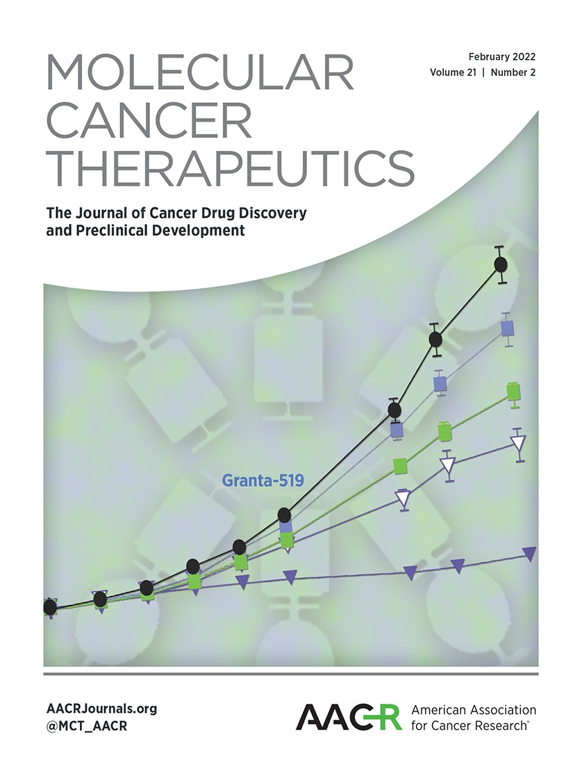
Co-targeting of MCL1 and BCL2 is highly effective in subsets of aggressive B-NHLOur data suggest that BCL2 protein might serve as a buffer for pro-apoptotic proteins released from MCL1 upon exposure to S63845 and induce resistance to S63845 (Fig. 2B). Thus, pharmacologic targeting of both, MCL1 and BCL2 proteins might represent an effective treatment strategy to overcome BCL2-induced resistance in aggressive B-NHL. The efficacy of S63845, venetoclax, or the combination of both agents (SV) was tested in vitro in selected DLBCL and Burkitt lymphoma cell lines and in vivo on a panel of six PDX models established from patients with treatment-refractory DLBCL (VFN-D1, VFN-D2, VFN-D9), Burkitt lymphoma (VFN-B2) or Richter syndrome (VFN-R1, VFN-R2; Fig. 6). All Burkitt lymphoma cell lines were resistant to venetoclax in vitro (i.e., displayed < 50% cell death after 24-hour exposure to 1 μmol/L venetoclax; Fig. 6B). In Burkitt lymphoma, the best in vitro efficacy (as assessed by % apoptotic cells) of SV combination was seen in UPF9T cell line characterized by the highest expression of BCL2 (Fig. 6B). Similarly, in BCL2-positive VFN-B2 model (a PDX model derived in parallel from the same patient as UPF9T), a better than additive effect of SV combination was observed in vivo (Fig. 6C and D; Supplementary Table S1). In DLBCL, SV combination induced additive to better than additive effects in BCL2-positive DLBCL cell lines and BCL2-positive PDX models of aggressive lymphomas (VFN-D1, VFN-D9, VFN-R1, VFN-R2), while only a modest or additive effect of the combination of both drugs was observed in BCL2-negative cell lines and VFND2, the BCL2-negative PDX model of DLBCL (Fig. 6A, C, and D; Supplementary Table S1). In summary, SV combination might overcome BCL2-induced resistance and enhance the activity of MCL1 inhibitors in BCL2-positive B-NHL.
 Figure 6.
 Co-targeting of MCL1 and BCL2 is highly effective in subsets of aggressive B-NHL. Cytotoxic effect of S63845, Venetoclax (VEN) or combination of both agents (S63845+VEN) in DLBCL (A) and Burkitt lymphoma (B) cell lines. Concentrations of S63845 and VEN were chosen based on known sensitivity of particular cell lines to S63845 and VEN. Colorful labels indicate level of BCL2 protein expression normalized to Actin. C, Antitumor efficacy of S63845, venetoclax (VEN) or combination of both agents (S63845+VEN) compared with untreated control animals (CTRL) in six PDX models of aggressive B-NHL. Graphs depict growth curves of subcutaneous tumors in the individual treatment cohorts. x axis displays days, where day 1 (D1) marks initiation of the therapy. y axis shows calculated tumor volumes in the individual treatment cohorts calculated from three perpendicular tumor dimensions (6–8 mice per cohort, means ± SDs). For details, see Materials and Methods. D, Western blot analysis of BCL2, BCL-XL, and MCL1 proteins in PDX models of aggressive B-NHL, including treatment-refractory DLBCL (VFND1, VFND2, VFND9), Burkitt lymphoma (VFNB2), or Richter syndrome (VFNR1, VFNR2).
DiscussionMCL1 is a promising therapeutic target for treatment of cancer, including lymphomas (25, 26). In this study, we investigated the mechanisms of sensitivity/resistance of aggressive B-NHL to recently developed highly specific MCL1 inhibitor S63845 using an extensive panel of established cell lines and PDXs. We demonstrated that a subset of DLBCL and majority of Burkitt lymphoma are dependent on anti-apoptotic MCL1 protein for their sustained survival. We showed that the level of BCL2 and MCL1 protein expression and occupational status of MCL1 are the key determinants of susceptibility to pharmacologic MCL1 inhibition. Finally, we proposed combined treatment approaches that could overcome BCL2-induced resistance and enhance the activity of MCL1 inhibitors in aggressive B-NHL.
We demonstrated that a subset of DLBCL and the majority of Burkitt lymphoma can be effectively targeted by S63845. Our results preclinically confirm previous studies that pointed out MCL1 as a critical survival molecule in both DLBCL and Burkitt lymphoma (17, 27). The concept of pharmacologic blockage of MCL1 is a promising strategy. Several small-molecule MCL1 inhibitors are currently being evaluated in prospective clinical trials in patients with various cancers (28). Importantly, conditional gene knockout studies demonstrated that MCL1 is essential for survival of several normal, non-transformed cell types, including hematopoietic stem cells or cardiomyocytes, raising the question of tolerability when targeting MCL1 (29–31). The current study, as well as previous studies showed that S63845 is well tolerated in vivo in murine PDX models of B-NHL (Supplementary Fig. S5; ref. 18). However, S63845 has an approximately 6-fold higher affinity for the human protein in comparison to murine MCL1 indicating that mouse models are not sensitive enough to reveal all potential toxic side effects (32). Early clinical trials evaluating safety profile of MCL1 inhibitors will be essential for further clinical development of the currently tested agents.
Our data demonstrate that BCL2 protein belongs to key modulators of sensitivity/resistance to S63845. Mechanistically, our data suggest that BCL2 likely serves as a buffer for pro-apoptotic protein BIM released from MCL1 upon exposure to S63845. Genetic alterations of BCL2, as well as high BCL2 protein expression are found in subsets of DLBCL and have been repeatedly associated with poor outcome (10, 33–35). In our dataset, the level of BCL2 protein expression was variable in DLBCL, with approximately 30% of the tested cell lines showing no detectable BCL2 protein. In accordance with the generally recognized molecular features of Burkitt lymphoma and in contrast to DLBCL, Burkitt lymphoma cell lines were either BCL2-negative or expressed low levels of BCL2 protein (Fig. 1B), possibly explaining their overall higher sensitivity to S63845 (14). BCL2 protein expression is routinely assessed in clinical practice and could serve as a predictive biomarker for selection of patients with DLBCL or Burkitt lymphoma who might benefit from MCL1 inhibitors. Some studies have shown that BCL-XL might induce resistance to several BCL2- or MCL1-targeting BH3 mimetics (36). In our dataset, BCL-XL was ubiquitously expressed in all tested DLBCL and Burkitt lymphoma cell lines and we did not find correlation between BCL-XL protein level and sensitivity to S63845 (Fig. 1B; Supplementary Fig. S3C).
In contrast to BCL2, we observed positive correlation between the level of MCL1 expression and sensitivity to S63845. We and others have shown that cells, whose anti-apoptotic BCL2 proteins are occupied by pro-apoptotic BH3 domain-only activators or effector proteins are “primed for death” and can undergo rapid apoptosis when exposed to BH3 mimetics (19, 24, 37). In this study, MCL1 protein in S63845-sensitive DLBCL and Burkitt lymphoma cell lines was occupied by BIM, BAK1, and NOXA (Fig. 3B). In such cells, S63845 displaces the pro-apoptotic activator BIM or effector protein BAK1 from MCL1 thereby triggering apoptosis. Such pattern of MCL1 binding specificity was, however, also observed in S63845-resistant Burkitt lymphoma cell line Raji (Fig. 3B), indicating that there might be other mechanisms mediating resistance to S63845 in cell lines that are primed for death on MCL1 (38, 39). Interestingly, in S63845-resistant cell lines, the MCL1 was occupied predominantly by NOXA (19). NOXA is a BH3-only protein that selectively binds to MCL1 and functions as a pro-apoptotic sensitizer. Consequently, NOXA released from MCL1 upon ex
留言 (0)