Vertebrae at the thoracolumbar junction: A quantitative assessment using CT scans
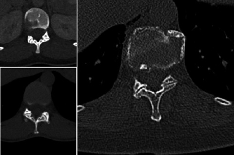
The thoracolumbar junction is often associated with traumatic injuries, due to its biomechanical instability. Reasons for this instability are currently still under debate; however, contributing factors such as the rapid change in spinal curvature and facet orientation from the thoracic to lumbar transition have been implicated. Normally, the superior facet orientation in the thoracic region is angled in a coronal plane, whereas vertebrae in the lumbar region have facets angled in the sagittal plane. Distinguishing between thoracic, lumbar, and transitional vertebrae at the thoracolumbar junction based on articular facet angles, using quantitative methods on CT scans has, to the authors' knowledge, not yet been reported in the literature. Therefore, this study aimed to evaluate whether quantitative measurements can be clinically applied and used to differentiate vertebrae at the thoracolumbar junction using CT scans and, additionally, to record possible cases of congenital defects or variations observed in the spine. A sample (n = 173) of CT scans representative of the Windhoek population in Namibia was retrospectively assessed using radio-imaging software. Measurements of the angle formed by the superior facets of the vertebrae at the thoracolumbar junction (T11-L1) were recorded. Based on the results of this study, quantitative morphometry of the superior facet of vertebrae can differentiate between thoracic, lumbar,. and transitional vertebrae at the thoracolumbar junction. All individuals with identified thoracolumbar transitional vertebrae (TLTV) in this sample had at least one other congenital anomaly of the spine.

留言 (0)