Multi‐modal biomicroscopic system for the characterization of cells with high spatial phase sensitivity and sub‐pixel accuracy
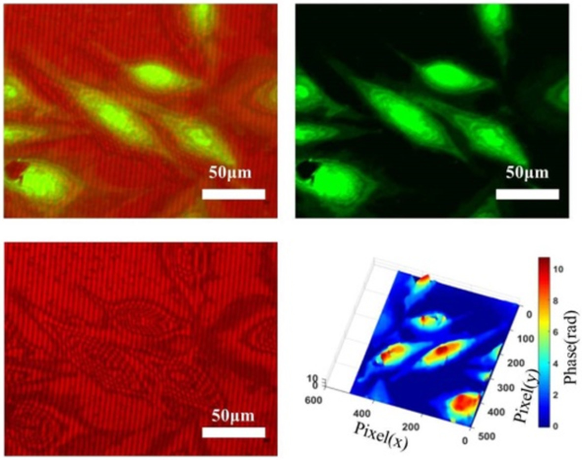
Multi-modal analysis is highly advantageous for various biomedical applications including cancer and brain studies. Simultaneous measurement of quantitative phase with sub-pixel accuracy and fluorescence image is difficult to achieve in single measurement. Conventionally, off- axis interferograms are analyzed using the Fourier-transform method which limits the accuracy of the phase maps by pixel size, and usually the location of the carrier peak is in sub-pixel. We report a multi-modal microscopic system consisting of high-resolution (HR) quantitative phase interferometer (QPI) to retrieve sub- pixel accuracy in phase imaging and an oblique-illumination based fluorescence imaging system which decouples the excited light from emitted signal light to avoid saturation of the camera, both integrated into a single unit. Here, highly-resolved phase maps are obtained using a two-step process. Firstly, using a speckle-free illumination which offers high spatial phase sensitivity. Secondly, using a hamming window for accurate estimation of original signal frequency information and HR discrete Fourier transform (DFT) which offers sub-pixel accuracy in phase measurements. HR-DFT has computational load of O(ABβ), where A×B is the size of the interferogram and β is the upsampling factor, making system computationally more robust and efficient compared to zero-padded FFT. The experiment is conducted on MG63 osteosarcoma and human mesenchymal stem cells( hMSCs) and their quantitative parameters are extracted with significantly improved accuracy. The average phase for MG63 cells and hMSCs; for nucleus is obtained to be 8.02rad ± 0.80rad and 4.29rad ± 0.43rad ,respectively and for cytoplasm is obtained to be 2.63rad ± 0.96rad and 1.73rad ± 0.57rad, respectively.
This article is protected by copyright. All rights reserved.


留言 (0)