Cicatricial Pemphigoid Brunsting‐Perry Variant Masquerading as Neutrophil‐Medicated Cicatricial Alopecia
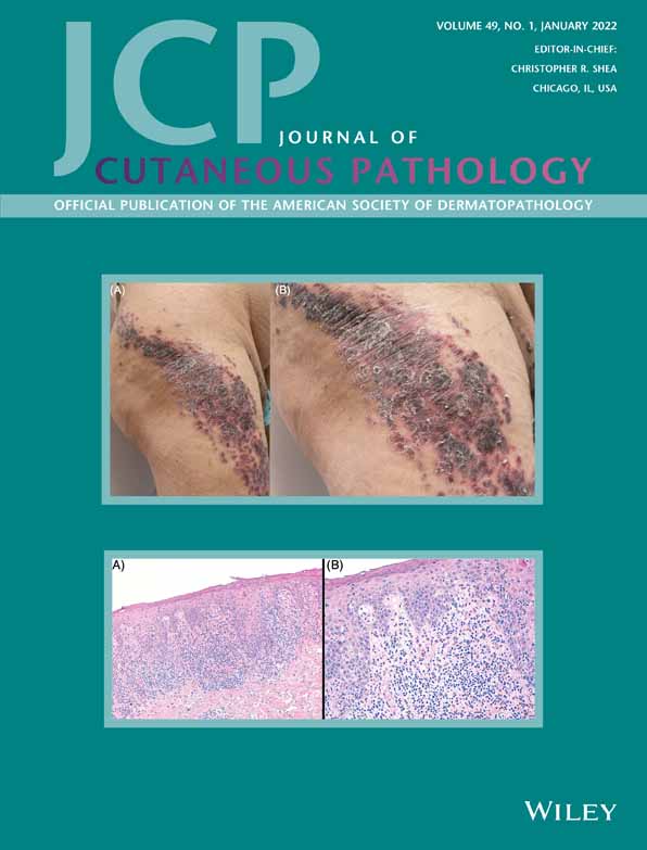
A 72-year-old male presented with scarring alopecia on the scalp vertex, multiple crusted plaques on the hairline, and a history of vesicular eruption on the face. The scalp showed crusted plaques with loss of follicular ostia. No follicular pustules or compound follicles were present. An initial transverse scalp biopsy showed perifollicular neutrophils, lymphocytes, and plasma cells along with dermal fibrosis. Focal epidermal/dermal and follicular/adventitial dermal clefts were apparent but were thought to be secondary to fibrosis, and the biopsy was interpreted to represent a neutrophil-mediated cicatricial alopecia. Concurrently, direct immunofluorescence (DIF) analysis demonstrated linear junctional deposition of IgG and C3. A repeat scalp biopsy revealed more prominent epidermal/dermal clefts, fibrosis, mixed infiltrate with neutrophils, lymphocytes, histiocytes and plasma cells and prominent follicular/adventitial dermal clefts with perifollicular neutrophils. Given the combination of clefts, perijunctional neutrophils, and positive DIF findings, it became clear that this eruption represented the Brunsting-Perry variant of cicatricial pemphigoid. Here, we illustrated that a neutrophil-rich form of cicatricial pemphigoid can masquerade as a neutrophil-mediated scarring alopecia. In evaluating a specimen suspected to be a neutrophil-mediated scarring alopecia, one should be alert to the presence of subepidermal and perifollicular clefting, and consider cicatricial pemphigoid.
This article is protected by copyright. All rights reserved.


留言 (0)