
記住我


The use of microscopes for the study of immune cells dates back to the 19th century with the German Pathologist Rudolf Virchow being one of the most prominent leaders in the field early on. It was him who coined the term “Zellularpathologie” (cellular pathology1), while the famous concept associated with this theory, “omnis cellula e cellula” (all cells are derived from previous cells) was probably not originally from him.2 Virchow was one of the earliest who observed by microscopy that inflammatory cells isolated from a punctured hydrocele were motile. Considering the inflammatory condition where they were harvested from and when looking at the images in his publication it might well be that he was looking at neutrophil granulocytes or macrophages at the time.3 Hence, this could be the first description of the motility of these cells. More well-known are the famous studies by the Ukranian scientist Ilya Metchnikoff 20 years later. Starting from observations of phagocyte recruitment to rose thorns in starfish, he then showed in amphibian larvae that an inflammatory trigger (both sterile and infectious) can recruit migratory and phagocytic leukocytes from the surrounding tissue and the blood to the inflammatory focus. From his microscopical observations, he hypothesized all necessary steps of what we would call today a chemotactic cascade4 as being required for the recruitment of migratory leukocytes from the circulation. An inflammatory stimulus acts on local phagocytes, these stimulate near-by endothelial cells which are key to recruit leukocytes from the blood.4 Metchnikoff also coined the term macrophage and “microphage,” the latter later becoming the neutrophil granulocyte.5, 6 Hence, intense motility, phagocytosis, and ultimately cell death as a result of these actions have all been shown to be key characteristics of innate cellular immunity as soon as investigators focused at the relevant cells with their microscopes. Almost 150 years later, model systems have changed fundamentally in terms of complexity and relevance for the human situation, but we are still fascinated by these features and continue to unveil novel secrets of innate immune function. Until today this has led to more than 400 000 papers in PubMed (more than 1% of all papers) dealing with either macrophage or neutrophil function, with microscopy being used in almost 34 000 of them.
Given its sheer volume, it is obviously impossible to provide a comprehensive review on all the available research. We have chosen instead to focus our overview on the analysis of neutrophil granulocytes and macrophages in sterile and infectious inflammation. Initially, research was restrained to simple and small model systems that allowed intravital inspection, permitting just glimpses into dynamic immune cell interactions in physiological and pathophysiological states. Intravital imaging and animal experimentation have grown a lot from the early days of Virchow and Metchnikoff. With present-day genetic tools, it is possible to optically label neutrophils in vivo in larger vertebrates including mammals. For instance, this is achieved by using mice where myeloid cell-specific promoters for genes such as Lysozyme (LysM)7 are employed to drive the expression of fluorescent proteins.
In addition to this rather frequently used but less specific system, we developed a mouse model using the promoter for the membrane protein Ly6G as a target for highly specific expression of the fluorescent protein tdTomato in neutrophil granulocytes. In addition, this mouse model hosts a specific knock-in allele expressing Cre recombinase upon Ly6G expression which allows the simultaneous modulation and visualization of neutrophils.8 This mouse model is termed “Catchup” and to date exhibits the most specific expression of fluorescence in neutrophils. In the meantime, a variant of this model with an inducible Cre recombinase has been generated that allows highly original studies into neutrophil lifetime and kinetics.9 Similar approaches have been employed for the targeting of macrophages and microglia, with the CX3CR1 promoter being a particularly widely used example.10
Also microscopy has developed fundamentally in the last 30-40 years. Present day high-end microscopes use lasers with their highly defined light emission for illumination and fast electronics for signal read out and highly performant computers for image generation and analysis11—what a difference from xylographs and descriptions that read like adventure stories from our scientific ancestors.
In the frame of this review, we will focus on confocal microscopy and its intravital-optimized derivative multiphoton microscopy.12 However, while intravital imaging is ideal to observe the activity of individual cells with high resolution, even achieving super resolution way beyond diffraction-limited optics,13, 14 it can only observe a relatively small region of interest (ROI, around 0.2–1 mm3) at the same time. Compared to the size of organs, which exceed one or more centimeters even in mice, a small ROI limits the conclusions that can be made from intravital movies alone. Hence, tissue level imaging is an essential addition to the microscopic armamentarium of present-day research. Here, light sheet microscopy (LSFM) has been of enormous value. Originally described in the early 20th century,15 it took until the last two decades that the technology had matured enough to allow wide-spread application in biological laboratories.16-18 Since then there has been an enormous number of studies with immune-related research being a strong focus.
Within this review, we will first introduce a particular method as listed above, thereby giving enough technical background to understand the principle, chances and limits of the approach, yet without digging too deep into complicated physics, which is dispensable for understanding its use in imaging innate immunity. Then, we will discuss our selection of studies that have made use of the described technology for the investigation of neutrophil and macrophage function. At the end, we will provide our vision of where the research should go next, both, technically and regarding the scientific challenges that we consider essential to be solved in the coming years.
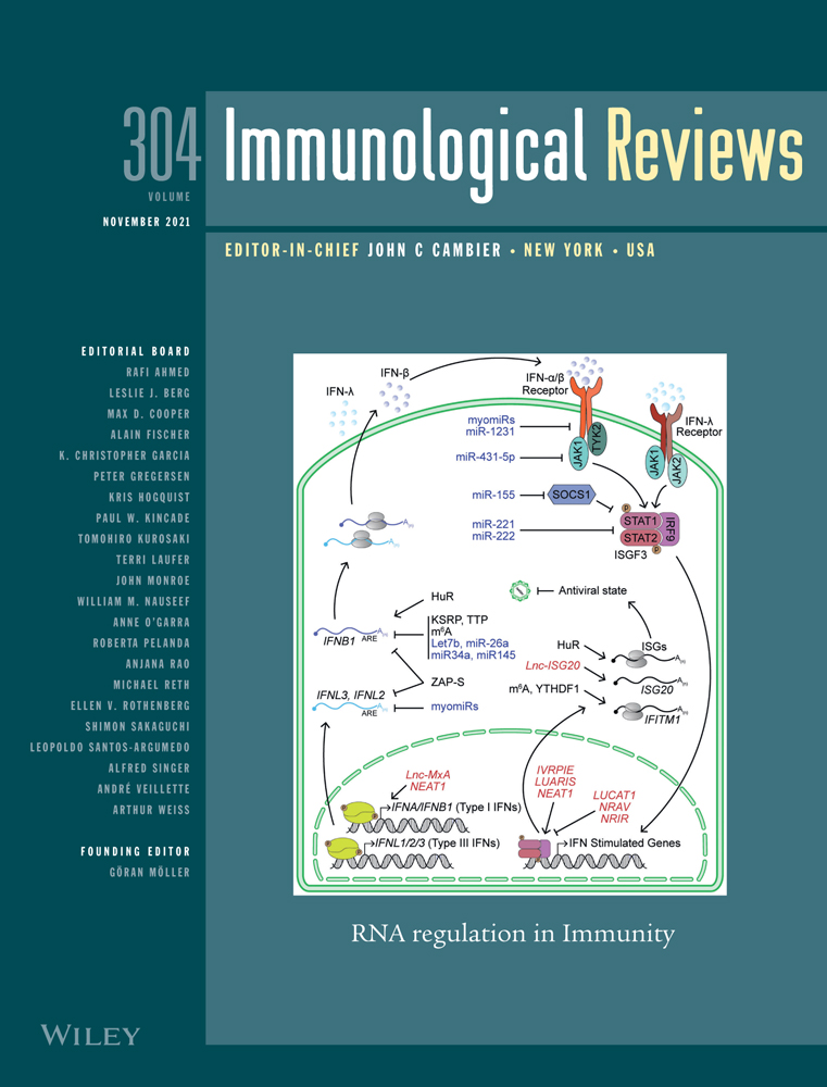
2 CONFOCAL MICROSCOPYConfocal microscopy is nowadays a well-established technique to address biological problems. Compared to conventional fluorescence microscopy, confocal microscopy is able to achieve high-resolution and -contrast images by blocking out-of-focus light in thicker specimens.19 Over time, this imaging technique has been constantly improved, but the fundamental design has remained unchanged. Laser light is focused by collimating and beam-steering optics, so that a small focal spot (its size is diffraction limited to minimum ~200 nm diameter) in the specimen is illuminated, thereby generating light emission from artificially introduced fluorescent molecules such as labeled antibodies, small molecules, or fluorescent proteins.19, 20 Also, the ability of fibrous proteins such as collagens to reflect light can be used for image generation.21 Fluorescent light emitted from the illuminated sample has to pass a pinhole, which is positioned in an optically conjugate plane in front of the detector and can be adjusted in size like the aperture in a photographic camera. The pinhole ensures that out-of-focus light is rejected and only signals from the focal plane are recorded.19, 20 When the pinhole is smaller than an Airy unit, which is defined as the best-focused spot of light in an optical system (Figure 1A,B), the resolution is ideally about 200 nm laterally and 500 nm axially.22, 23 As a big advantage of confocal microscopy, the pinhole allows to observe thin optical Z-sections in a 3-D sample without physically cutting it (Figure 1C). By making a series of images in different depths (a Z-stack), it is thus possible to generate highly resolved tomograms of a cell or tissue analogous to a computer tomogram in humans but with diffraction-limited resolution24 (Figure 1D). When such Z-stacks are performed repeatedly on live samples it is possible to generate 3-D videos of migrating cells, which is a powerful approach to investigate the dynamic pericellular probing of cells migrating in tissues.25-28 In addition, the tomograms can later be reconstructed into a 3D model, which helps to understand the spatial arrangement of labeled targets and allows for colocalization analysis of different molecules.29, 30

Fluorescence microscopy approaches for imaging innate immune cells. (A) Exemplary photography of a street light demonstrating the Airy pattern. Enlargement of the indicated white box with schematic representation of local intensity maxima (red arrows, left; red rings, right). Camera settings: 28 mm, ISO12800, 1/15-s exposure, f/16. Image size: 9504 × 6336 pixels. (B) Schematic drawing of light diffraction at a circular aperture, resulting in an airy disk pattern with decreasing intensity from the central region to the outer diffraction rings. (C) Comparative optical resolution of human neutrophils (CD15, green; CD66b, red; DAPI, blue) imaged by widefield and confocal laser scanning microscopy. Scale bars = 5 µm. (D) Optical sectioning by confocal microscopy enables the 3-dimensional reconstruction of a scanned human neutrophil and volumetric rendering of its cellular components (CD15, green; CD66b, red; CD11b, white; DAPI, blue). Scale bars = 2 µm. (E) Intravital TPLSM enables the visualization of immune cells in the living organism. Here, neutrophils (CatchupIVM, red) and macrophages (Cx3cr1-EGFP, green) are simultaneously detected at the skull exterior skin (SHG, exterior view) and in the bone marrow of the calvaria (SHG, interior view). Scale bars = 50 µm. (F) Light-sheet fluorescence microscopy is a powerful tool for three-dimensional analysis of whole organs. Here, the spatial distribution of different macrophage populations (Cx3cr1cre:R26-tdTomato, green; F4/80, blue) within a knee joint and adjacent subchondral bone (tissue autofluorescence, gray) as well as their localization with respect to the surrounding blood vessel network is shown (CD31, red). Scale bars = 200 µm
We started in the late 90ies and early 2000s to use confocal microscopy and 3D collagen matrices to spatially visualize the migration behavior, signaling, and cell-environment interactions of neutrophils, dendritic cells, and lymphocytes.27, 28, 31, 32 This phase also included our first attempts to study T cell activation in lymph nodes in vivo,33 a key step for the laboratory, although not focusing on innate immunity.
Though neutrophils have a pivotal role in the first-line defense against pathogens,34 some bacteria such as the stomach lining bacterium H. pylori can reside in a neutrophil-rich environment without destruction. Confocal images revealed the redirection of destructive superoxide anions into the extracellular milieu thereby preventing NADPH oxidase destruction inside the phagosome.35 Neutrophil studies with confocal microscopy moreover significantly contributed to our understanding of bacterial killing mechanisms. Next to phagocytosis and enzymatic degradation, confocal images of neutrophils stained for neutrophil elastase, nuclear DNA, and H2A-H2B-DNA complexes showed the formation of neutrophil extracellular traps (NETs) as a novel suicidal defense mechanism, in which neutrophils externalize their DNA as sticky glue rod in an explosive process to ensnare pathogens such as bacteria36 or fungi extracellularly. In line with this, we were able to show that NET formation is a highly dynamic process, which is especially pronounced against hyphae of the pathogenic fungus Aspergillus fumigatus and thus presumably counteracts the spread of the fungus.37 In another study, we made the surprising discovery that neutrophils are able to sense the dimensionality of their environment and this has dramatic consequences for their efficiency to fend off different fungal pathogens. Here, neutrophils efficiently phagocytosed fungal pathogens that typically infect tissues like Candida albicans, in 3-D but not in 2-D environments, while the same cells could phagocytose the more surface-restricted inhaled pathogen A. fumigatus best in 2-D and not so efficiently in 3-D.38 The mechanisms behind this environmental sensing still remain obscure.
The release of NETs is a highly organized process that was able to be separated into three distinct phases (t1: chromatin expansion with intact nucleus, t2: chromatin expansion until cell membrane, t3: ruptured membrane and NET release) with live-cell confocal microscopy.39 NETs were further shown by the help of confocal microscopy to degrade pro-inflammatory mediators. Here, we could prove that neutrophil activation and NET formation during eye closure and opening ameliorate the inflammation at the ocular surface.40 Another anti-inflammatory mechanism of neutrophils was also proven to act by reprogramming of macrophages via NF-κB.41 In contrast to their many protective functions, neutrophils can also have adverse effects, for example, in controlling cancer growth, which was characterized using confocal microscopy. The excessive formation of NETs after radiation therapy was shown to induce radio resistance of bladder cancer.42 Other studies had similar findings that revealed the contribution of neutrophils to tumor progression by NET formation.43 Furthermore, unwanted effects of NETs include impaired vascular remodeling and revascularization after ischemic stroke44 and formation of immunothrombosis in COVID-19 acute respiratory distress syndrome.45 Additionally, NETs can also exert regulatory influence on other immune cells, such as macrophages. For example, in a sepsis model, NETs were shown to induce macrophage pyroptosis and thus exert a pro-inflammatory influence on the course of infection.46 However, in contrast to this pro-inflammatory influence, it has been shown recently that NETs can also exert anti-inflammatory regulatory functions on other immune cells by enhancing the phagocytotic activity of macrophages.47 While the formation of NETs by neutrophils has been intensively studied since its discovery in 2004, it has only recently become known that other immune cells also possess this mechanism. For example, macrophages can eject extracellular traps that, like NETs, exert antibacterial and inflammation-regulating functions. Accordingly, in addition to its cell-specific designations, this mechanism is now also commonly referred to as "ETosis" and its immune cell spanning regulatory functions are being studied in diverse disease contexts.36, 48, 49 Next to ex vivo studies, confocal microscopy can also be used for in vitro studies, which are important to, for example, understand TNFα-mediated trafficking of neutrophils into and within lymphatic vessels during acute inflammation.50 Unsurprisingly, given the enormous importance of NETs for a multitude of physiological and pathological conditions, current approaches also include the development of automatic detection and quantification methods for NETs by confocal microscopy for the diagnosis and measurement of therapy efficiency.51
Hence, while obviously being a powerful discovery tool, the downsides of confocal microscopy are a narrow field of view (< 1 mm diameter), which results from objectives that feature a high numerical aperture.52 Due to point illumination and pinhole, only a part of the specimen can be imaged at a certain time. Two different approaches have been made to solve this problem. A confocal laser scanning microscope (CLSM) performs raster-scanning of the entire area of observation to generate a virtual 2-D image pixel by pixel in the controlling computer. While this scanning process is fairly time intensive, CLSMs are highly sensitive since they use photomultiplier tubes (PMTs) or hybrid detectors (HyD), for detection of emitted light. To speed up confocal imaging, spinning disc confocal microscopes (SDCM) scan multiple points at the same time. This is achieved by using a constantly spinning Nipkow disc, which has hundreds of pinholes that are arranged in a spiral. Signals have to be collected in the entire field of view, so that CCD cameras are mainly used in SDCMs for image generation.19, 53 The Kubes group has intensively used SDCM in live mice to study neutrophil function in vivo. For example, they showed the infiltration of neutrophils in inflamed tissue based on inflammatory signal recognition54 and demonstrated that neutrophils are essential for generating blood vessel channels for revascularization in necrotic tissue.55 Others have shown how the interaction between neutrophils and nonclassical monocytes in the circulation of kidneys triggers glomerular inflammation56 and that the Btk signalosome is essential for the recruitment of neutrophils to sites of tissue necrosis.57
3 INTRAVITAL TWO-PHOTON MICROSCOPYConventional confocal laser scanning microscopy is able to generate movies from innate immune cells such as monocytes and macrophages in mice in vivo,58, 59 but it is difficult. A severe limitation is the relatively shallow tissue penetrance due to light scattering. This can be overcome to a large part by two-photon laser scanning microscopy (TPLSM). Consequently, TPLSM has become a powerful tool for studying biological function in live tissue thereby offering many advantages over conventional imaging techniques. In comparison with one-photon fluorescent microscopy techniques, fluorescent dyes or proteins in TPLSM are excited not by a single photon but by two photons of lower energy, which provide the required excitation energy upon near simultaneous absorption that effectively combines the energies of both photons.60-63 Thus, the required wavelength to excite a given fluorophore in TPLSM is red-shifted to roughly two times its excitation wavelength in one-photon excitation.64-66 Since it is using excitation with red-shifted wavelengths, which are less subject to scattering by tissue structures, TPLSM can penetrate tissues much more deeply than one-photon microscopy, thereby reaching several hundreds of µm in native samples.12 Further, due to the nature of its generation, which requires extremely high photon fluxes, 2-photon excitation only occurs in a limited spherical volume of the sample that is defined by the refractive features of the microscopy optics. As a result, and opposed to confocal microscopy, which generates a complete hour-glass-shaped double cone of light along the entire illumination path in the sample, TPLSM is inherently confocal (without the need for a pinhole). This minimizes phototoxicity and bleaching of fluorophores above and below the focal spot. Therefore, TPLSM is well suited for long-term imaging of live cells without compromised tissue viability. The method typically employs Titanium:Sapphire (Ti:Sa) crystal-based pulsed lasers that feature wavelength tunability from ~700 - 1100 nm providing two-photon excitation of fluorophores in the ultraviolet to deep red region of the light spectrum.67-69 In addition, using a non-linear optical process called second harmonic generation (SHG) TPLSM allows to visualize unlabeled structures formed by molecules with anisotropic fiber-like conformations, such as collagen and myosin70 (Figure 1E).
TPLSM became widely available in the early 2000s and immunologists quickly adopted this technique to investigate dynamic cell functions in vivo.71-73 Consequently, intravital imaging has made considerable contributions to our knowledge of how the immune system acts under different conditions. Early neutrophil studies showed the ability of parasites to hijack infiltrating neutrophils for body invasion74 or how an uncontrolled neutrophil infiltration wreaks havoc in the CNS following viral infection.75 The neutrophil responses to bacterial skin infection were highly reminiscent of the old Metchnikoff descriptions, yet with methods of the 21st century.76 We could show how neutrophils, which are essential for the defense against a fungal lung infection, contain the invader by phagocytosis.37 But neutrophils are also key cells under conditions of sterile inflammation. It was in fact in a model of sterile skin necrosis that a particular feature of neutrophil tissue invasion was described, a collective and self-enhanced migration pattern referred to as “neutrophil swarming”.77, 78 This swarming principle appears to underlie neutrophil tissue invasion in general, also under conditions of infection with various pathogens, including bacteria, fungi, and parasites.79 Swarming of neutrophils into inflamed tissues requires a series of sequential phases: (a) initial chemotaxis of individual neutrophils close to the damage, followed by (b) amplified chemotaxis of neutrophils from more distant interstitial regions, leading to (c) neutrophil clustering.79 TPLSM again helped to identify the molecular players of this swarming behavior. In mouse models of sterile injury and infection, Lämmermann et al showed a critical role for an intercellular signal relay among neutrophils mediated by leukotriene B4.78 Furthermore, very recently it was shown that GPCR desensitization acts as a negative feedback control mechanism to stop permanent neutrophil migration in swarm aggregates.80 It is highly interesting that resident macrophages appear to use their membrane processes to shield small tissue lesions from direct neutrophil contact, which would otherwise induce a massive swarming response.81 Hence, swarming is an important reaction to tissue damage, yet needs tight control to avoid overt collateral injury.
In fact, when re-assessing observations made in a different context we and others might have observed that swarming or at least a phenotypically highly reminiscent group migration also underlies more physiological responses of neutrophils in the bone marrow to a systemic trigger with the hematopoietic cytokine granulocyte colony-stimulating factor (G-CSF82-84). Using intravital TPLSM, we demonstrated that G-CSF stimulates emergency mobilization of neutrophils from the bone marrow to peripheral sites in a CXCR2-dependent manner, where the entry of individual cells into the circulation occurred on exit hotspots in the bone marrow in a highly coordinated manner reminiscent of swarming.82, 83 TPLSM was also essential for us to demonstrate that neutrophils early and massively invade the brain following stroke.85 Here, the interaction with local phagocytes seems essential for control, as brain-invading neutrophils are phagocytosed by resident microglia to contain the additional damage86, 87 and failure to do so exacerbates the outcome of the insult.88
Neutrophils also cooperate with monocytes and platelets in the induction and propagation of venous thrombosis as it was demonstrated by the Massberg group.89 In addition to elucidating diverse neutrophil-specific mechanisms, intravital TPLSM is also excellent for understanding more basic cellular processes. It was recently shown that ion efflux from the endocytic pathway is an essential mechanism in macrophages to regulate pinocytosis of extracellular fluid.90 The analysis of macrophages in their physiological environment could also help to understand how they migrate into tissues and infiltrate tissue lesions. Intravital TPLSM allowed to demonstrate that macrophages can infiltrate sterile tissue lesions in visceral organs very rapidly via a nonvascular route. In a sterile liver injury model, recruitment of peritoneal macrophages occurred by direct migration from the peritoneal cavity and was dependent on the alarmin ATP and CD44. This mechanism, which deviates from the classical recruitment pathway via vascularization, allows a much faster recruitment of macrophages to the tissue damage and thus a faster restitution.91
Cancer is another form of sterile inflammation. During the last decade, a high intratumoral frequency of tumor-associated neutrophils (TAN) was established as a strong predictor of poor clinical outcome in the majority of solid tumor entities92, 93 and we identified the immunosuppressive function of neutrophils as one potential mechanism for this.94 Other mechanisms include angiotropism95 or a type of cellular chaperoning for metastasizing cancer cells in the circulation.96
Our development of the Catchup mouse8 allowed the targeted observation of unperturbed TAN in the living mouse during very early tumor establishment in vivo.97 In a model of skin-based tumorigenesis, we showed that peritumoral and intratumoral TAN differ strongly by their motile behavior. Intratumoral TAN displayed prolonged tumor-associated persistence and reduced motility compared with peritumoral TAN, whose velocity increased with tumor progression. Furthermore, the selective pharmacological blockade of CXCR2 receptors effectively inhibited the recruitment of TAN into peritumoral regions, while intratumoral infiltration was only transiently attenuated.98 Indeed, successful cancers seem to be characterized by their ability to recruit neutrophils via production of CXCR2 ligands.99 Therefore, the efficient blockage of neutrophil infiltration in tumors might be a promising target for future studies. For this one could establish an initial in vitro high-throughput screen searching for compounds with the potential to inhibit the directed migration of neutrophils into tumors. Potential hits worth additional investigation could then be tested for their activity in vivo using intravital imaging. Beyond neutrophils also macrophages massively infiltrate tumors and can regulate both tumor cell dynamics and their migratory behavior.100, 101 Thereby, contact with macrophages promotes the formation of pseudopodia-like extensions in tumor cells and more rapid tumor cell migration compared with non-macrophage-associated cancer cells.101 Collectively, intravital imaging has provided key insights into tumor-immune interaction and continues to do so.
4 TISSUE LEVEL IMAGING WITH LIGHT-SHEET MICROSCOPYWhile intravital TPLSM is clearly a highly relevant technology to study neutrophil function in vivo, it suffers from a severe limitation, namely a very small field of view (FOV). A typical intravital TPLSM experiment observes a cube of tissue with a volume comprising less than one per mill of the involved organ in mice. Hence, in order to understand innate immunity on a global scale, tissue level or mesoscopic imaging is required.
LSFM is a technique that allows mesoscopic tissue imaging by utilizing a sheet of light for excitation, obtained by focusing the laser beam into a single plane within the sample by, for example, using a cylindrical lens.11, 18 This sheet of light allows signal acquisition of the entire plane instead of a single point, thus increasing the recording speed significantly. The other key feature of LSFM is the fact that the image acquisition is obtained by separate optics which are arranged perpendicularly to the plane of the light sheet. Modern LSFM systems use highly sensitive digital cameras to detect the fluorescent section through the sample that is illuminated by the light sheet.16, 102 Since only a part of the sample is illuminated, LSFM also shows minimized phototoxicity and photobleaching effects, as these are restricted to the illuminated plane of the sample. Similarly, the planar illumination also causes lower scattering effects than for example widefield microscopy, and hence, the acquired images have higher contrast. Acquiring a larger plane also means a larger field of view, which in return requires a lower numerical aperture (NA). As the resolution of a system is dependent on the NA, LSFM systems come with lower resolution than for example confocal microscopy.11
A key requirement for LSFM of tissues is high transparency of the sample. Naturally occurring transparent samples such as zebrafish or Drosophila larvae can be imaged directly and even as live animals103, 104 but vertebrate organs are naturally opaque and hence are not suitable for direct LSFM investigation. Therefore, organs have to be treated with a mixture of various chemicals in order to make them optically transparent in a process called clearing.105, 106 Originally invented in the early 20th century by the German anatomist Spalteholz,107 there has been an enormous development of techniques for organ clearing within the last 10-15 years, which we will not detail here. The reader is referred to several excellent reviews on the issue that appear regularly in updated fashion.105, 108 Once an organ is optically clear its tomographic reconstruction by LSFM offers phenomenal new insights into its function (Figure 1F).
LSFM was essential for our discovery of a previously unknown blood vessel system, the trans-cortical vessels (TCVs), which provide a very rapid direct transport route for blood and neutrophils (and likely all other blood-resident immune cells) into and out of the bone marrow. Further analysis of the LSFM data even showed that these TCVs are indeed the main transport route for blood in long bones.83, 109 Based on the 3-D capacity of LSFM, we also could identify a novel type of macrophages, the so-called lining macrophages, which play a major role in inflammatory joint disease. The application of this imaging method has enabled us to demonstrate that these macrophages form a tight shield around the synovial cavity to prevent the invasion of inflammatory neutrophils and thereby exhibit features of endothelial cells such as tight junctions.29 The Hidalgo laboratory has used LSFM in a seminal paper to show that there is a novel type of cardiac macrophage that is essential for mitochondrial homeostasis.110 In two other studies, they demonstrated that tissue-infiltrating neutrophils control the local status of organs111 and tissues in turn are responsible for functional reprogramming of the infiltrating cells.9 In our own studies, the whole-organ aspect of mouse hearts subjected to ischemia-reperfusion injury revealed a massive infiltration of neutrophils to the injured regions112 while a multimodal imaging approach in which we combined positron emission tomography of lungs with LSFM of the same organs later showed the dramatic impact of the loss of neutrophils on defense against Aspergillus infection in the lung.113 Studies in cleared whole-mount mouse brains exposed to ischemia-reperfusion injury revealed the delayed occlusion of preferential small-sized (<10 µm diameter) microvessels in previously ischemic areas in the subacute stroke phase114 and the angiogenic response of microvessels to a mesenchymal stromal cell-based restorative therapy in the subsequent stroke recovery phase,115 the latter of which depended on neutrophils. We also showed that blood-derived neutrophils are indispensable for angiogenesis in the periinfarct ischemic brain in that model.115
The power of in vivo LSFM in zebrafish was recently harnessed in a study showing the impact of CDK9 inhibitors on neutrophil injury of the inflamed heart.116 Meanwhile, LSFM might now even be possible in live mice using near-infrared dyes to stain the relevant cells, tissues, and blood.117 Combinations of the various techniques described above for multimodal imaging also bear enormous potential for new discoveries.11, 83 What is certainly a future field worth investigating is the use of modeling approaches to understand the physiology of organs better. We have shown that it is possible to generate complex 3-D reconstructions of blood vessel systems in organs and demonstrate the transport of single neutrophils in individual vessels.83 Based on the available 3-D data on the blood vessel tree, it would be highly interesting to apply blood flow modeling to the real data and thereby get an understanding of the volume flow and cellular transport in such systems.118, 119 In the future, this might also allow to assess the impact of interventions on such processes on the level of entire blood vessel trees.
5 WHERE NEXT? NOVEL SHORES FOR THE IMAGING OF INNATE IMMUNITYAs seen, we have learned a tremendous amount on the biology of innate immune cells since the initial description of their motility more than 150 years ago. Even though we are certainly not in a position to look into the crystal ball for the next 150 years, we would nevertheless like to offer a few suggestions here that could provide some impulses for research, at least for the next decade.
Next to NET formation neutrophils express a whole set of antipathogenic mechanisms such as phagocytosis or the production of reactive oxygen species (ROS).120 Hence, to understand their function better, imaging these processes in vivo would be very helpful. As such, novel developments that allow imaging of ROS in vivo are highly relevant.121 Also, tools to detect and study neutrophils in human patients would be desirable. Initial attempts to use radioactively labeled antibodies against CD66b have not found their way into routine application,122 and more recent developments with neutrophil-targeted nanoparticles for positron emission tomography are not yet available for use in humans.123 Also, the presence of neutrophil subsets is a relevant issue that requires further attention, since such subsets appear to be related to disease conditions.124 In the closing chapters, we also detail two additional issues, one experimental and one applied, that deserve to receive more focused research in the years ahead.
5.1 Optogenetics for modulation of neutrophil migrationOptogenetic tools became popular in the last years for controlling brain activity by altering the activation status of neurons.125 This technique has since then not only been improved to alter cell signaling in excitable cells but also in other non-excitable cell types such as T cells126 and macrophages.127 There are many ways to manipulate cell behavior by optogenetic tools. Some of them include the regulation of the mitochondrial membrane potential to remotely influence T cell effector functions at the tumor site128 as well as controlling of calcium influx into the cytosol of T cells to manipulate cell arrest and adhesion.129 It is conceivable that optogenetic tools could also be used in neutrophils to study their various functions and their contribution to tumor development. Since we previously discovered that tumor-associated neutrophils (TANs) are heterogeneous and probably contribute differently to tumor progression,98 it would be of high interest to further characterize them on site. It is now known that the motility of intratumoral TANs is in general low, while peritumoral TANs increase their motility with tumor progression.98 Since neutrophil migration is controlled via intracellular Ca2+ levels, it would be possible to induce migration in intratumoral TANs by activating store-operated Ca2+ entry (SOCE).130 By using genetically encoded Ca2+ actuators (GECAs), SOCE could be selectively induced with light.131 To achieve GECA expression, for example, in T cells, target cells are retrovirally transduced and injected intravenously into recipient mice.132 However, handling of neutrophils in vitro is fairly difficult and leads to apoptosis and change in phenotype.133 Therefore, retroviral neutrophil transduction is no option. Transgenic mouse strains that constitutively express GECAs in neutrophils could prospectively solve this problem. Such studies would help to decipher TAN subset functions and to understand how immobilized intratumoral TANs influence the tumor milieu, so that new approaches for neutrophil-specific tumor treatment would become possible.
Moreover, optogenetic manipulation of calcium signaling could also shed light on age-related decrease of neutrophil function. Since neutrophils from elderly people were found to feature decreased calcium influx,
留言 (0)