Neurosensory anatomy of Varanopidae and its implications for early synapsid evolution
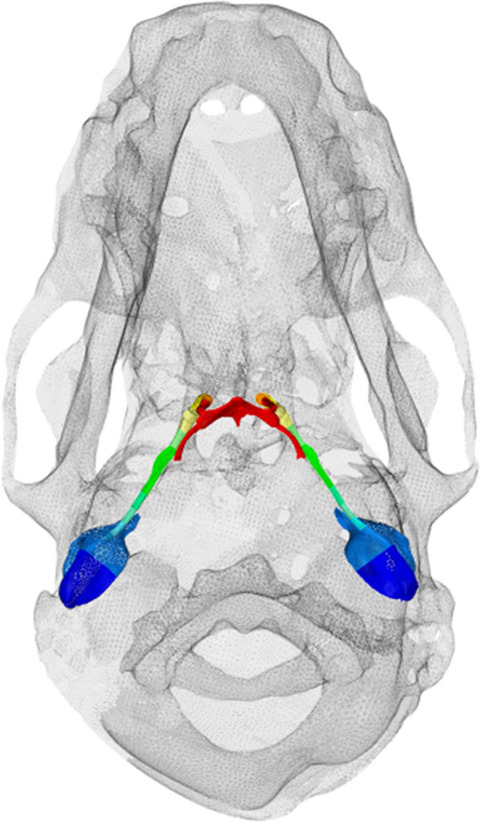
Varanopids are a group of Palaeozoic terrestrial amniotes which represent one of the earliest-diverging groups of synapsids, but their palaeoneurology has gone largely unstudied and recent analyses have challenged their traditional placement within synapsids. We utilized computed tomography (CT) to study the virtual cranial and otic endocasts of six varanopids, including representative taxa of both mycterosaurines and varanodontines. Our results show that the varanopid brain is largely plesiomorphic, being tubular in shape and showing no expansion of the cerebrum or olfactory bulbs, but is distinct in showing highly expanded floccular fossae. The housing of the varanopid bony labyrinth is also distinct, in that the labyrinth is bounded almost entirely by the supraoccipital-opisthotic complex, with the prootic only bordering the ventral portion of the vestibule. The bony labyrinth is surprisingly well-ossified, clearly preserving the elliptical, sub-orthogonal canals, prominent ampullae, and the short, undifferentiated vestibule; this high degree of ossification is similar to that seen in therapsid synapsids and supports the traditional placement of varanopids within Synapsida. The enlarged anterior canal, together with the elliptical, orthogonal canals and enlarged floccular fossa, lend support for the fast head movements indicated by the inferred predatory feeding mode of varanopids. Reconstructed neurosensory anatomy indicates that varanopids may have a much lower-frequency hearing range compared to more derived synapsids, suggesting that, despite gaining some active predatory features, varanopids retain plesiomorphic hearing capabilities. As a whole, our data reveal that the neuroanatomy of pelycosaur-grade synapsids is far more complex than previously anticipated.

留言 (0)