Cytological Scoring for Pancreatic Specimens Obtained by Endoscopic Ultrasound‐Guided Fine Needle Aspiration
Objective
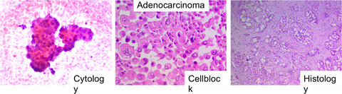
Cytological diagnosis of pancreatic specimens obtained by endoscopic ultrasound-guided fine needle aspiration (EUS-FNA) is often challenging because of the small sample size or well-differentiated adenocarcinoma with weak cytological atypia. Therefore, the sensitivity and specificity of cytological diagnosis for pancreatic cancer should be improved. Hence, we aimed to clarify the utility of cytological scoring to distinguish malignant from benign lesions for the cytological diagnosis of pancreatic EUS-FNA specimens.
Methods
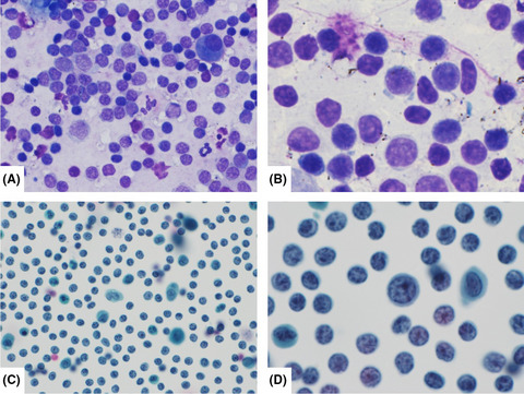
Seven reviewers, including four cytotechnologists and three medical doctors, evaluated 20 morphological indices in pancreatic specimens obtained by EUS-FNA (malignant, n=111, benign, n=31). Statistical analyses were performed using Fisher’s exact test, logistic regression analysis, the area under the receiver operating characteristic curve, and Youden index.
Results
Among the 20 indices, there was a high incidence rate (> 40%) of the following 13 indices in malignant cases: irregular structure, hyperchromatic nucleus, irregular cell polarity, unclear cell boundaries, nuclear membrane thickening, anisonucleosis, overlapping, irregular nuclei, high nuclear/cytoplasmic ratio, binding decline, the simultaneous appearance of malignant and benign cells, enlarged nucleoli, and background necrosis. When we diagnosed pancreatic specimens using these 13 cytological indices, the cutoff value of 8/9 showed the highest Youden index (0.950) as well as high sensitivity and specificity in distinguishing malignant from benign specimens (98% and 97%, respectively).
Conclusion
Thirteen cytological indices showed high sensitivity and specificity in differentiating malignant and benign lesions using pancreatic EUS-FNA samples. All 13 indices were important for diagnosing malignancy in the pancreatic cytology smear of EUS-FNA. Further validation studies are required.


留言 (0)