
記住我


The oral cavity is a diverse and dynamic environment. The three domains of life (Archaea, Bacteria, and Eukarya) along with viruses constitute the human oral microbiome. The ratio of prokaryotic organisms to human cells is reported to range from 1:1 to 10:1 while viral-like particles surpass this prokaryotic ratio and lie closer to 100:1.1 The formation of our commensal microbiome begins shortly after birth and develops continuously throughout our lifetimes.2 Within this ecological unit, there is compositional variation of the human microbiota between body sites as a result of distinct selective pressures. For example, the taxonomic and genomic composition of a microbial community on the coronal portion of a tooth is more similar among individuals than those of the same individuals’ tongues.3
Coexistence with our diverse microbiome equips us with crucial biological functions and traits to protect us from invasion by transient or pathogenic microorganisms. Despite how stable the oral landscape may be, rupture of this symbiosis contributes to oral diseases, such as dental caries, periodontitis, and oral mucosal diseases, and is implicated in a number of systemic diseases.4-7 Traditionally, investigations describing these oral diseases have focused on a bacterial or fungal etiology despite the advancement of viral metagenomics and cultivation methods, which have demonstrated viruses to be drivers of oral blisters, ulcers, and tumors.8 More recently, extensive reviews have detailed the intimate topographic relationship between common herpesviruses and known periodontopathogens and their potential role in the development of periodontal diseases.9
1.2 Periodontal inflammationWithin clinical dental practices, treatment is generally centered on dental and periodontal diseases. Periodontitis continues to be a leading chronic polymicrobial inflammatory condition that affects over 30% of the adult population. Periodontal health can be disrupted when there are shifts in the complex interactions between the microbiota in a biofilm state and the host immune defenses. This dysbiosis can arise from modifications of biological, social, and environmental factors, such as diabetes, alimentary habits, or smoking. Although bacteria are considered the primary etiologic factors in the initiation of periodontitis, host factors influence the progression of periodontitis. Thus, although plaque is necessary for the development of periodontal disease, on its own it is insufficient to drive all the destructive processes that are seen.
Viruses as potential drivers of periodontal disease have been implicated in studies using murine models of disease and human oral subgingival plaque, where virome composition and diversity differ with disease progression.10, 11 Additionally, characterizing the oral virome cannot be limited to eukaryotic viruses without taking into account the most abundant entity, namely, prokaryotic viruses. Prokaryotic viruses, more commonly known as bacteriophages, are the natural predators of bacteria. After utilizing the bacterial machinery for replication, bacteriophages become lytic or lysogenic and consequently may influence bacterial composition and genetic diversity.12 Bacteriophages have a more defined role in the management of biofilms, which in periodontitis has revolved around the more established periodontopathogens, such as those bacterial members of the red complex (Porphyromonas gingivalis, Treponema denticola, and Tannerella forsythia) and the “adhesive” bacterium Fusobacterium nucleatum. Successful bacteriophage-based antibiofilm strategies in animal models have been employed in other systemic conditions as the growth of antibiotic resistance limits the efficacy of standard of care treatment.13 While several bacteriophages have been isolated within the oral cavity and subgingival plaque, the compositional shift that has been described in inflammatory conditions has yet to be linked to changes within the periodontopathogens they infect.
Understanding the effects that certain risk factors have on the development of periodontitis enables better prevention and treatment of periodontitis.14, 15 Consequently, identifying potential etiologic viruses and unraveling their roles in health and periodontitis becomes crucial for improving the outcomes of periodontal therapy. The purpose of this review is to describe how eukaryotic and prokaryotic viruses interact with periodontopathogens and the host immune response, and to examine their potential role in periodontal therapy.
2 EUKARYOTIC VIRUSESAny ecosystem absent of viruses would look foreign or cease to exist. Similarly, human evolution has been influenced greatly by these obligate parasites. Approximately 10% of identifiable functional elements and more than 50% of the “dark matter” in the human genome sequence have been linked to retroviruses’ ability for viral genome integration.16 The long-standing presence of the human virome thus becomes essential to our development. Despite these benefits, the mention of viruses connotes morbidity and mortality, as eukaryotic viruses have the potential to promote cellular degradation, dysregulation, and carcinogenesis.
Compulsory to the life cycle of viruses is the production of messenger RNA that can be translated by host ribosomes. Variable and specialized viral replication strategies, which are derived from environmental pressures that eukaryotic viruses face, drive human disease states. Because of the oral cavity's exposure to the external environment (eg, diet, mechanical abrasion) and variable physiologic states (eg, pH, salivary composition), a diverse and highly individualized oral microbiome has developed.17 Decoding the human oral virome will characterize the environmental stresses that modulate host-viral interactions18 and the composition of bacterial and eukaryotic members in the oral cavity. Members of the human herpesvirus and human papillomavirus families cause the most common primary viral infections of the oral cavity. Below, we describe a number of these putative eukaryotic viruses that have an established role in systemic diseases and oral cancers, but are now being associated with periodontitis.
2.1 Herpesvirus familyHerpesviridae are large DNA viruses that are highly host specific but have the potential to cross host species barriers.19 More than 100 herpesviruses have been identified; however, only eight of them routinely infect primarily humans and these are divided into three subfamilies, alpha-, beta-, and gamma-herpesviruses because of their replicative cycle and range of host infectivity. All herpesviruses can establish latent infection within specific tissues, which are characteristic for each virus. Latency and active replication have been demonstrated to occur within various microenvironments of the oral cavity and the different cell types that comprise the periodontium. Herpesviruses are sensitive to a number of environmental stresses that lead to reactivation of viral replication, cellular lysis, and clinical symptoms. Along with the associations between marginal periodontitis and two herpesviruses, Epstein-Barr virus and cytomegalovirus, the involvement of these herpesviruses is implicated in the inflammatory process of periapical bone destruction.20, 21 Additional studies associate lytic proteins with activation of cellular signaling pathways, such as Notch signaling and increased pro-inflammatory cytokine expression, which promote receptor activator of nuclear factor kappa-B ligand transcription and consequently osteoclastogenesis, leading to bone resorption.22, 23 Similar mechanisms may appear in periodontitis, thus this review will briefly illustrate the proposed contributions of Epstein-Barr virus and cytomegalovirus in the development of periodontitis.
2.1.1 Epstein-Barr virusEpstein-Barr virus is classified as part of the gamma-herpesviruses subfamily because of its highly restrictive host range. Persistent infection and latency of Epstein-Barr virus occurs in epithelial cells of the oropharynx and in beta-lymphocytes. It is estimated that more than 90% of adult humans present with a latent infection of Epstein-Barr virus and are subject to Epstein-Barr virus reactivation causing classic mononucleosis and several life-threatening diseases.24, 25 The mechanisms that promote and regulate the reactivation of Epstein-Barr virus are still being uncovered and are critical for potential prophylactic treatment.
Salivary transmission of Epstein-Barr virus indicates a site of productive infection in the oral cavity, either directly from epithelial cells or from beta-lymphocyte infection.26 Cycling between these cell types is mediated through a glycoprotein complex (glycoprotein H/glycoprotein L/glycoprotein 42) and allows Epstein-Barr virus to travel from the oral cavity to the peripheral blood in asymptomatic carriers.27 Several studies have demonstrated elevated Epstein-Barr virus DNA present in salivary samples in patients with lymphoproliferative disorders and immuno-suppressive conditions.28, 29
Similar studies have indicated that the levels of Epstein-Barr virus in saliva may reflect the status of periodontal inflammation, because a significantly elevated difference of Epstein-Barr virus levels in patients with periodontitis was recorded compared with healthy patients.30-32 In a number of studies, the gingival crevicular fluid of patients with periodontitis reflects a similar association of Epstein-Barr virus with periodontal inflammation.33, 34 However, these findings have been countered by contrasting results that indicate a lack of association between Epstein-Barr virus and periodontitis.35-37
The role of Epstein-Barr virus in the pathogenesis of periodontitis is not devalued by the lack of association reported from these studies. Instead, they highlight novel associations between Epstein-Barr virus and periodontitis that warrant further research. For example, one study demonstrated that visfatin is associated with P. gingivalis in patients with chronic periodontitis, but did not find a significant increase in Epstein-Barr virus in the same patients.37 However, the investigators did note that, regardless of periodontal inflammation status, an increase in Epstein-Barr virus levels denoted an increase of gingival crevicular fluid visfatin levels. Visfatin participates in immunity and inflammation by modulating the production of inflammatory mediators, which is suggested to be a link to a variety of metabolic conditions and the pathogenesis of periodontitis.38 Production of visfatin occurs in periodontal ligament cells and fibroblasts and is stimulated by the presence of periodontal pathogens such as P. gingivalis.39 The interactions between P. gingivalis and Epstein-Barr virus in periodontal pockets have been emphasized in other studies as potentially accelerating the destructive nature of periodontitis.40-42 Thus, exploring the function of visfatin and its interactions may lead to a more definitive role for Epstein-Barr virus in periodontitis.
Latency of Epstein-Barr virus has been reported in the gingival epithelium and subgingival plaque in healthy individuals.43 In addition, these tissues follow the trend of elevated levels of Epstein-Barr virus in periodontal inflammation that has been described in salivary and gingival crevicular fluid samples of patients with periodontitis.43-45 Subgingival plaque is required for the initiation of periodontal inflammation. As periodontal inflammation is sustained, the periodontal pockets of patients increase in depth as a result of periodontal connective tissue being degraded from the release of collagenases and dysregulation of the host immune response. In one study, Japanese periodontal patients with deepened pockets (≥5 mm) had subgingival plaque composed of higher detection rates of Epstein-Barr virus (66%) than shallow pockets (48%) and healthy pockets (45%) in patients with chronic periodontitis.44 Within these same patients with chronic periodontitis, coinfection of Epstein-Barr virus with P. gingivalis was detected in 44% of deep pockets, while shallow and healthy pockets had coinfection rates of 14% and 13%, respectively.44 Another study highlighted the ability of Epstein-Barr virus to modulate the host immune response, as, compared with shallow or healthy sites, deep pockets with increased levels of Epstein-Barr virus DNA also demonstrated elevated levels of monocyte chemoattractant protein-1, a known chemokine released in Epstein-Barr virus-infected tumors, within periodontal epithelial cells.43 Interestingly, 38% of deep pockets without Epstein-Barr virus DNA also demonstrated elevated levels of monocyte chemoattractant protein-1, which could be explained by the ability of P. gingivalis to regulate expression of this chemokine.46 However, in deep pockets with a coinfection of Epstein-Barr virus and P. gingivalis, monocyte chemoattractant protein-1 levels were consistently higher than epithelial cells infected with just one pathogen. All of the studies described demonstrate that the presence of Epstein-Barr virus may exacerbate periodontal inflammatory conditions by promoting pro-inflammatory response in infected cells, either directly or indirectly.
In addition to the synergistic effects seen in coinfected periodontal pocket sites, periodontopathogens may interact with Epstein-Barr virus to cause reactivation of these viruses within the periodontium. The life cycle stage which the Epstein-Barr virus is at during detection is determined by measuring the expression of latency transcripts (Epstein-Barr nuclear antigen 1, Epstein-Barr nuclear antigen 2, latent membrane protein 1, and latent membrane protein 2), transactivator BamHI Z fragment leftward open reading frame 1, and lytic transcripts. A potent lytic inducer of Epstein-Barr virus via the activation of BamHI Z fragment leftward open reading frame 1 is the short chain fatty acid, butyric acid, which is seen in high concentrations within the gingival crevicular fluid of periodontally inflamed pockets.47 The high concentration of butyric acid may be attributed to its fermentation from periodontopathogens such as P. gingivalis and F. nucleatum.48 The close association between Epstein-Barr virus and these butyric acid-producing periodontopathogens suggests that tissues of the periodontium and the microbiome associated with it provide Epstein-Barr virus with an environment that facilitates its replication and latency. Regulation of the life cycle of Epstein-Barr virus within the periodontium thus becomes critical, not only for the progression of periodontitis, but for other Epstein-Barr virus-related malignancies.
2.1.2 CytomegalovirusCytomegalovirus is classified as part of the beta-herpesviruses subfamily because of its long replicative cycle and restricted host range. Individuals with cytomegalovirus can potentially transmit the virus through bodily secretions, such as breast milk, saliva, blood, and urine. The seroprevalence rate of cytomegalovirus is approximately 83% in immunocompetent adults and 31% in children aged 0-7 years.49, 50 Vertical or transplacental transmission of cytomegalovirus may occur and results in congenital cytomegalovirus infection in approximately 35% of primary maternal cytomegalovirus infections.51 Depending on the stage of pregnancy during which cytomegalovirus is contracted, congenital cytomegalovirus may result in a range of conditions in the neonate, such as hearing loss, mental retardation, and microcephaly.52 Significant morbidity or mortality can occur in immunocompromised individuals from an active cytomegalovirus infection but is self-limiting in immunocompetent adults.
Cytomegalovirus residence and viral production occurs within epithelial cells, fibroblasts, endothelial cells, monocytes, and T lymphocytes.53 Comparable with Epstein-Barr virus, salivary levels of cytomegalovirus are used as a diagnostic marker for systemic conditions aggravated by cytomegalovirus54 and it has been detected in the saliva, gingival crevicular fluid, and subgingival plaque of individuals with periodontal inflammation.55-57 A recent meta-analysis of 26 studies with periodontal patients calculated statistically significantly increased odds of periodontitis with the detection of subgingival cytomegalovirus (odds ratio 5.31; 95% confidence interval 3.15-8.97).58 Lack of cytomegalovirus prevalence and association to disease states have also been indicated,59 while one study found elevated levels of cytomegalovirus in healthy controls compared with periodontally diseased sites.60
It has been suggested that herpesviruses, along with certain gram-negative periodontopathogens, are associated with more severe forms of periodontal disease, such as aggressive periodontitis.61-63 A study of 34 Sudanese adolescents examined the subgingival plaque of 17 adolescents with localized aggressive periodontitis and 17 adolescents with no clinical attachment loss.64 Four putative periodontopathogens (Aggregatibacter actinomycetemcomitans, P. gingivalis, T. forsythia, and T. denticola) and two herpesviruses (Epstein-Barr virus and cytomegalovirus) were detected in the subgingival plaque samples via loop-mediated isothermal amplification. Detection of these microorganisms was seen in both aggressive periodontitis and healthy samples although there was a significant association of A. actinomycetecomitans, P. gingivalis, and cytomegalovirus with aggressive periodontitis.64 However, A. actinomycetemcomitans demonstrated the highest association with aggressive periodontitis (odds ratio 38.0; 95% confidence interval 3.9-373.1) and the strongest dual infection association belonged to A. actinomycetemcomitans with cytomegalovirus (odds ratio 39.1; 95% confidence interval 2.0-754.6).
Another similar case-control study, which used subgingival plaque samples to determine the presence of periodontopathic bacteria and herpesviruses in 100 Jamaican adolescents, found that the strongest association with aggressive periodontitis was in a dual infection with P. gingivalis and cytomegalovirus (odds ratio 51.4; 95% confidence interval 5.4-486.5).65 The same dual infection also had the strongest association in attachment loss (odds ratio 3.9; 95% confidence interval 1.3-12.0).65 Thus, dual infection with cytomegalovirus may allude to additive or synergistic effects of herpesviruses in aggressive periodontitis-diseased sites.
The course of aggressive periodontitis is multifactorial as it is dependent on genetic factors, microbial composition, and the host response. Bacterial pathogens such as A. actinomycetemcomitans and P. gingivalis share the ability of cytomegalovirus to invade and infect epithelial cells and may benefit from cytomegalovirus modulation of the immune defenses by upregulating chemotaxis and inhibiting apoptosis of neutrophils.61, 66 Cytokine production can be altered in periodontal tissues as innate cytokine production of interleukin-1-beta and tumor necrosis factor-alpha were elevated in cytomegalovirus-infected gingival tissues, and interleukin-8, monocyte chemoattractant protein 1, macrophage inhibitory protein 1-alpha, and macrophage inhibitory protein-1-beta followed an elevating expression pattern.67 Upregulating bacterial virulence has been described in cytomegalovirus-infected renal transplant patients as cytomegalovirus infections promote expression of ASA, a plasmid-encoded surface protein that increases enterococcal adherence to renal epithelial cells.68 Furthermore, a theory has been proposed that early infant cytomegalovirus infection in tissues surrounding tooth germ may alter tooth morphology, which may increase the susceptibility to development of periodontitis after complete development.69
3 ANTIVIRALSThe innate antiviral immune response of periodontal tissues also alludes to the presence and involvement of certain herpesviruses in both healthy and periodontitis patients. Interferons are a family of cytokines that possess immunomodulatory and antiviral properties that are critical for dampening immunopathic mechanisms.70 Interferons are rapidly produced after pattern-recognition receptor and toll-like receptor stimulation in natural killer cells, dendritic cells, and monocytes.71 Type III interferons share the same antiviral effects as other interferons but are produced more abundantly at mucosal sites by epithelial and myeloid cells in response to viral infections.72
Gingival tissue also displays expression of interferon-gamma as one study measured interferon-gamma mRNA transcripts from healthy, chronic periodontitis, and aggressive periodontitis patients, and found significantly elevated interferon-gamma mRNA expression in chronic periodontitis and aggressive periodontitis gingival samples.73 The findings of this study, however, cannot be used to implicate the presence of herpesviruses within the periodontium as we have discussed. Additionally, the study lacked any attempt to detect herpesviruses. It is critical to address that type III interferons can be induced by toll-like receptors expressed on mucosal plasma membranes and detect bacterial products, such as lipopolysaccharides.74 For example, impairment of negative regulation of type I interferon expression occurred in gingival epithelium in a murine model that was repetitively inoculated with P. gingivalis.75 This dysregulation and elevated levels of interferons induced alveolar bone loss as type I interferon overstimulated receptor activator of nuclear factor kappa-B ligand expression from cluster of differentiation 4 and T cells.75
However, another study measured a significant difference in interferon-gamma levels and detected several herpesviruses in the gingival crevicular fluid of patients with chronic periodontitis. Of 30 patients with chronic periodontitis, 50% had detectable levels of one of four herpesviruses (ie, Epstein-Barr virus, cytomegalovirus, herpes simplex virus-1, and herpes simplex virus-2), and these 15 patients demonstrated significantly lower interferon-gamma levels than those patients with chronic periodontitis who were herpesvirus-negative.76 The investigators in this study suggested that interferon-gamma levels were inversely correlated with increased detection of herpesvirus, but they did not quantify the level of herpesvirus in each herpesvirus-positive patient with chronic periodontitis to evaluate this association. Despite the lack of association from this study, the antiviral effects of type III interferons have been well documented for herpesviruses.77, 78 Not surprisingly, however, herpesviruses such as Epstein-Barr virus have developed mechanisms to evade the effects of the host immune response, including interferons. Early lytic protein-2 and latent membrane protein 1 can suppress toll-like receptor and Janus kinase-signal transducer and activator of transcription signaling that promotes interferon production and binding.79, 80 The potential immunoregulation of interferon expression and function via herpesviruses needs to be further clarified before a definitive association between active herpesvirus infection and interferon levels can be made.
Considering the increased evidence of a synergistic relationship between herpesviruses and bacterial periodontopathogens, additional forms of periodontal therapy have been proposed. Type I interferons are considered a “standard of care” in patients with hepatitis C or hepatitis B infections but this immunotherapy does not come without any systemic effects, as interferon induces hyperactivity in the host response.81 Therefore, full characterization of interferon therapy and immunomodulation of herpesviruses should be documented prior to the use of specific autoimmune drugs.
Adjunctive antimicrobial regimens administered to enhance mechanical calculus removal in classical treatment of chronic periodontitis have been well established. Minocycline, in particular, has been administered as a systemic oral antibiotic or locally via minocycline microspheres, and has been proven to be more effective than other tetracyclines.82 Interestingly, the clinical potential of minocycline is not limited to only antibiotic activity, as it has also been described as having anti-apoptotic, immunomodulatory, and antiviral effects. The antiviral effects of minocycline are demonstrated through in vitro inhibition of HIV reactivation and lytic transformation through its ability to reduce the activation of monocytes and their permissiveness to viral infection.83
Although not described in this review, the immuno-suppressive state of HIV-positive individuals may predispose them to increased herpesvirus viral loads and dampened immune host response, which putative periodontopathogens utilize to increase the risk of initiation of uncontrolled periodontal inflammation.84 A study conducted in South Africa on HIV-infected children and adolescents with either gingival recession or localized aggressive periodontitis observed that children who started antiretroviral therapy earlier in life and were still in treatment were less likely to display gingival recession or localized aggressive periodontitis. Instead, clinical periodontal conditions were associated with antiretroviral therapy duration (odds ratio 0.9, 95% confidence interval 0.83-0.97) and immuno-suppression and/or virologic failure (odds ratio 1.77, 95% confidence interval 1.06-2.96).85 Management of systemic viral infections thus become critical in the clinical therapy of immuno-suppressed patients presenting with periodontal inflammation. Antiviral agents are effective in treating systemic conditions in active herpesvirus infections. An in vitro study of cytomegalovirus-infected gingival tissues illustrated that viral replication can be halted in the oral mucosa with the treatment of a nucleoside analog, ganciclovir, as used with other in vivo anti-cytomegalovirus therapies.86 Other antivirals have been utilized in periodontal patients, as one case report documents a definitive clearance of excessive Epstein-Barr virus levels and improved clinical presentation following valacyclovir (500 mg twice daily for 10 days) within a patient with chronic periodontitis who had not responded to conventional nonsurgical therapy.87 The improvement seen from this case report and the lowered prevalence of periodontitis in immuno-suppressed adolescents receiving antiretroviral therapy may indicate the potential use of antivirals in a specific subset of periodontal patients.
4 ORAL PHAGEOME 4.1 Bacteriophage classificationsProkaryotic viruses that infect bacteria are known as bacteriophages and they are the most abundant biological entities known to date.88 A general rule regarding bacteriophages is that they are present in all environments in coexistence with their highly specific bacterial hosts. Interestingly, bacteriophages constitute a larger known portion of the human virome than their eukaryotic counterparts, despite limited taxonomic sequences in current genomic databases.89 Similar to eukaryotic viruses, taxonomic classification of bacteriophages is based on several properties, including molecular composition of the viral genome (ssDNA/dsDNA, ssRNA/dsRNA), the structure of the viral capsid, the presence of the viral envelope, host range, and shared genomic sequencing.90 Bacteriophages are classified into 19 families and are characterized in regard to their morphology and size; however, although approximately 96% of bacteriophages are tailed, filamentous and pleomorphic morphologies have also been described.91
Bacteriophages are distinct not only because of their abundance but also because of their highly developed life cycle of predation and lysogenic conversion, which regulates bacterial populations and biodiversity. Initial interaction between bacteriophages and their host occurs randomly by Brownian motion, dispersion, diffusion, or flow.92 Adsorption involves the recognition and attachment to a highly unique region of the bacterial cell wall, capsule, surface receptor, or appendages, and dictates the host range of bacteriophages, which typically involves only a single host species or strain.93 Pressure-driven ejection of viral genetic material and auxiliary proteins into the host cytoplasm occurs through a conformational change in the multi-protein tubular apparatus that attaches to and penetrates the host cell membrane.94 Following successful genomic infiltration, virulent bacteriophage replication occurs using the host cell’s machinery if host intracellular conditions are unfavorable, in a process called the lytic cycle. Alternatively, a temperate bacteriophage may enter a dormant state if conditions within the host cell are favorable, in a process known as the lysogenic cycle.
Our understanding of the physiologic significance of the human phageome is still incomplete because most viral genomes cannot be taxonomically classified or linked to a specific bacterial host. Prior to the emergence of the field of viral metagenomics, a very limited toolkit for the direct observation and counting of bacteriophages using transmission electron microscopy techniques or plaque assays existed. High-throughput sequencing technologies have permitted a comprehensive characterization of bacterial communities and are now illuminating the diversity and membership of the human virome in health and disease.95, 96 The human gut microbiota has been one of the most surveyed human sites. Sequencing demonstrates that the gut phageome composition and dynamics vary with age97 and signal inflammatory conditions through altered bacteriophage community richness, specifically affecting enteric bacteriophages belonging to the Caudovirales order.98
4.2 Prevalence of bacteriophages in the oral cavitySimilar findings associated with the gut phageome are reflected in viral metagenomic studies of saliva, dental plaque, or oral swabs from both healthy individuals and those patients in a periodontally diseased state. The abundance of bacteriophages approximated in the gut phageome (109 virus-like particles per gram of human feces)99 are comparable with salivary (108 virus-like particles per milliliter of fluid)100 and dental plaque samples (1010 per milligram of plaque).101 Despite the abundance of identified bacteriophages within the oral cavity, viral contigs from salivary and dental plaque samples from healthy individuals reveal that only a small percentage of these bacteriophages are constant between individuals.100, 102, 103 Differences by sex in the oral virome have been previously reported,104 however, in a large study of 72 healthy Spanish young adults, the investigators did not see a significant difference by sex.103 Although the oral phageome has not been characterized in children, a study with salivary samples taken at different time points spanning 60 to 90 days in a healthy adult population demonstrated conservation of viral contig reads, suggesting that the human virome in adults is stable within individuals.100
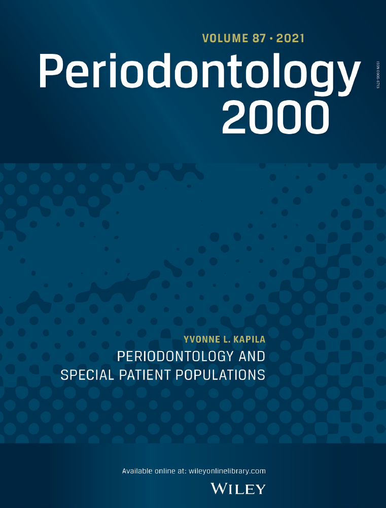
Biogeographic sites of the oral cavity play an important role in the viral contigs that are recognized. The lower recognizable viral contigs found in dental plaque compared with salivary samples can be attributed in part to the limited viral sequence homologues in databases used to identify viral genomes.102, 105 This limitation has led various researchers to lower the homologous sequence thresholds used, to analyze viral-like particles without purification, or to report only bacteriophage families rather than species. However, across studies, a high abundance of oral bacteriophages belonging to the Caudovirales order (Figure 1) has been documented, along with a shift in the composition of the well-described Caudovirales families (Siphoviridae, Myoviridae, and Podoviridae) in periodontal disease states.11, 102, 103

The majority of identified bacteriophages within the oral cavity pertain to the Caudovirales order, which contains three different families of tailed bacteriophages, all with an icosahedral capsid along with a linear dsDNA genome that is injected into bacterial hosts after penetration of cell membrane with tail. A, Myoviridae have a long contractile tail; B, Siphoviriadae have a long, noncontractile tail; and C, Podoviridae have a short tail
Variations in bacteriophage diversity and richness in gut inflammatory conditions consistently demonstrate homogenization of species composition in individuals in diseased states.106, 107 Periodontal inflammation mirrors the decreased species richness and diversity in plaque samples in diseased tissues compared with healthy tissue.11 Specifically, the temperate bacteriophage, Siphoviridae, was the most abundant family in healthy and diseased individuals for all sample types, while subgingival plaque from diseased pockets demonstrated the greatest shift in abundance for the virulent phage, Myoviridae.102 The dominance of temperate phages, such as Siphoviridae, is commonly found in health and is believed to be involved in host bacterial fitness and attenuation of bacterial virulence.108 Meanwhile, the surge of Myoviridae abundance in these diseased pockets may imply that a lytic cycle shifts the collective virulence of the complex biofilm that these bacteriophages are embedded in. However, these associations may currently be limited because of a lack of sequence data and need further validation by determining specific bacteriophage-host interactions.
4.3 Bacteriophages of oral pathogensAs bacteriophages play a role in bacterial population control and DNA transfer, bacterial hosts have developed an adaptive defense mechanism within their genome, known as clustered regularly interspaced short palindromic repeats, along with associated proteins systems to protect against bacteriophages. Investigators have turned to the heterogeneous diversity of spacers within clustered regularly interspaced short palindromic repeats to trace the history of bacteriophage attacks and to match those bacteriophages or conjugative plasmids containing this sequence array that naturally infect the bacteria.109 Sequencing of clustered regularly interspaced short palindromic repeats and associated proteins systems within the dental plaque of four periodontally healthy individuals allowed for identification of various bacteriophages at a species level, including a high abundance of Streptococcus prophages (UCN34 and IS7493), Actinomyces bacteriophage AV-1, Streptococcus bacteriophage DP-1, Enterobacteria bacteriophage P7, and Enterobacteria bacteriophage lambda.101 Parallel to other studies, identification of bacteriophages using clustered regularly interspaced short palindromic repeats and associated proteins systems demonstrated that bacteriophage ecology is characteristic for each individual, despite approximately 50% of contigs remaining without homologous sequences.101
Clustered regularly interspaced short palindromic repeats and associated proteins systems act as a catalog of bacteriophage-host interactions and demonstrate how these bacterial hosts become resistant to certain bacteriophages. In the same study described above, a unique finding in one individual's phageome included a transposon encoding tetracycline resistance within the Enterobacteria phage P7.101 P7 belongs to the Myoviridae family and is frequently described in coexistence with bacteriophage P1 carrying ampicillin or colistin resistance genes and extended spectrum beta-lactamase genes.110 In a recent review, bacteriophages identified against their respective oral bacterial inhabitants were surveyed and exhibited a high volume of bacteriophages infecting the early colonizers for dental plaque formation, such as Actinomyces and Streptococcus species.111
Fewer bacteriophages infecting putative periodontopathogens have been described but those isolated suggest their involvement in pathogenesis. The arsenal of virulence factors for bacteria implicated in more aggressive forms of periodontitis, such as A. actinomycetemcomitans, may be attributed to temperate Myoviridae that, in vitro, transfers antibiotic resistance genes, induces serotype conversion, and stimulates production of leukotoxins.112, 113 Another identified temperate Myoviridae bacteriophage infecting T. denticola, designated as φtd1, was found as integrated prophage DNA and upregulated expression of prophage genes during biofilm growth.114 Curiously, unlike the extensively documented herpesvirus-P. gingivalis relationship,41 no viral contigs or clustered regularly interspaced short palindromic repeats spacers have been isolated to suggest a bacteriophage that infects P. gingivalis.115
4.4 Bacteriophage-host interactionsAs optimization of metagenomic databases progresses steadily with increased viral sequence reads and contigs captured, more homologous sequences become available to identify resident oral bacteriophages with their bacterial hosts. Currently, genomic similarity between different bacteriophages paired with limited sequence data may result in an underestimation or misidentification of bacteriophages, as studies have described a fraction of the contigs that are unable to be classified.116 This observation should be taken into consideration with regard to both metagenomic and species-specific bacteriophage studies, as the reported bacteriophages may reveal a wider host range of virulent factors than described earlier.
留言 (0)