Non-Meckel small-bowel diverticula are rare, and the frequency of autopsy cases was reported by Edwards as 9 out of 2820 cases (0.31%). According to Edward’s locus minoris resistentiae theory, increased intestinal pressure causes a hernia-like prolapse of the mucosa and submucosal tissue out of the intestinal wall at the mesenteric vascular penetration zone, where the muscular layer is underdeveloped, often resulting in multiple pseudodiverticula in the mesenteric attachment zone [1]. Perforation occurs mainly in the mesenteric leaves of the small intestine, resulting in a mesenteric abscess. In our case, diverticula and perforations were also observed on the mesenteric border. We summarized the differences between Meckel’s diverticulum and non-Meckel's small-bowel diverticulum (Table 1).
Table 1 Comparison of Meckel’s diverticulum and nonMeckel’s small-bowel diverticulumAs with colonic diverticula, small-intestinal diverticula are often asymptomatic, but they may cause life-threatening acute complications, such as bleeding, volvulus, obstruction, diverticulitis, and perforation, which can lead to major diagnostic and therapeutic problems [4]. In particular, the mortality rate of diverticular perforation is high, ranging from 21 to 40%, and is closely related to delays in diagnosis and older age [5, 6].
Owing to their rare incidence, there is no clear treatment strategy for small-bowel diverticular perforations. Because of the difference in the pathogenic bacteria, we assume that the same treatment protocols as for upper gastrointestinal perforation cannot be applied. Therefore, we collected cases of small-intestinal diverticular perforation since 2000–2023 and summarized recent trends in treatment strategies (Table 2) [7,8,9,10,11,12,13,14,15,16,17,18,19,20,21,22,23,24,25,26,27,28,29,30,31,32].
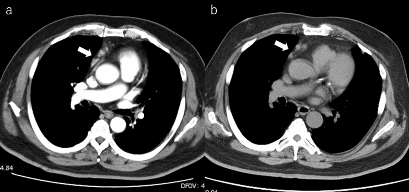
Table 2 Summary of previous reports and our cases since 2000 to 2023The clinical findings vary widely and patients visit the hospital with various concerns. Abdominal pain varied according to its location, severity, and progression. CT plays a major role in the diagnosis of diverticulitis in the small intestine. Localized and asymmetric thickening of the small-intestinal wall and inflammation or abscess of the periportal adipose tissue are the diagnostic criteria for small-intestinal diverticulitis, and when added to these findings of extraintestinal gas, a diagnosis of perforation or permeation can be made [33]. Non-Meckel’s small-bowel diverticula are often multiple, and the presence of diverticula in the normal small intestine may also help in the diagnosis of small-intestinal diverticulitis. In Table 2, however, there are scattered cases in which an exploratory laparotomy is selected even when CT is taken. One reason may be that small-bowel diverticular perforation is a rare disease not mentioned in the differential. The non-surgical management of perforated small-intestinal diverticula is a relatively new concept. When perforation of a small-intestinal diverticulum causes localized peritonitis and the patient remains stable, non-surgical management, such as antibiotics, bowel repose, and CT-guided aspiration of localized intraperitoneal collections, may avoid the need for surgery [18, 21, 23, 24, 31, 34]. On the other hand, emergency surgery is performed when there is remote air from the inflamed diverticulum. Patients successfully treated conservatively are often discharged from the hospital relatively early (2–6 days) [18, 21, 23, 24, 31]. Considering the high mortality rate associated with diverticular perforations of the small intestine, conservative treatment should be provided in limited cases. Even when conservative treatment is selected, surgery should be considered immediately in patients who do not improve after a few days of conservative treatment. In the early stages of the perforation, as in Case 1, it is impossible to determine whether the inflammation stays in the mesentery or spreads. A repeat CT scan may confirm the spread of extraintestinal gas and worsening of inflammatory findings.
We have summarized the main surgical techniques for non-Meckel’s small-bowel diverticular perforation (Table 3). Segmental intestinal resection with primary anastomosis is the most common procedure for perforating diverticula in the small intestine. Other surgical techniques, such as simple closure, invagination, and excision of the perforated diverticulum, should be abandoned because of their high mortality rate [35]. When the diverticula extend over the long intestinal tract, resection should be limited to the perforated or inflamed portion to avoid short-bowel syndrome. The presence of a retained diverticulum should be recorded for future reference in light of case reports of recurrent small-intestinal diverticular perforation after surgery [31, 36].
Table 3 Major surgical techniques for diverticular perforation of small intestine and number of cases in review


留言 (0)