
記住我


External apical root resorption (EARR) is one of the undesired biological effects of an orthodontic treatment.[1-3] Permanent loss of root structure is originated by excessive pressure, which leads to capillary collapse, reduction in blood flow, and override of the repair capacity of supporting tissues. These, in turn, lead to the formation of necrotic areas, known as hyaline zones, and activation of the inflammatory response, activating cellular and molecular mechanisms that promote EARR.[1,2,4-7]
Formation of hyaline areas during orthodontic treatment is unavoidable, but the incidence and severity of EARR are variable,[7-9] ranging from 26% to 100% as a function of biological, mechanical, and molecular factors, as the orthodontic movements are not the only factor that influences the onset of EARR.[7,10-13]
Different investigative approaches associate the following factors with a risk of developing EARR: age, gender, nutrition status, medication, systemic diseases, genetics, oral habits, malocclusion, tooth type, root morphology, history of dental trauma, previous orthodontic treatments, pulp vitality, infections, and inflammations are the most relevant biological factors.[2,3,6,11,14-18] Mechanical factors include the type of orthodontic appliances, orthodontic movement type, extractions, treatment time, and level of force magnitude.[6,19-27]
In addition, several biomarkers are related to the progress of EARR as a response to the orthodontic dental movement. Tissue tension generates tissue structural reorganization that releases neurotransmitters, growth factors, and cytokines, which have been attempted to study in-depth by analyzing the gingival crevicular fluid (GCF) that constitutes their releasing medium.[4,7,16,28-30]
Interleukins (ILs) are a complex of cytokines or low-molecular weight proteins that act as messengers and are physiologically secreted during the bone remodeling process in response to local stress (IL 1 β, IL-6, IL-7, IL-8, TNFα, IL-4, IL-10, IL-13, IL18, and Interferon-γ). The human genome codifies around 50 ILs and associated proteins. However, their association with EARR has not been conclusive, as data convergence has not been obtained. [7,8,10,16,29-34]
Diagnosis of EARR depends on early detection using routine radiographies. Root shortening begins between the second and fifth treatment weeks, but such change will be only visible in panoramic or periapical radiographs 3 or 4 months after the beginning of the orthodontic treatment. As these diagnostic aids underestimate the extension and produce negative false cases, the “gold standard” to diagnose EARR is cone-beam computed tomography (CBCT) because it accurately detects EARR without the existing limitations of other techniques. CBCT offers high-resolution structural analysis in the three planes of space and superimposition elimination, which provide high sensitivity and specificity in the identification of these types of conditions. [2,3,10,35-37]
Consequently, multiple clinical and orthodontic variables are associated with EARR. However, investigations show contradictory results, so careful analysis is necessary due to the high heterogeneity within original studies. Therefore, the main objective of this umbrella review was to analyze current evidence on orthodontically induced EARR to identify clinical and molecular factors associated with this condition.
MATERIAL AND METHODS DesignAn umbrella review was performed, beginning with a Population, Intervention, Comparison, and Outcome (PICO) question. A search strategy, inclusion criteria, and quality assessment with analysis of results were carried out. The study’s protocol was inscribed in the PROSPERO (International Prospective Register of Systematic Reviews) database (CRD42020198971).
Search strategyFour electronic databases were used: PubMed, Science Direct, Scopus, and Cochrane. Systematic reviews and meta-analyses were identified using the terms orthodontic AND root resorption. The search was conducted, including articles from 2015 to 2020. To guarantee the exhaustivity of the protocol, an additional search using thesaurus terms and different word combinations was performed. This addition search included (((((“orthodontal”[All Fields] OR “orthodontic”[All Fields]) OR “orthodontical”[All Fields]) OR “orthodontically”[All Fields]) OR “orthodontics”[MeSH Terms]) OR “orthodontics”[All Fields]) AND ((“root resorption”[MeSH Terms] OR (“root”[All Fields] AND “resorption”[All Fields])) OR “root resorption”[All Fields]))))).
Inclusion and exclusion criteriaThe PICO question used in the current work according to the main objective was as follows: (P) patients from different ages, genders, and ethnicities; (I) previous orthodontic treatment; (C) during orthodontic treatment; and (O) what clinical and molecular factors are associated with the incidence of EARR? Systematic reviews and meta-analyses performed on human subjects and published between 2015 and 2020 were included in the study. Exclusion criteria included other types of investigations (analytical, clinical, guidelines, review articles, letters to the editor, opinion articles, and observational studies).
The following inclusion criteria were applied to the title and abstract reading:
Search terms in the title or abstract
Publications in human subjects
Systematic review or meta-analysis.
Article selectionA total of 124 potentially eligible articles were identified. After duplicate elimination, 118 articles remained, which were then screened for title and abstract. One hundred articles were not related to the topic and were discarded. The remaining 18 articles were read and analyzed and eight were subsequently discarded because the studied population was animals. Ten articles (four systematic reviews and six systematic reviews with meta-analysis) were selected for data analysis and validation using the Preferred Reporting Items for Systematic Reviews and Meta-Analyses (PRISMA) and Admeasurement Tool to Assess Systematic Reviews (AMSTAR-2) guidelines. The flow of article selection is shown in [Figure 1].
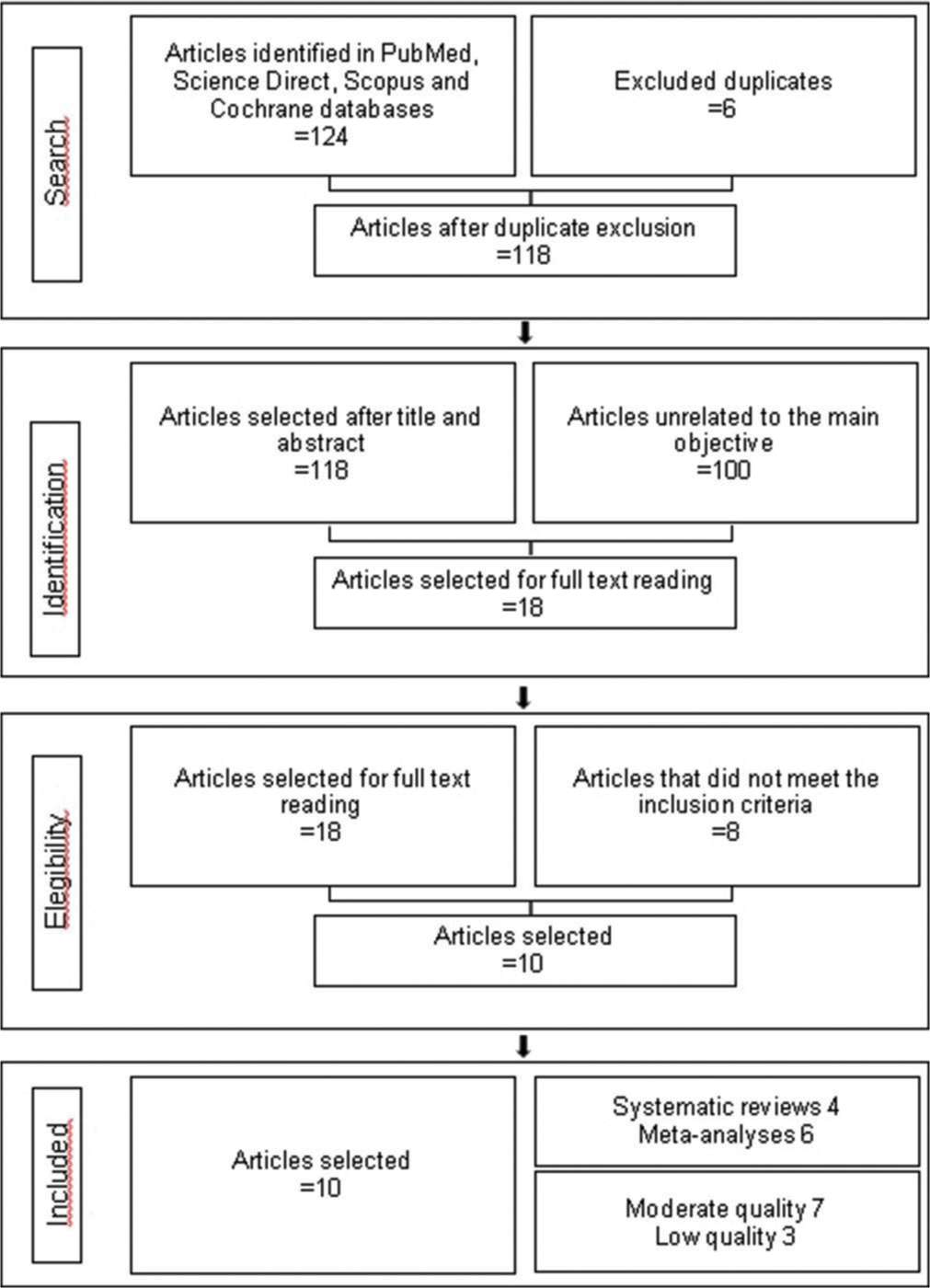
Export to PPT
Critical analysisThree independent investigators assessed the validity of the selected articles. The PRISMA and AMSTAR-2 guidelines were used to verify their quality. A calibration process was then performed, and a 90% concordance index was obtained.
All the evaluations were performed using the PRISMA checklist, which applied the identification, screening, selection, and inclusion phases from the guidelines using 27 questions. Then, the AMSTAR-2 guideline was applied to assess the quality of the articles, and four levels of quality were obtained: high, moderate, low, and critically low.
The risk of bias, classified as low, high, or undefined, was assessed for every single article. In addition, whether heterogeneity was reported was also established. A descriptive analysis of the main characteristics of the included revisions was carried out.
RESULTS Quality assessment of systematic reviews and meta-analysesQuality assessment of the ten included articles in this umbrella review is shown in [Table 1]. According to the AMSTAR-2 guideline, seven studies were classified as having moderate quality and 3 as low quality. According to the PRISMA guideline, the articles with the best scores were investigations performed by Nowrin et al.[4], Gandhi et al.[2], Yi et al.[37], and Fang X. et al.[38]. Out of the 27 items included in the PRISMA checklist, only one article matches all the criteria and the score is over 18 points. Besides, nine out of ten articles report high heterogeneity and the remaining article does not report it.
Quality assessment instruments reported in these studies are the Grading of Recommendations, Assessment, Development, and Evaluations, which was used in three articles; the Strengthening the Reporting of Genetic Association Studies statement in two articles; the Risk of bias in Non-randomized Studies-of Interventions tool in 1 study, the Methodological index for non-randomized studies index in 1 work, the Quality Assurance International certification in 1 article, the PRISMA guideline in 1 article, and the Methodologic Scoring System adopted by Roscoe et al. in 2015 in the remaining article.[6]
Table 1: Assessment and quality control of selected articles
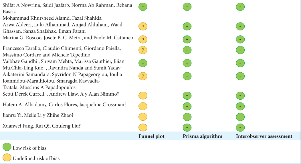
Article Prisma guideline information qualification Amstar-2 guideline general quality Heterogeneity Quality assessment instrument used in the study Shifat A Nowrina[4]Cochrane evaluation of individual studies identified sources of bias [Table 2] and assessed the presence of a prism algorithm, the report of interobserver evaluation, and the presence of a funnel plot in each study. The results of this study showed that all investigations exposed the use of the prism algorithm, resulting in a low risk of selection bias. Assessment of interobserver concordance was reported in all articles, leading to a low risk of bias. Due to the presence of a funnel plot, only two studies showed a low risk of report bias, and it was undefined or unclear in the remaining articles as some doubts about the results arose.
Table 2: Cochrane assessment for individual studies
With the purpose of reducing systematic errors, possible bias in the included articles was identified. Due to the high variability among ethnic groups, a selection bias may be found in genetic studies, as genotypes vary according to ethnicity. Samples of fewer than 100 patients were considered a potential bias. The difference in precision among radiographies and measurement and quantification methods was also considered a measurement bias. The absence of control groups, lack of high-quality prospective studies, non-homogeneity at the GCF collection time, different methods to collect this fluid, and measurement of applied force magnitude were other biases.
Main characteristics of reportsThe main characteristics of articles included in this umbrella review compared different clinical and molecular variables that increase the risk of developing EARR during the orthodontic treatment. Out of ten investigations, four were performed in Asia, two in North America, one in South America, two in Europe, and one in Oceania.
Most investigations are randomized controlled clinical trials performed in human subjects under orthodontic treatment of about to begin one. Sample size was established according to each investigation and most works only include 3D diagnostic aids. For qualitative studies (systematic reviews), the minimum number of included studies was 2, and the maximum was 30. For quantitative studies (meta-analysis), the minimum number of included studies was 3, and the maximum was 16.
As for age, the minimum age to participate in the studies was 8 years, and the maximum was 75 years. Only six papers reported gender information, and more females than males were included. Diagnostic aids ranged from lateral cephalograms, panoramic, occlusal radiographs, periapical radiographs, and scanning electron microscopy. However, 80% of the authors preferred the CBCT as the measurement method due to its high precision. Samples from GCF were obtained using filter papers, paper strips, and micropipettes, and analyses were performed using techniques such as Taqman, Ncoi sequencing, and enzyme-linked immunosorbent assay (ELISA) immunoassays, among others. Only two types of orthodontic appliances were reported (clear dental aligners or fixed orthodontic appliances). For the volumetric measurement of the EARR, the following methods were reported: linear measurements, radiometric, millimeters, and voxel 3D. Four studies classified EARR as light, moderate, and severe. As for biological factors, one study analyzed ethnicity and found a higher prevalence in Caucasian and Hispanic populations than in Asians. As for patient type (children, adolescents, or adults), the most severe form of EARR was found more frequently in adults than adolescents [Table 3]. Immunological factors were referred to as variables such as genetic polymorphism expression and the presence of cytokines, ILs, RANK, OPG, dentin phosphoprotein (DPP), dentin sialoprotein (DSP), and alkaline phosphatase (ALP), among other biomarkers. All these factors and the main conclusions are summarized in [Table 4].
Table 3. Main characteristics of included systematic reviews and meta-analyses. Description of individual studies
Article Average age Gender Radiographies Treatment type Root resorption quantification method Association between genetic polymorphisms and external apical root resorption: A systematic review and meta-analysis 8.0-55.4 years NR Lateral cephalogram, panoramic, CBCT, occlusal, periapical "Treatment type" confounding factor was not considered Radiographic analysis Association of Orthodontic Clear Aligners with RootTable 4: Contributing factors to the development (EARR) according to results provided by systematic reviews and meta-analyses
Category Factor Description Article Mechanical factors Force magnitude Under the same mechanical stress, many subjects exhibit low and some severe ERR 1 there are positive correlations between ERR and continuous forces 3 there is a positive correlation between ERR and an increase in the orthodontic force magnitude regardless of force direction 7 Treatment duration The higher the treatment time, the higher the root resorption 3 a pause in dental movement is beneficial to reduce ERR 3,7,10 a reduction in root resorption was observed in patients who received orthodontic treatment in two phases 7 Movement direction or type Buccal inclination was associated with ERR in the Bucco cervical and linguoapical regions 3 buccal root torque was associated with ERR in the Bucco apical and palatocervical regions 3 distal inclination was associated with ERR in the distal aspect of the apical and middle thirds and in the mesial aspects in the cervical third 3 extrusion movement was associated with increased resorption on the distal surfaces 3 There are positive correlations between ERR and intrusive forces 3.7 Type of orthodontic device Transparent aligners do not reduce the risk of developing ERR, although the incidence and severity might be reduced 2.1 similar results are observed when patients are treated with light forces using aligners or brackets 3 teeth subjected to super elastic NITI arch wires show higher ERR 3 differences were not found in the prevalence or severity of ERR when straight-wire appliances were compared with standard appliances 7 class-II elastics is a risk factor for ERR 8 meta-analysis results suggest that self-ligating brackets are better than conventional brackets at protecting maxillary central incisors against ERR 9 Biologic factors Age No age predilection was found 2 Race ERR is higher in Caucasians and Hispanics than Asians 2 Gender Proportion of ERR cases was higher in females than males 8 No predilection of ERR for males or females 2 Tooth type The highest root resorption was found on the maxillary lateral incisors followed by maxillary central incisors and canines 2,6,8 The highest ERR was observed in the anterior maxilla followed by the anterior mandible, posterior mandible and posterior maxilla 6 Root morphology Root morphology (abnormal shape, long and narrow roots) is associated with ERR 8 Provious extractions Orthodontic treatment involving extractions are more associated with reduction in root size 6 Dental extraction to resolve severe dental crowding may be considered a risk factor for ERR 10 Pulp vitality An increase in ERR in endodontically treated teeth after orthodontic treatment was not observed 8 No difference was found in the degree of ERR between endodontically treated teeth vs contralateral vital teeth 8 Endodontic treatment in males exhibited a significant increased ERR 8 Molecular factors Expression of genetic polymorphisms IL-1B (+3954) polymorphism is considered a promising gene to predict ERR 1 Patients who are homozygous for allele 1 of the IL-1B (+3954) gene have a 95% probability of developing ERR > 2mm 1 Cytokines Levels of IL-4, IFN-γ and GMCSF are higher in light ERR cases 4 Dentin matrix protein (DMP - 1) is not a useful biomarker because it is not possible to differentiate between its physiological and pathological activities 4 Dentin phosphoprotein (DPP) is a relatively useful biomarker for ERR diagnosis 4 RANKL concentration in the GCF is higher in patients with light and severe ERR 4 There are higher concentrations of DPP, DSP and IL-6 in patients with severe ERR 4 Overall alkaline phosphatase (ALP) activity increased with higher rates of tooth movement at 150 g of force 4 Cytokine levels are different depending on sampling sites and occurring time 4 DISCUSSIONDue to the absence of pathognomonic signs, EARR is casually detected in routine panoramic radiographs. However, underestimating its severity may lead to permanent loss of important root structure since other risks patients are subjected to from their biological background, their interaction with the environment, and factors related to the mechanics employed in orthodontic treatment are mostly unknown. Results presented in this umbrella review may be used as a foundation to develop more solid investigations on this topic.
In 2019, Currel et al.[1] analyzed the degree of root resorption in teeth subjected to orthodontic treatment considering mechanical factors such as type of device and orthodontic force magnitude and direction. It was found that continuous forces increase EARR regardless of magnitude and direction. Bracket type, ligation, and archwire sequence did not influence the severity of EARR.[1]
In 2019, Samandara reported that root shortening is significantly increased after orthodontic therapy and confirmed that heavy forces, extractions, and anterior teeth with abnormal root morphology are factors that increase the prevalence and severity of this condition. Similar results are reported by Fernandes stating that orthodontic therapy with extractions increases the risk of EARR by 70% and also considering other variables, such as increased overjet and long dilacerated roots.[3,39]
Harris et al.,[40] Barbagallo et al.,[27] Cheng et al.,[25] and Paetyangkul et al.[36] concurred that there is a directly proportional relationship between force and EARR and that the type of orthodontic movement is a significant mechanical factor as forces intensify on certain areas of the root according to the orthodontic action. For instance, pressure accumulates on the root apex during intrusion movements, thus increasing the risk of EARR in that zone. During extrusion movements, EARR is more frequently found on the cervical third toward the mesial and distal, which are the areas where pressure accumulates. However, it is important to mention that extrusion movements have 4 times lower resorption probabilities than intrusions.[41]
The type of appliance used in the orthodontic treatment is another variable that may influence the behavior of the root resorption process. Conventional brackets have been compared with self-ligating ones to determine whether significant differences are found between both bracket types in the incidence of EARR . Yi J. et al.[37] established that self-ligating systems may produce lighter forces during aligning movements since no ligature is needed, which may produce a protective effect for maxillary central incisors that are most vulnerable. However, Aras et al. concluded that it is not possible to suggest the superiority of one system over another due to the lack of investigations that follow solid methodologies to identify the exact differences between both systems. [37,42]
Regarding molecular factors, articles suggest that EARR has an important genetic component. Homozygous patients for allele 1 of IL-1B exhibit 5–6 times higher risk of developing EARR >2 mm than other groups. Data show that allele 1 in the IL-1B gene, known for reducing the production of IL-1 cytokine, significantly increases the risk of resorption. In addition, it has been suggested that EARR is a complex condition influenced by many different factors that are important to know to understand the contribution of environmental factors, such as habits and biomechanics.[4,43]
The search for EARR biomarkers intensified after finding dentin specific proteins (DPP-and DSP) that are byproducts found in the GCF, even though such proteins are not routinely released within the periodontal ligament space. ELISA immunoassays were analyzed by James et al. and later confirmed by Balducci et al.,[44,45] who identified and quantified these proteins in patients under orthodontic treatment. Dentin matrix protein-1 (DMP-1) was found in large quantities in the GCF as it is eliminated from bone and dentin during resorption processes. However, based on the results of the current work, DMP-1 is not dentin-specific, and its presence may be explained not only because of EARR but also due to the remodeling process during the orthodontic movement. As such, it is not an adequate biomarker of this condition as it is not possible to differentiate between its normal and pathological activities. Likewise, DSP protein was found in control groups, so no consensus is reached in the scientific literature to classify them as exact molecular biomarkers of EARR as they are not exclusive of dentin and are expressed in the osseous tissue. Their presence in the GCF may be explained by physiological remodeling processes, which are increased in patients under orthodontic treatment.[43,46]
Perinetti et al. assessed the activity of ALP in the GCF to evaluate its utility in the diagnosis of EARR during orthodontic treatment. These authors observed a significantly higher ALP activity in tension sites compared to compression zones, which increases as the force increases. However, this finding only reflects the biological activity of such compound in the periodontium during the dental movement and must be further studied, as proposed by Tarallo et al. in 2019.[7,33,47]
As for dental pulp status and root resorption, Alhadainy et al. and Huzar et al. concurred that the endodontic treatment does not seem to increase root resorption as no significant differences are found between vital and endodontically-treated teeth.[13,48]
CONCLUSIONDifferent factors or individual characteristics are paramount to define the risk of root resorption. The dental professional must carry out a comprehensive medical record of patients, including their background, to make the best treatment decision possible.
Biomarkers such as I-1B, I-6, I-4 ILs, and DPP are potential indicators of root resorption, and such molecules might be used to establish the individual risk and/or reach an early diagnosis of EARR to reduce the negative impact of this condition on orthodontic treatments.
留言 (0)