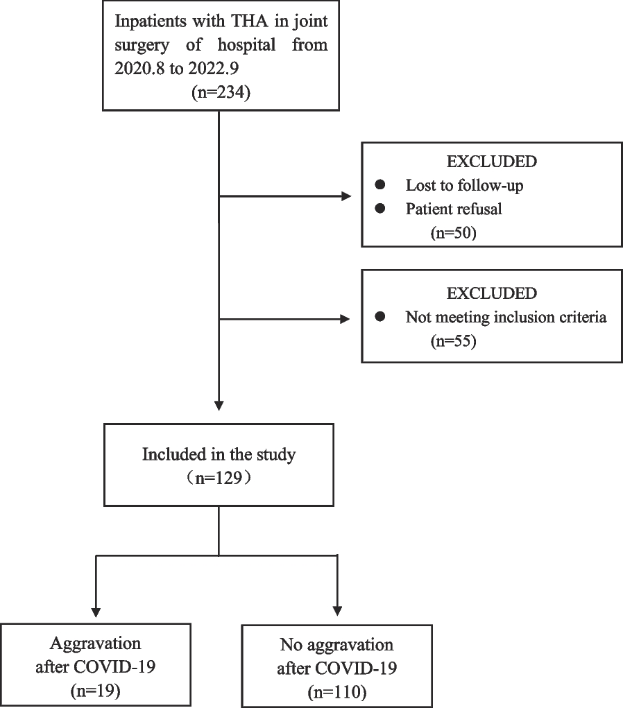
Study population
We identified patients diagnosed with knee OA and treated at our institution (a regional tertiary hospital) between January 1st 2015 and January 1st 2018 using the hospital information system. Written informed consent was obtained from all patients once they have administered in our hospital that their medical data may be used for scientific analysis but will be kept anonymous. Patient data were collected on May 2nd 2023; authors did not have access to information that could identify individual participants during or after data collection.
The current study was done on knee level. To be eligible for inclusion, knees had to meet the following criteria: they belonged to patients aged over 40 years, were reported to have knee pain, were confirmed to have radiographic knee OA (Kellgren & Lawrence (KL) grade > = 2) through radiographs and had magnetic resonance imaging (MRI) films available. The year when the patient underwent their initial MRI examinations following the diagnosis of knee OA was defined as the baseline for this study. Knees were excluded if they had injuries resulting in structural damage after the baseline assessment, had other knee joint conditions or forms of arthritis, lacked radiographs or MRI exams, or if the patient had scheduled knee surgery at the baseline evaluation. In cases where a patient had bilateral knee OA, we selected the knee that was first diagnosed with OA. If both knees were diagnosed at the same clinical visit, we randomly selected one for inclusion in the study.
Baseline patient data
After a patient was included in the study, their baseline clinical data and imaging films were retrieved from the information system. The baseline clinical data consisted of demographic information (age, sex, BMI (body mass index), education, affected side of knee OA and OA in other joints) and pain level of the past week was assessed using the numeric rating scale (NRS) score (0–10, 0 indicates no pain; patients were asked when receiving MRI examinations). Two researchers (J.Z. and Z.W.), who were unaware of the patients’ clinical information, independently reviewed weight-bearing knee radiographs taken at baseline (or before) and assigned scores using the KL classification system [9]. In cases where there were discrepancies, the two researchers held a consensus meeting to resolve differences and reach agreements.
MRI scan and BML
All the knees were imaged using a 3.0T MRI unit in the sagittal plane (resolution 1.5; slice thickness 2 mm) in our hospital. Two researchers (J. Z. and Z.W.), who were blinded to the patients’ clinical information, independently reviewed the baseline T1 and T2 MRI films. They assigned BML grades according to the standardized MRI Osteoarthritis Knee Score (MOAKS) system [10].
In the MOAKS system, a BML is defined as an ill-defined trabecular bone signal that appears hypointense on T1-weighted imaging and hyperintense on T2-weighted fat-saturated imaging. The researchers assessed BMLs in each subregion, which consisted of six subregions for the femur, two for the tibia and two for patella, as specified by the MOAKS system. BMLs, including cysts, were graded on a scale of 0 to 3 based on the percentage of subregional volume involved: grade 0 indicated no BML, grade 1 indicated involvement of less than 33% of the subregional volume, grade 2 indicated involvement of 33–66% of the subregional volume, and grade 3 indicated involvement of more than 66% of the subregional volume. If multiple BMLs were present in a single subregion, their volumes were combined into a single percentage. We proceeded to determine the Max BML grade for the medial, lateral, patellofemoral, and total compartments, respectively. This was determined by selecting the highest grade among all corresponding subregions. Building upon this, we created BML burden grades by summing the Max BML grades of the medial, lateral and patellofemoral compartments. As a result, the BML burden grade ranged from 0 to 9. However, among included knees, there was no knee graded 8 and 9, and prevalence of grade 5 (2 knees), 6 (1 knee) and 7(1 knee) was very low. Therefore, we incorporated grade 5–7 into grade 4 group. Additionally, we assigned Presence BML grades for each compartment: an Presence BML grade of 0 indicated the absence of BML, while a grade of 1 indicated the presence of BML.
Outcome measure
The primary outcome is incident knee surgery within 5 years from baseline. At the 5-year mark, we reached out to all patients and inquired whether they had undergone any knee surgery specifically for OA in the knee that had been subject to the baseline MRI. The knee surgery included total/unicompartment knee replacement, arthroscopic procedures and high tibial osteotomy. The exact surgery indication for each individual could not be reached. In our hospital, knee surgery was indicated for OA patients when conservative treatments, such as medication, physical therapy, and lifestyle modifications, have failed to provide adequate relief from symptoms, and the patient’s quality of life is significantly impacted by knee pain and dysfunction. The first surgery was used if multiple surgeries were done on one knee. To verify this information, we cross-referenced patient medical care records in our hospital system.
We used radiographic progression indicators as the secondary outcomes to further support the role of BMLs in disease development. We identified patients who had received radiographic exams at 4 to 6 years from baseline and created radiographic progression indicators according to KL classification system, i.e., KL progression (KL progressed 1 grade or more vs. no change). Knees with baseline KL grade 4 were excluded.
Statistical analysis
We tabulated descriptive statistics to summarize the characteristics of the subjects. We built logistic regression models to assess the relationship between baseline Max BML/ BML burden/Presence BML grades and the incident surgery over the 5-year period for medial, lateral, patellofemoral and total compartments, respectively. Crude odds ratios (ORs) and adjusted ORs (adjusted for baseline age, sex, BMI and KL grade) were calculated, as well as their 95%CIs. The reference group for comparison was baseline Max BML/BML burden/Presence BML grade 0. The co-variates were chosen according to previous literature that those could be confounding factors [11]. We used similar logistic regression models to assess the relationship between baseline Max BML/ BML burden/Presence BML grades and KL progression.
Next, we determined the positive and negative predictive values (PPV and NPV) of baseline BML grades for predicting the incidence of surgery over the 5-year period. To do this, we applied multiple cutoffs on the Max BML grades to obtain dichotomized categories, i.e., Max BML grade 1 or higher, Max BML grade 2 or higher and Max BML grade 3. We did this for all the compartments separately and did the same for the BML burden grades.
Knees with baseline characteristic data missing were imputed by the data from prior clinical visits (e.g., age, sex and BMI). While 78 knees had missing values in baseline NRS score were excluded from the descriptive analysis. There was no missing value in the primary outcome measure (surgery information) as we reached out to patients, and most were also available in the healthcare information system. All statistical analyses were performed using the SPSS software (IBM, Chicago, USA). Statistical significance was defined as P < 0.05.


留言 (0)