Cell survival rate assessment
Tanshinone IIA (CAS No. 568-72-9) was purchased from Yuanye Biotechnology Co., Ltd (Shanghai, China). The purity of Tanshinone IIA was confirmed to be more than 98% by high-performance liquid chromatography. Primary bone marrow MSCs were plated in a 96-well plate at a density of 5 × 10³ cells/well for 24 h and further treated with 0, 10− 9, 10− 8, 10− 7, 10− 6 or 2 × 10− 6M Tanshinone IIA/dimethyl sulfoxide (DMSO) solutions for another 24 h. For H2O2 treatment, cells were plated in a 96-well plate at a density of 2 × 10³ cells/well for 24 h and were pretreated with 2 × 10− 6M Tanshinone IIA or DMSO for another 24 h. Then, the medium was discarded, and the cells were incubated with 0, 50, 100, 200, 400, 500, 600, 800, or 1000µM H2O2 for 24 h. The cell survival rate was assessed with an enhanced cell counting kit-8 (C0041, Beyotime Biotechnology, Shanghai, China), and the absorbance at 450 nm was recorded by a microplate reader.
Osteogenic differentiation assay
Bone marrow cells from long bones of 4-week-old C57BL/6 mice were collected and plated in αMEM supplemented with 10% fetal bovine serum and 1% penicillin–streptomycin. After 7 days of culture, the suspended cells were removed, and the adherent cells were collected and plated in a 12-well plate at a density of 8 × 10⁴ cells/well with osteogenic differentiation medium containing 50 mg/L vitamin C, 10nM dexamethasone, and 10mM β-glycerophosphate in complete αMEM. The cells were incubated with different concentrations of Tanshinone IIA with or without 500µM H2O2. The culture medium was refreshed every 3 days. After 10 days or 21 days culture, the cells were fixed with 10% buffered formalin for 15 min and stained with 1-step NBT/BCIP substrate solution (34,042, Thermo, Waltham, Massachusetts, USA) for 30 min or 0.1% Alizarin red (A5533, Sigma, St. Louis, Missouri, USA) aqueous solution for 30 min.
Real-time reverse transcriptase polymerase chain reaction (RT–PCR) assay
Primary bone marrow MSCs were plated in a 6-well plate at a density of 2 × 10⁵ cells/well with osteogenic differentiation medium. Cells were treated with either 500µM H2O2 or 500µM H2O2 and different concentrations of Tanshinone IIA for 72 h. Total RNA of the cells was extracted using an RNA Extraction Kit (B0004D, HifunBio, Shanghai, China) and reverse transcribed to cDNA using an RT reagent kit (RR407, Takara Bio, Japan). PCR was performed using a TB Green Premix Ex Taq II kit (RR820, Takara Bio, Japan). The primers used for specific mRNAs are listed in Table 1.
Table 1 PCR primers for specific genesWestern blotting assay
C3H10T1/2 cells provided by the National Collection of Authenticated Cultures, Chinese Academy of Sciences were plated in a 6-well plate at a density of 8 × 10⁴ cells/well for 24 h and were pretreated with 0, 10− 8, 10− 7, or 10− 6M Tanshinone IIA for 24 h. After incubation in 500µM H2O2 for another 24 h, the cells were collected, and total proteins were extracted on ice using radioimmunoprecipitation assay lysis buffer (P0013B, Beyotime Biotechnology, Shanghai, China). The protein concentration was detected using an enhanced bicinchoninic acid assay kit (P0010, Beyotime Biotechnology, Shanghai, China). Western blot analysis was conducted as previously described [18]. The following antibodies were used: anti-cleaved caspase 3 (ab214430), anti-pro caspase 3 (ab32499), anti-superoxide dismutase 1 (SOD 1, ab13498), anti-heme oxygenase-1 (HO-1, ab68477), anti-catalase (CAT, ab16731) from Abcam (Cambridge, UK) and anti-Nrf2 (12721), anti-β-actin (8457), anti-Keap1 (8047), anti-Bcl2 (3498) and anti-Bax (5023) from Cell Signaling Technology (Danvers, Massachusetts, USA). A chemiluminescent horseradish peroxidase substrate kit (WBKLS0500, Millipore, Billerica, Massachusetts, USA) was used for the electrochemiluminescence detection assay.
Detection of antioxidant enzymes
C3H10T1/2 cells were plated in a 12-well plate or 6-well plate for 24 h. After pretreatment with different concentrations of Tanshinone IIA or DMSO for 24 h, the cells were incubated in fresh medium supplemented with 500µM H2O2 for another 24 h. For ROS detection, the cells were incubated in αMEM containing a 10mM 2,7-dichlorodihydrofluorescein diacetate (H2DCFDA) fluorescence probe (287810, Sigma, St. Louis, Missouri, USA) for 30 min, and the fluorescence intensity was measured by flow cytometry (Accuri C6, BD Biosciences, Franklin Lakes, New Jersey, USA). SOD and CAT activity in cells was detected using relative activity test kits (SOD, S0101S; CAT, S0051; Beyotime Biotechnology, Shanghai, China).
Immunofluorescence staining
C3H10T1/2 cells were plated in a 48-well plate and cultured with 0, 10− 8, 10− 7, or 10− 6M Tanshinone IIA for 24 h, followed by culture in fresh medium supplemented with 500µM H2O2 for another 24 h. After fixation with 4% formaldehyde solution at 37℃ for 15 min, the cells were incubated with an Nrf2 antibody (12721, Cell Signaling Technology, Danvers, Massachusetts, USA, 1:500) overnight at 4℃. Cells were further incubated in CoraLite594-conjugated goat anti-rabbit IgG (SA00013-4; Proteintech, Wuhan, Hubei, China; 1:200) for 1 h at 37℃ in the dark. Antifade mounting medium with 4,6-diamidino-2-phenylindole (DAPI) was used for nuclear fluorescence staining (H-1200, Vector Laboratories, San Francisco, California, USA).
Animals
The study was conducted according to the guidelines of the Declaration of Helsinki, and approved by the Institutional Animal Care and Use Committee of Longhua Hospital, Shanghai University of Traditional Chinese Medicine (2019-N051). Three-month-old female C57BL/6 mice (21–23 g body weight) ordered from Lingchang Biotech (Shanghai, China) were housed at 22˚C with a 12 h light/dark cycle, with 4 or fewer mice in each cage, and were provided ad libitum access to food and water in the specific pathogen-free animal experiment center of Longhua Hospital.
Grouping and model establishment
Following a 1-week acclimatization phase, the mice were randomly assigned to the sham, model, gel, and Tanshinone IIA groups using a random number table. Mice in the model, gel, and Tanshinone IIA groups underwent bilateral ovariectomy, and the rest underwent sham operations. Three months after the surgery, all mice received a mid-shaft transverse osteotomy fracture on the left tibias, and the tibias were fixed with 0.5-mm-diameter intramedullary metallic pins. Anesthesia was induced in mice by isoflurane inhalation through an inhalation anesthesia machine before surgery.
Hydrogel preparation and intervention
The injectable hydrogel was prepared as previously described [19]. A filtered 40 mg/ml dextran/phosphate buffered saline (PBS) solution containing 4µM Tanshinone IIA/DMSO or an equal volume of DMSO and 20 mg/ml filtered chitosan/PBS solution was prepared. When setting up the fracture model, the dextran/PBS solution and chitosan/PBS solution were mixed at a 1:1 ratio, and 20 µl of semisolidified hydrogel was injected into the bilateral bone marrow cavities of the fracture ends before fixing the tibias with pins. Mice in the Tanshinone IIA group were injected with a hydrogel containing 2µM Tanshinone IIA, and mice in the gel group received a hydrogel containing DMSO.
Sample harvest
Mice were sacrificed at the corresponding time points. Mice were anesthetized with isoflurane in an induction chamber, followed by intraperitoneal injection of pentobarbital sodium (5.4 g/kg body weight). The left tibias (6 from each group per time point) were fixed in 10% buffered formalin and subjected to micro-computed tomography (micro-CT) scanning. After decalcification, dehydration, and embedding, 4 μm thick serial sections of the tibias were cut for histomorphometric evaluation. Another set of 5 tibias from each group was harvested 28 days postfracture and stored at -80°C for biomechanical testing.
Biomechanical test
The tibias were equilibrated with saline at room temperature before assessment. A three-point mechanical bending test was performed on the left tibias to assess the maximum force and yield displacement with a mechanical testing instrument (ElectroForce 3200 Series III, TA Instrument, New Castle, Delaware, USA).
Threedimensional (3D) reconstruction analyses
The fractured tibias were subjected to X-ray imaging and micro-CT scanning (vivaCT 40, Scanco Medical AG, Brüttisellen, Switzerland) successively at a voltage of 55 kV and a current of 72µA. The integration time was 300ms, and the slice increment was 10 mm. The bone volume (BV, mm3), total volume (TV, mm3), bone mineral density (BMD), trabecular bone number (Tb.N), trabecular bone thickness (Tb.Th), and trabecular bone separation (Tb.Sp) of the callus were measured.
Histological evaluation
For morphometric analysis, midsagittal sections of the tibias were dewaxed and stained with Alcian blue/hematoxylin solution and orange G solution. After dehydration, clearing, and mounting, images of the sections were captured by a virtual slide system (VS120-S6-W, Olympus, Japan).
TdT-mediated dUTP Nick End Labelling (TUNEL)
C3H10T1/2 cells were plated in a 48-well plate at a density of 1 × 10⁴ cells/well for 24 h and were pretreated with 0, 10− 8, 10− 7, or 10− 6M Tanshinone IIA for 24 h. After incubation in 500µM H2O2 for another 24 h, cells were fixed with 4% formaldehyde at 37℃ for 30 min. For the animal study, midsagittal sections of the callus were dewaxed and rehydrated. TUNEL staining was performed using a staining kit (C1088, Beyotime Biotechnology, Shanghai, China), and cell apoptosis was observed using a fluorescence microscope and were analyzed with an image Pro Plus 6.0 software (Media Cybernetics, PA, USA).
Immunohistochemical staining
After dewaxing and rehydration, sections were treated with 3% H2O2 solution for 15 min at 37 °C, followed by the antigen repair with sodium citrate solution for 15 min at 95 °C. The sections were incubated in primary Nrf2 antibody (16396-1-AP, Wuhan Sanying, Wuhan, China, 1:100) overnight at 4 °C. The sections were then incubated in horseradish peroxidase labeled goat anti rabbit IgG (A0208, Beyotime Biotechnology, Shanghai, China) for 40 min. After staining with diaminobenzidine and counterstaining with hematoxylin, the images were captured by by a virtual slide system (VS120-S6-W, Olympus, Japan), and were analyzed with an image Pro Plus 6.0 software (Media Cybernetics, PA, USA).
Statistical analyses
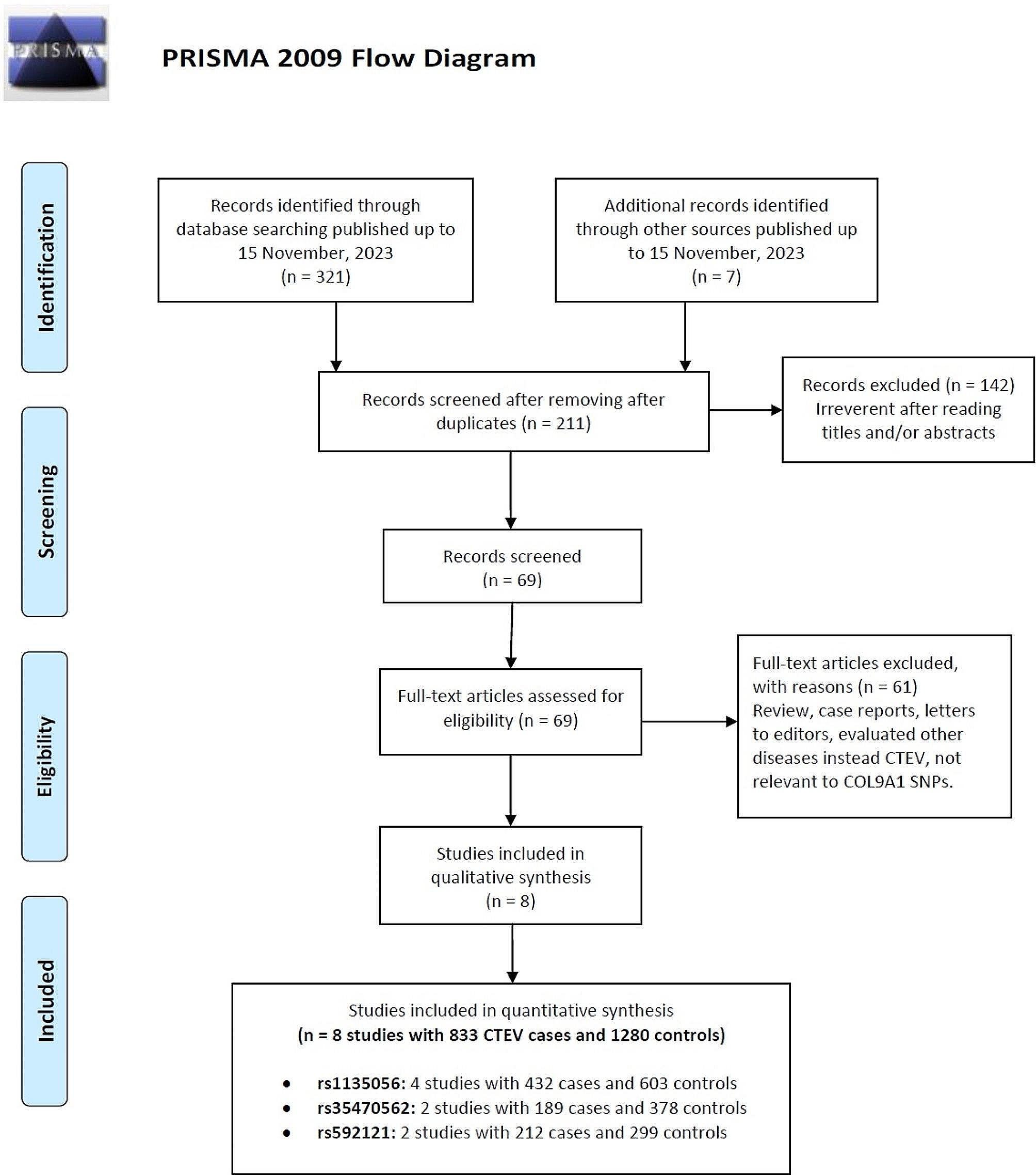
All of the data are presented as the means ± standard deviations. Statistical analyses and graph generation were performed using GraphPad Prism 8 (GraphPad Software, San Diego, California, USA). The normality of the data was assessed by the Shapiro‒Wilk test, and the homogeneity of variance was tested. For data with homogeneity and normality, statistical significance among multiple groups was assessed by one-way analysis of variance (ANOVA). Otherwise, the Kruskal‒Wallis H test was used. A P value < 0.05 was considered to indicate statistical significance.


留言 (0)