The patient had previously undergone treatment with VEGFR inhibitors, including bevacizumab. However, during follow-up, there were no indications of proteinuria or hematuria, and renal function remained within normal limits. Due to disease progression, furqinitinib was introduced to the treatment protocol. Approximately two weeks after initiating furqinitinib therapy, the patient developed hand-foot skin reactions, hypertension, proteinuria, and other adverse effects, clinically manifesting as nephrotic syndrome. There were no systemic manifestations of TMA, and both medical history and laboratory analyses did not support diagnoses of thrombotic thrombocytopenic purpura (TTP) or hemolytic uremic syndrome (HUS). Renal pathology suggested thrombotic microangiopathy induced by anti-VEGF medication. Given the temporal relationship and direct association with furqinitinib use, the patient's renal impairment was attributed to furqinitinib-induced renal-limited thrombotic microangiopathy.
Thrombotic microangiopathy presents as a pathological syndrome featuring microangiopathic hemolytic anemia, thrombocytopenia, organ involvement, and dysfunction. Its etiology encompasses thrombotic thrombocytopenic purpura and hemolytic uremic syndrome [1]. Additionally, secondary thrombotic microangiopathy can arise from various factors, including infection, autoimmune diseases, solid organ transplantation, radiation, disseminated malignancies, and anti-tumor drugs.
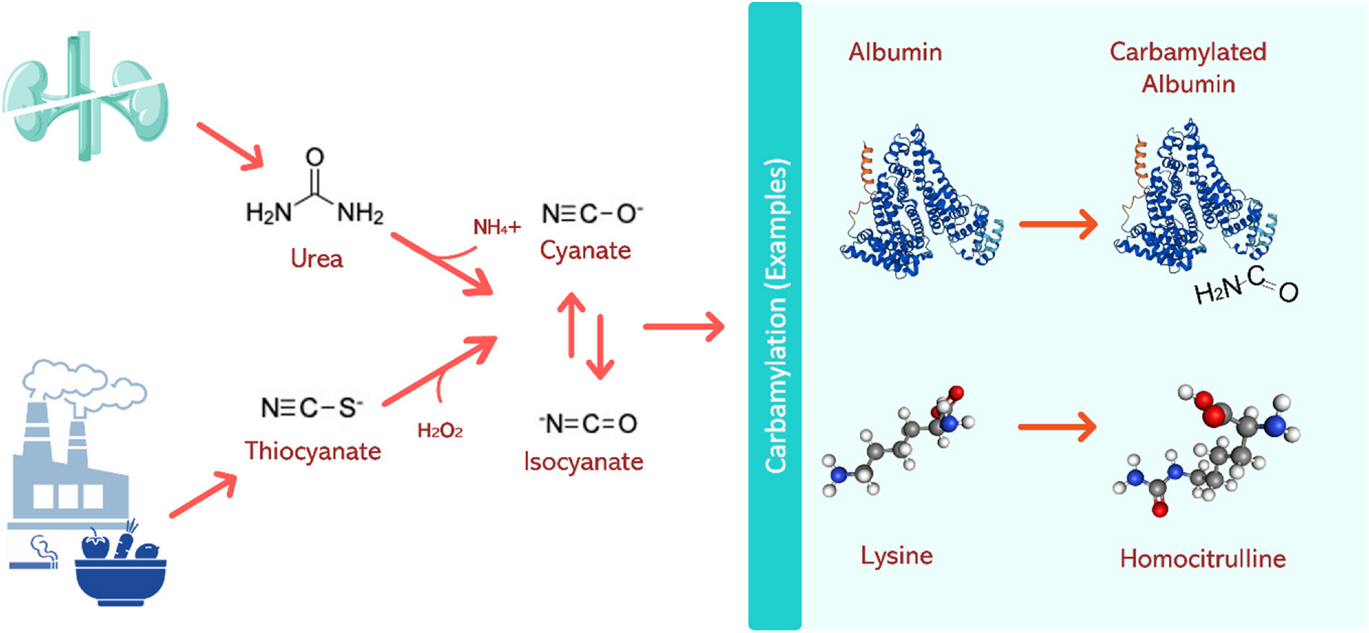
TMA induced by anti-tumor drugs can be categorized into two types: Type I TMA, induced by anticancer drugs, encompasses all chemotherapy regimens (such as Streptomyces C, Gefitinib, etc.), which potentially contribute to long-term kidney damage, increased incidence, and mortality rates. Type II TMA, induced by anti-tumor drugs, includes anti-VEGF drugs. VEGF plays a crucial role in maintaining the physiological function of glomerular endothelial cells, podocytes, and renal tubular epithelial cells. Excessive or deficient VEGF signaling has been shown to negatively impact both the structure and function of podocytes [2]. Within podocytes, VEGF signaling plays a crucial role in organizing the actin cytoskeleton. Proper signaling also provides a trophic survival signal via Akt (protein kinase B) and regulates cell cycle function through Ras/Raf interactions. Additionally, appropriate VEGF stimulation suppresses Nuclear Factor Kappa-light-chain-enhancer of activated B cells-mediated targets of inflammation and the activation of the renin–angiotensin–aldosterone system. In endothelial cells, VEGF signaling contributes to nitric oxide production and vasodilation, offering a trophic signal for endothelial survival and function. Furthermore, VEGF signaling regulates Di-Acyl Glycerol-Kinase-Epsilon, and its disruption can lead to thrombotic microangiopathy. Consequently, significant inhibition of this pathway can result in podocyte effacement, inflammation, and nephrotic syndrome by disrupting the podocyte cytoskeleton. Moreover, there is a clear association with thrombotic disorders and hypertension, stemming from endothelial cell dysfunction, dysregulation of clotting, and disruption of nitric oxide synthesis [3].
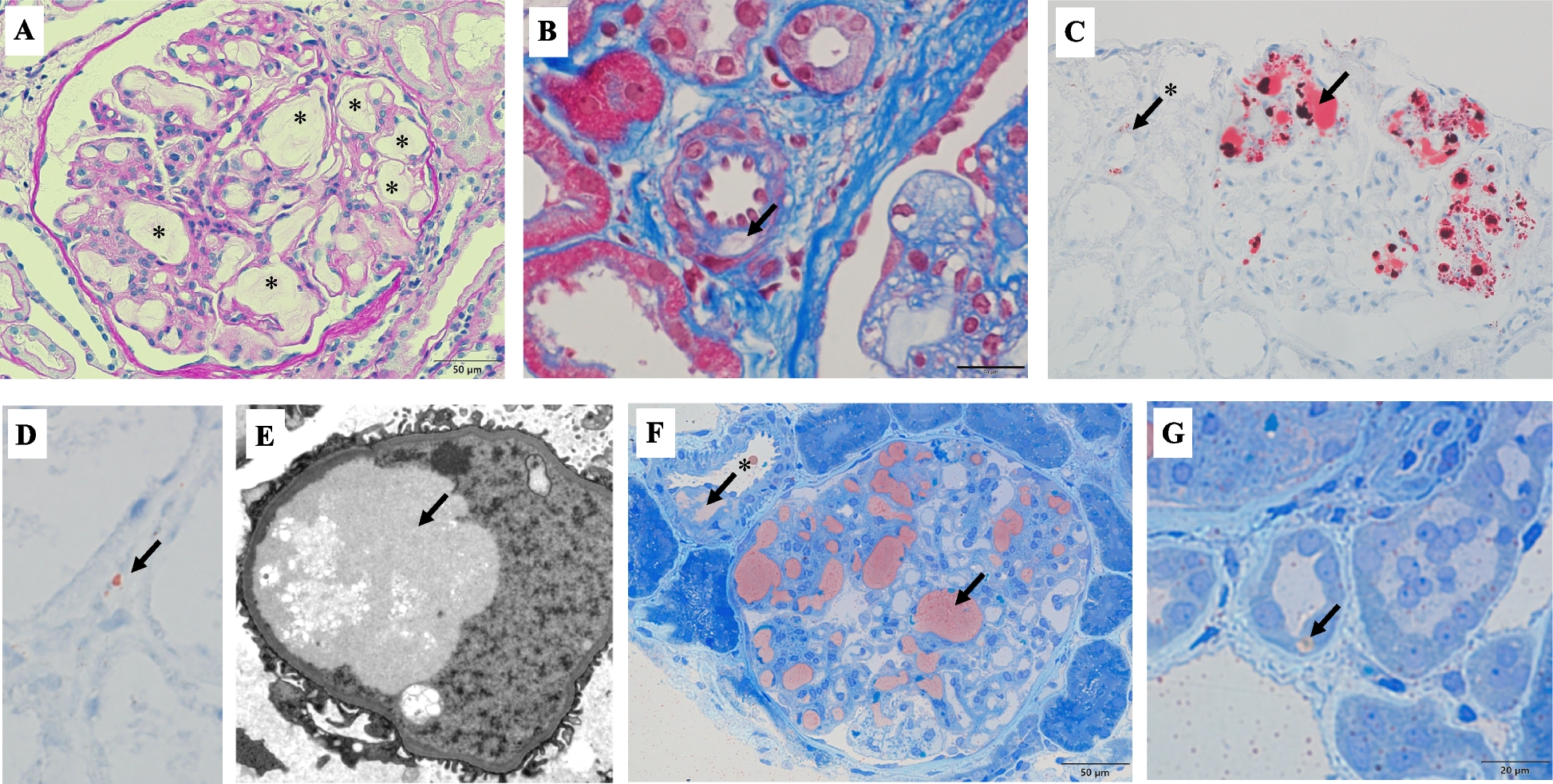
The kidney diseases associated with VEGF pathway inhibitors commonly manifest in two pathological categories: thrombotic microangiopathy, confined to renal tissues and devoid of abnormal ADMATS13 activity or complement gene mutations [4], and a subset of patients experience amelioration upon drug cessation. Angiotensin-converting enzyme inhibitors serve as the primary therapy for kidney-restricted TMA; however, efficacy is limited in cases with substantial proteinuria within the nephrotic syndrome spectrum. Marco Stortz [5] et al. documented a case of kidney-restricted TMA induced by bevacizumab monoclonal antibody, resulting in systemic TMA subsequently alleviated by steroid pulse therapy and plasma exchange. Rituximab administration has shown promise in relieving proteinuria in non-responsive patients to drug discontinuation [6]. The patient presented with hypertension preceding proteinuria, and the blood pressure once reached as high as 200/120 mmHg, with negative results for renal pathological immunofluorescence, multiple serum autoantibodies, and complement levels, ruling out immunologically mediated TMA at present.In light of hepatoprotection, glucocorticoids and biologics were not pursued. Following fruquintinib cessation and sacubitril/valsartan addition, serum albumin increment, urine protein reduction, serum creatinine normalization, edema alleviation, and renal function improvement were observed at the 5-month follow-up. It is hypothesized that furquidone-induced malignant hypertension may result in elevated mechanical shear forces, resulting in endothelial damage and rupture of the glomerular capillaries in the kidneys. This pathological process presents as capillary endothelium loosening, substantial widening of the affected area, and segmental changes resembling MPGN. Concurrently, activation of the renin-angiotensin system contributes to the development of TMA.Necessitating further investigation into complement pathway activation, cell-mediated immunity, and other mechanisms. Additionally, podocyte diseases, encompassing Minimal change disease (MCD) and focal segmental glomerulosclerosis (FSGS), represent another TMA variant induced by antitumor drugs. Receptor tyrosine kinase inhibitors (RTKIs) inhibit RelA activity, potentially leading to increased c-mip expression in podocytes, which disrupts the podocyte cytoskeleton and foot process fusion [7]. Hence, RTKIs predominantly instigate MCD or FSGS in certain patients. Clinical presentations encompass substantial proteinuria, potentially progressing to nephrotic syndrome, with a majority of patients typically experiencing amelioration post cessation of the medication. The renal pathology of this patient revealed podocyte fusion of nearly 60%, which was related to the use of anti-VEGF drugs. Despite not meeting the diagnostic criteria for podocytopathy, the impairment of the glomerular filtration barrier can precipitate substantial proteinuria. Moreover, there have been documented instances of VEGF pathway inhibitors provoking acute interstitial nephritis, acute tubular necrosis, crescentic glomerulonephritis, and proliferative immune complex glomerulonephritis [8].
Fruquintinib, a VEGFR inhibitor independently developed in China and approved by the CFDA, exhibits potent and highly selective inhibitory activity against all three isoforms of VEGFR (VEGFR-1, 2, 3), thus concomitantly suppressing tumor angiogenesis and lymphangiogenesis. As a third-line therapy for metastatic colorectal cancer, it significantly extends patients' survival duration [9]. Notably, fruquintinib demonstrates pharmacological efficacy at lower blood concentrations compared to other VEGFR inhibitors, thereby enhancing safety profiles. However, akin to other VEGFR inhibitors, hypertension and proteinuria represent common adverse reactions to fruquintinib. Eremina et al. [10] documented six cases of renal-limited thrombotic microangiopathy induced by bevacizumab usage. Upon cessation of the medication, improvements were observed in blood pressure, proteinuria, and renal function, suggesting the reversibility of these bevacizumab-induced side effects. Nonetheless, not all patients experience such favorable outcomes. Prolonged and substantial proteinuria may lead to irreversible renal damage, making medication discontinuation sometimes the most prudent course of action. For individuals who fail to respond to discontinuation, treatment modalities such as glucocorticoids, rituximab, and plasma exchange may be considered; however, the efficacy of these interventions remains uncertain [11].
Therefore, when employing anti-VEGF medications in clinical practice, it is crucial to acknowledge the role of VEGF as a pivotal signaling molecule synthesized by podocytes. These medications commonly induce proteinuria and hypertension as adverse effects. Therefore, regular monitoring of proteinuria and blood pressure is imperative for all patients undergoing anti-VEGF drug therapy. Prompt cessation of the drug is warranted if there is onset or progressive aggravation of hypertension, hematuria, proteinuria, etc. Additionally, if circumstances permit, renal biopsy should be promptly conducted to facilitate early detection of thrombotic microangiopathy. Furthermore, enhancing communication and collaboration among multidisciplinary teams including oncology, nephrology, and pharmacy is essential to actively advance the interdisciplinary realm of tumor nephropathy. This approach aims to foster early detection and timely management of nephrotoxicity induced by antitumor medications, thereby ameliorating the prognosis of malignant tumor patients and augmenting their quality of life.


留言 (0)