Leaving varices untreated may lead to significant bleeding, so intervention is often necessary. Esophageal varices associated with portal hypertension resulting from cirrhosis are common. In those cases, local endoscopic treatment such as endoscopic variceal ligation therapy (EVL) and endoscopic variceal sclerotherapy (EIS) are performed. Radiological interventions (IVR), such as balloon-occluded retrograde transvenous obliteration aimed at addressing the underlying cause of increased venous pressure may also be performed to prevent variceal bleeding. When treating colonic varices, it should be borne in mind that the risk of severe complications, particularly perforation, is much higher in the lower gastrointestinal tract than in the upper gastrointestinal tract. Moreover, the risk of recurrence would be high if the local endoscopic treatment, such as EVL or EIS, is performed with ignoring the underlying cause of the varices. In our case, elevated venous pressure in the IMV caused by an AVF in the IMA region was the underlying factor. In this present case, the cause of the transverse colonic varices was the elevated venous blood pressure due to the AVF. Therefore, we considered that excising the AVF could lead to an improvement in the varices. Regarding the other treatment, IVR may be one of the options in the point of view of invasiveness compared to surgical treatment, however coil embolism can cause post-procedural complications, including intra-abdominal bleeding [7] and intestinal ischemia [4], emphasizing the need to consider changes in blood flow after treatment. Moreover, in this case, the diameter of the fistula was relatively large and the distal portion of the AVF was in close proximity to the colon, so blood flow could impair if an embolus flowed into peripheral blood vessels. Regarding surgical treatment, ligating or resecting the AVF alone using surgical clips was also difficult because the distal side of the AVF was close to the marginal blood vessels of the sigmoid colon and was likely to cause ischemic complications on the colon.
AVF in the IMA region is a rare entity, and its causes have often been unclear in the small number of cases reported. Acquired factors, including trauma, postoperative complications, and cancer, are cited, with postoperative iatrogenic causes being more prevalent. Congenital cases are known as colonic arteriovenous malformations, where an arteriovenous shunt forms in an abnormal vascular network called a nidus. Although a nidus was not pathologically identified, the absence of a history of intestinal surgery or trauma in the present case made it difficult to determine whether the condition was derived from the congenital or acquired reason.
In general, IMV typically drains into the splenic vein or the portal vein. Although there are some reports of a collateral circulation developing between the IMV and the middle colic vein in the context of portal hypertension, however, in our case, the radiological examination did not reveal the direct flow of the contrast from the IMV to the portal or splenic vein. Therefore, it is presumed that the IMV originally drained into the middle colic vein. This anatomical feature with a specific hemodynamics likely led to the formation of varices in the splenic flexure area.
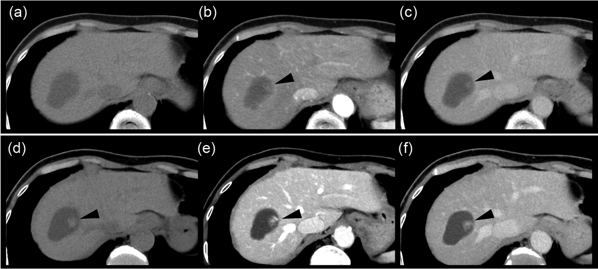
Whether to excise the area of the intestine with varices is a matter of debate. In this case, the variceal region on the colon was not excised because the goal was to decrease venous pressure after excision of the AVF. Use of ICG fluorescence imaging was important in this case for real-time visualization of vascular dynamics during surgery. The usefulness of ICG during resection of arteriovenous malformations has been reported [8]. ICG imaging confirmed successful exclusion of the AVF and subsequent reduction in venous pressure, providing direct evidence of the efficacy of surgical intervention. This technique not only enhanced the precision of the procedure but also potentially reduced the risk of postoperative complications by immediate identification of vascular abnormalities. Postoperative imaging analysis and endoscopic observation confirmed disappearance of the varices, suggesting the effectiveness of the reduction in venous pressure achieved by resection of sigmoid colon and mesentery with AVF.
A search of the PubMed database from 2013 to 2023 using the key terms “arteriovenous fistula”, “colon”, and “arteriovenous malformation” yielded 25 case reports on inferior mesenteric AVF. We briefly reviewed these cases, with inclusion of our present case (Table 1). While reports exist of cases presenting with AVF and varices, in those instances both were present in the cecum and were excised together [9]. Our present case is the only instance in which a colonic varix was treated by excising an AVF located at another site. The median patient age was relatively young at 56 years (range 24–81). Twenty-one of the 26 cases were in men, indicating a male predominance. The most common primary symptoms were abdominal pain and diarrhea, with many cases suspected to be ischemic colitis. A presumed cause was recorded in eight cases, four of which were post-colectomy for colon cancer and one post-hysterectomy, suggesting a significant proportion of postoperative cases. Treatment involved bowel resection in 14 cases, colostomy in one, and embolization in ten. In a case, surgery was scheduled after embolization [10]. A further case was treated conservatively [5]. Five of 9 patients who underwent embolization experienced recurrence of symptoms, ischemia, or rupture of varices, leading to subsequent surgical intervention [4, 7, 11,12,13]. There have been reports of embolization material deviating peripherally in AVFs with a diameter of ≥ 8 mm [4], suggesting that even when embolization is considered, surgery may ultimately be necessary.
Table 1 Literature review of the inferior mesenteric arteriovenous fistulaThe uniqueness of this case lies in the fact that the AVF in the IMA region was the cause, and sigmoidectomy was performed to treat transverse colonic varices with preserving the transverse colon. Furthermore, changes in blood flow were confirmed intraoperatively using ICG fluorescence imaging. The postoperative course also allowed observation of the healing process of the varices over time, making this a rare report.


留言 (0)