AIHA is an uncommon autoimmune disorder that produces the antibodies against red blood cell antigens, leading to hemolytic anemia. Although corticosteroid therapy is the primary treatment modality and often yields positive outcomes, many patients eventually become steroid-dependent, making cessation challenging; some patients even require alternative therapeutic approaches. In cases of AIHA refractory to conventional medical management, splenectomy is the recommended course based on its high efficacy rate. After splenectomy, approximately two-thirds of patients exhibit either complete or partial remission in the short term. Furthermore, there is evidence suggesting that a significant portion of these patients will experience sustained remission, negating the need for further medical interventions for prolonged durations [2]. Because of the improved laparoscopic techniques, LS has become the prevalent surgical procedure for patients with hematological disorders necessitating splenectomy [3]. In the presented case, LS was chosen as a secondary intervention for AIHA refractory to standard treatments. The post-LS surgical outcomes were satisfactory until the event of disease recurrence.
The primary objective of splenectomy in hematological conditions is the comprehensive removal of all splenic tissues, including both the main spleen and any accessory spleens. According to the findings presented by Maskal et al., splenectomy resulted in short-term complete remission in 61% of the patient cohort. However, this remission rate diminished to 39% at the 1-year follow-up, with 22% of the patients exhibiting symptomatic recurrence [5]. The incidence of recurrence attributable to the presence of accessory spleens remains indeterminate. Notwithstanding, accessory spleens are detected in 10–30% of the population at autopsy, suggesting a potential impact on the therapeutic outcomes of splenectomy procedures [5, 6]. Targarona et al. underscored the importance of meticulous care during LS for hematological disorders to circumvent parenchymal rupture, cell spillage, and inadvertent retention of accessory spleens, which could culminate in surgical failure [7]. While some studies suggest that preoperative CT scans might be redundant for the detection and localization of accessory spleens, given their ostensible visibility adjacent to the spleen during laparoscopy [8, 9], Gigot et al. contended that accessory spleens might elude complete identification via laparoscopy alone [4]. In the presented case, the accessory spleen evaded detection during the initial LS and remained challenging to discern during the subsequent LEAS, despite a meticulous search. Consequently, we suggest there should be exhaustive identification of accessory spleens using imaging techniques prior to LS. Tools such as Levovist-enhanced US, CT scanning, Tc-99 m denatured red blood cell scintigraphy, and SPIO-enhanced MRI have demonstrated efficacy in detecting accessory spleens [10].
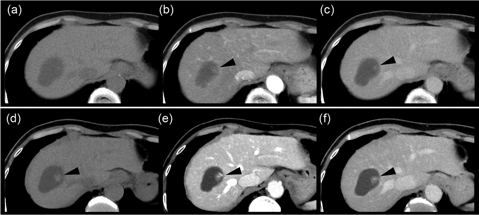
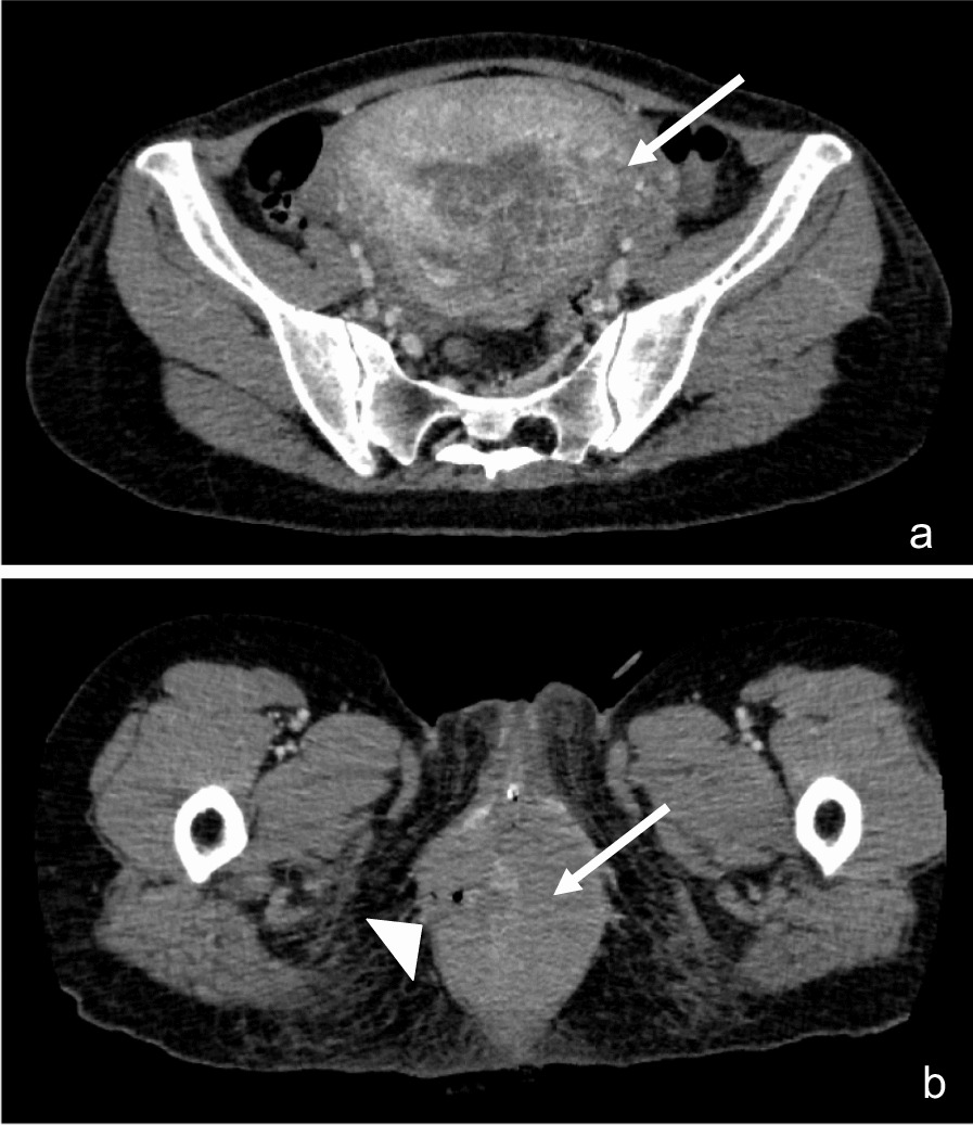
During LEAS, an accessory spleen, ensconced within adipose tissue, presented challenges via laparoscopy in our patient. Supplementary techniques such as employing a handheld gamma probe and preoperative CT-guided methylene blue injection have been recommended to enhance laparoscopic explorations and ensure that no accessory spleens remain undetected [11, 12]. Remarkably, in our instance, intraoperative US proved invaluable in delineating an accessory spleen, despite infrequent mention in existing LEAS literature. Intraoperative US offers a straightforward, safe, and effective approach to locate accessory spleens, proving to be particularly beneficial in scenarios marked by previous surgical adhesions or when the accessory spleens are shrouded by adipose tissue, as observed in our patient.
In our case, the recurrence of AIHA was linked to the presumed compensatory hypertrophy of the accessory spleen that wasn't removed during the initial LS procedure. Certainly, the recurrence of AIHA due to compensatory hypertrophy of the accessory spleen is less prevalent in the medical literature compared to conditions, such as ITP and other hematological disorders [13, 14]. There exists a singular case report addressing LEAS in the context of AIHA [15], but it lacks an in-depth exploration. Our patient's journey—undergoing LEAS following AIHA relapse post-splenectomy and then exhibiting symptom alleviation—adds a significant case to the existing literature.
Given the insights from our case, we describe three key recommendations:
Concurrent Resection: When performing splenectomy for AIHA, there should be a conscious effort to locate and excise accessory spleens concurrently to minimize the risk of recurrence.
Thorough Investigation for Recurrence: In cases where AIHA manifests again following a splenectomy, an intensive evaluation targeting the presence of an accessory spleen is essential. The act of resecting such an accessory spleen may offer alleviation from hemolytic manifestations.
Utility of Intraoperative US in LEAS: Particularly when the accessory spleen is concealed by adipose layers, intraoperative US emerges as a practical tool during the LEAS procedure to ensure accurate detection.


留言 (0)