Main reagent
Myocardial cell line H9c2 (National Biomedical Experimental Cell Resource Bank, USA), DMEM medium (Gibco, USA), Fetal bovine serum (FBS, Hyclone, USA), X-tremeGENE9 (Roche, Switzerland), double luciferase reporter gene detection system kit (Sigma, Germany), overexpression of miR-200b-3p vector (Genechem, Shanghai), ELISA kit (Mlbio, Shanghai), Polyclonal antibodies (Abcam, USA), isoproterenol (Sigma, Germany).
Collection of sample tissues
The subjects were 100 patients diagnosed with heart failure at Yongchuan Hospital of Chongqing Medical University. The inclusion criteria were as follows: (1) adults over 18 years old, (2) diagnosed with heart failure (clinically using the Framingham standard or echocardiography), and (3) received follow-up care in the heart clinic for at least three months. The exclusion criteria included severe respiratory diseases, chronic inflammation, organic heart disease, and hyperthyroidism. The normal control group consisted of 50 age-matched healthy subjects without cardiovascular disease or metabolic disorders. This study received approval from the Medical Ethics Committee of Yongchuan Hospital of Chongqing Medical University. The peripheral blood of the patients was collected and separated into serum and blood cells. The blood cells were stored at -80 °C after adding the appropriate RNAlater solution.
RNA-seq analysis
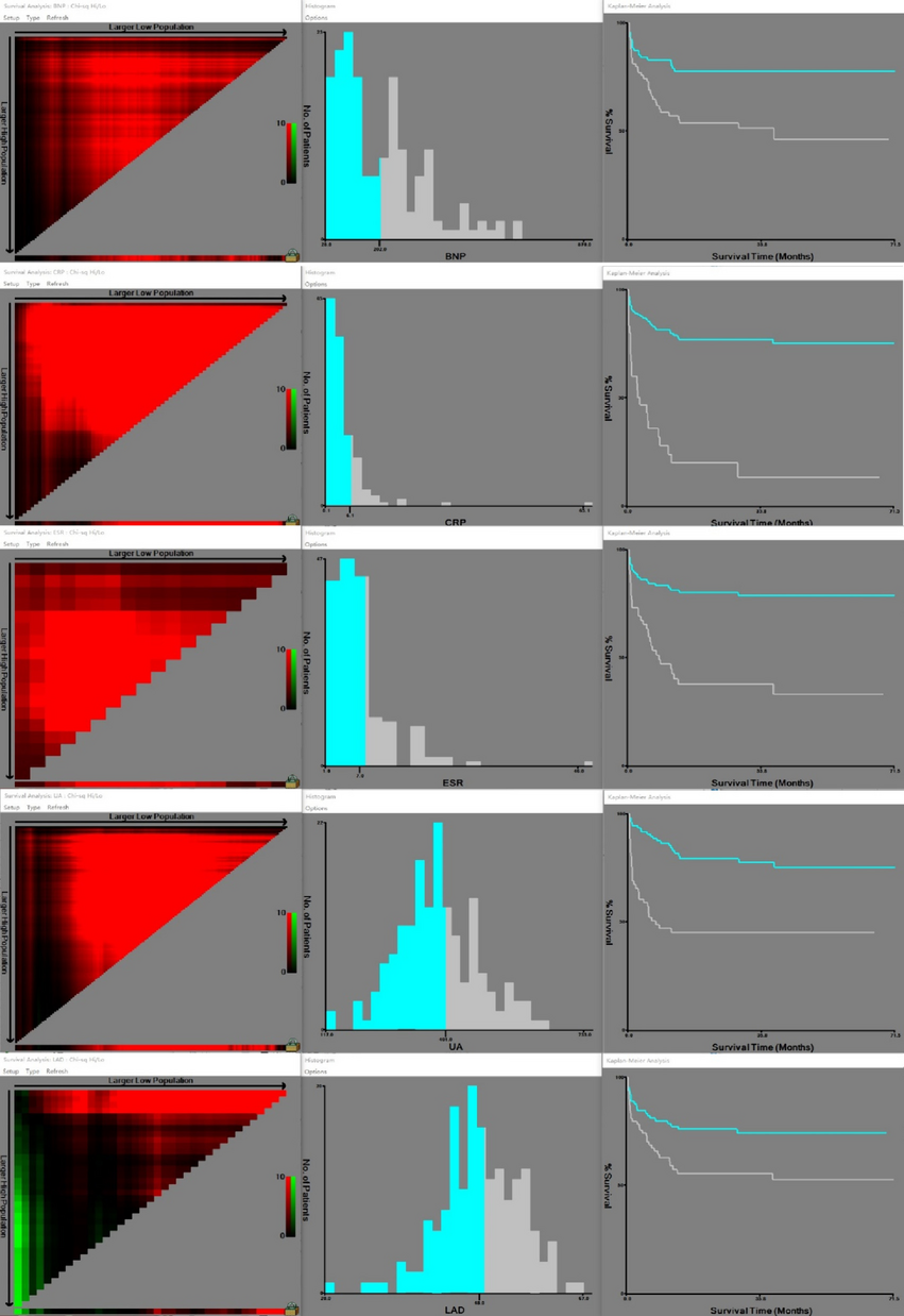
RNA extraction from blood cells was performed using TRIzol reagent. Following quality inspection, a cDNA library was constructed. After passing the quality control, the Illumina HiSeq4000 platform was utilized for high-throughput sequencing. The sequencing process and result analysis were conducted by Beijing Nuohe Zhiyuan Company. Specifically, |log2(fold change, FC)| > 1 and P < 0.05 were used as cutoffs to screen differentially expressed genes. An online website was employed to create heat maps. The KOBAS software was utilized to conduct KEGG pathway enrichment analysis on the genes associated with each distinct set of differentially expressed transcripts, and the screening threshold for significant differences was set to P < 0.05.
Cell culture and experimental grouping
H9c2 cardiomyocytes were cultured in DMEM medium containing 10% FBS and Penicillin-Streptomycin (80 U/ml) at 37 °C and 5% O2. For the experimental setup, four distinct groups were established: the control group, ISO group, ISO-miR-NC group, and ISO-miR-200b-3p group. The control group utilized culture medium as the control and added 50 µM ISO. Following the instructions of the X-tremeGENE9 kit, the expression vector was transfected into H9c2 myocardial cells. After 24 to 48 h of cell culture, well-growing cells were selected for subsequent experiments.
QRT-PCR experiment
QRT-PCR was conducted following the protocols outlined in the SYBR fluorescence quantitative assay kit instructions. The QRT-PCR reaction was set for 40 cycles with an initial denaturation at 94 °C for 5 min, followed by denaturation at 94 °C for 20 s, annealing at 60 °C for 1 min, and signal collection at 60 °C. To perform relative quantitative analysis, internal references such as GAPDH and U6 were utilized. The 2−ΔΔCT method was employed for data analysis in this study.
Bioinformatics prediction and double luciferase experiment
The binding sites of miR-200b-3p and ZEB1 were predicted using the online gene prediction website StarBase database. These cells were divided into four groups, namely ZEB1 (WT) + miR-NC group, ZEB1 (WT) + miR-200b-3p group, ZEB1 (MUT) + miR-NC group, and ZEB1 (MUT) + miR-200b-3p group. The dual luciferase reporter vectors were transfected into cells, and cell lysate lysis and 10 µL LAR II reagent were added. As an internal reference, sea kidney luciferase was utilized, and the firefly luciferase activity was measured using an enzyme labeling instrument.
Western blot experiment
Total protein was extracted from cell samples. The protein concentration was detected using the BCA kit. Load 40 µg protein samples per lane (used for detecting NLRP3 and ZEB1) were separated using 10% SDS-PAGE. Under the action of an electric field, proteins were transferred to the PVDF membrane. Seal the membrane with skim milk, dilute the primary antibody to a certain volume, and incubate it overnight at 4 °C. After washing, incubate with the corresponding secondary antibody for 2 h, and expose the target band in a chemiluminescent solution. Quantitative analysis of protein bands was performed using ImageJ software. Calculate the relative quantification of proteins using GAPDH as a control.
ELISA experiment
Dilute the standard substance and detect antibodies according to the method described in the ELISA kit. Add cell supernatant and incubate for 1 h. Subsequently, add the enzyme-labeled reagent and incubate for 50 min. Next, add the substrate chromogenic agent and incubate for 15 min. Finally, terminate the reaction, gently shake, zero with a blank, and measure the OD values at 450 nm of each well in sequence using a microplate reader. Calculate the levels of various inflammatory factors based on the standard curve and sample OD values.
Statistical analysis
SPSS 22.0 software was utilized to analyze all experimental data. Continuous data were represented using the format mean ± standard deviation (x ± s). To compare between two groups, we employed the t-test. For comparing multiple groups, we utilized one-way ANOVA. Finally, for pairwise comparisons, we employed the LSD-T test. We consider P < 0.05 to represent a statistically significant difference.


留言 (0)