
記住我


The BA is a common trunk formed by the union of the left and right vertebral arteries. It begins at the medullary-pontine sulcus and becomes narrowly related with the basilar apophysis of the occipital bone and the pontine cistern [48]. It has a somewhat tortuous course, as it slithers across the basilar sulcus, a midline vertical sulcus on the ventral aspect of the pons where the artery normally runs. Along its course it provides several pontine branches that irrigate numerous structures of the pons. Additionally, there are crucial collaterals to be found, which will be detailed shortly. At its end, the Basilar Artery terminates at a bifurcation right in front of the pontomesencephalic sulcus, point in which it gives off its two symmetrical terminal branches: the left and right posterior cerebral arteries, at the level of the interpeduncular cistern [25]. This division is also known as Basilar Apex or “Basilar tip”, which may be situated at the exact same level, above, or beneath the superior border of the dorsum sellae, towards the prepontine cistern. In cases where the basilar bifurcation lies above the dorsum sellae or below it, they are designated as high-riding or low-riding basilar tips, respectively, having different implications regarding the surgical approach of choice [7, 11, 25, 35]. Basilar lesions, such as injury due to arterial wall dissection can lead to a broad assortment of gravely severe outcomes, ranging from many brainstem alternate syndromes to the dreaded Locked-in Syndrome [10, 12, 16]. The BA has also an utmost importance when treating stroke or aneurysms via endovascular procedures [26, 32].
The branches of the BA are anterior inferior cerebellar artery (AICA), posterior cerebral artery (PCA), superior cerebellar artery (SCA), and pontine perforating branches which are responsible for supplying numerous and crucial pontine nuclei.
Anterior inferior cerebellar artery (AICA)The AICA is the first important collateral of the BA. It courses across the central portion of the cerebellopontine angle (CPA) and is part of one of the three complexes described by Rhoton [38, 39]: AICA is intimately associated with the pons, the lateral recess of the fourth ventricle, the middle cerebellar peduncle, the VI, VII and VIII cranial nerves, the cerebellopontine fissure, the cerebellar petrosal surface and the conspicuous cerebellar-pontine cistern [50]. Its major deep venous group lies in the cerebello-pontine fissure, accompanied by veins that receive drainage from the middle cerebellar peduncle. From the lower part of BA, it originates proximate to VI CNs origin and then runs posterolaterally anterior to VI, VII and VIII CN. It often makes a loop that runs across the internal auditory meatus. It commonly divides into a rostral and a caudal trunk, near the VII–VIII CN complex [13, 43, 44].
When involved in a stroke or lesion, should it happen, certain syndromes can occur: such as the Millard-Gubler or the Foville Syndrome, that include VII and VIII ipsilateral palsy and contralateral sensory-motor deficit. Even more, ataxic hemiparesis can be found, when the cerebellum blood flow is compromised. From this artery can also arise complex wide neck aneurysms prone to be treated surgically through intraoperative clipping [43], being high-risk procedures. The AICA is divided into four segments and each of these segments may include more than one trunk, depending on the level of bifurcation of the AICA.
Anterior pontineIn relation with the VI CN, the anterior pontine segment of AICA extends from its arising point at the BA to the medullary olive.
Lateral pontineThe lateral pontine segment of AICA extends to the anterolateral region of the pons, in which it continues its course towards the CPA. This segment can be further subdivided into three parts: premeatal, meatal and postmeatal, depending on the proximity and relation with the internal acoustic meatus. It gives rise to several nerve-related branches including the labyrinth or labyrinthine artery, the recurrent perforating arteries (in the parafloccular space or on the cisternal surface of the middle cerebellar peduncle [42], and the subarcuate artery. Moreover, specific branches for the cranial nerves from VI to the XI can be found, overlapping with blood flow sources as diverse as those coming from the PICA or even the ascending pharyngeal artery [9, 15].
FlocculonodularThe flocculonodular segment of AICA begins where the AICA passes the flocculus towards the CPA, until it finally reaches it, along with the cerebellopontine fissure and middle cerebellar peduncle. Perforating arteries can also be found nourishing the brainstem or cerebellum with the blood flow provided from this segment.
CorticalIt supplies the petrosal cerebellar surface and the floccular area. It bifurcates into superior and inferior branches. The superior branch, anastomoses with cortical branches of the SCA. The inferior branch, anastomoses with cortical branches of the PICA at a point that varies according to whether the AICA expresses cortical dominance over the latter or, on the contrary, the PICA dominates over the former.
Superior cerebellar artery (SCA)This artery originates 2 or 3 mm below the basilar bifurcation, travels immediately beneath the III CN, goes round the pontine-mesencephalic union, and passes below the IV CN and above the V CN in all cases. Afterwards, it reaches the cerebellar-mesencephalic fissure and finally heads towards the vermis and the tentorial cerebellar surface. SCA is part of the upper complex, and relates with the upper half of the fourth ventricle’s roof, the superior cerebellar peduncle, the cerebellar-mesencephalic fissure, the tentorial surface of the cerebellum, and the III, IV and V cranial nerves [13, 53]. As for venous anatomy of the upper complex, its key deep vein rests within the cerebello-mesencephalic fissure, as a functional tandem with veins that drain the superior cerebellar peduncle. The midbrain is frequently the portion of the brainstem to be related if this vessel is somehow subject to injury or occlusion. We point out as mere examples the Weber, the Claude or the Benedict syndromes (which involve ipsilateral III cranial nerve paralysis and contralateral weakness or ataxia). Although, SCA aneurysms are rare, their management alternatives are not well delineated and need to be tailored accordingly. As stated, there is an increasing role of endovascular treatment for all aneurysms, especially for aneurysms of the posterior circulation. Nevertheless, in some situations (wide base, dysmorphic features) coiling is not feasible, thus their surgical management has its own distinctive complexity and requires careful planning. When ruptured, these dysmorphic aneurysms must be treated urgently either via endovascular surgery or intraoperative clipping [24]. Four segments of SCA can be recognized as follows:
Anterior pontomesencephalicThe anterior pontomesencephalic segment of SCA extends from its origin in the BA to the anterolateral portion of the mesencephalon.
Lateral pontomesencephalicThe Lateral pontomesencephalic segment of SCA extends from the lateral portion of the mesencephalon until it enters the cerebellar-mesencephalic fissure, closely related to the fifth nerve.
Cerebellar-mesencephalicThe cerebellar-mesencephalic segment of SCA courses through the homologous fissure, along with IV CN.
CorticalThe cortical segment of SCA commences where the SCA leaves the fissure to reach its destination territories, the vermis and the tentorial cerebellar surface.
SCA’s collateral branches are perforating, pre-cerebellar and cortical [38]. Perforating collateral branches is accountable for irrigation the tegmentum, the interpeduncular fossa, the cerebral peduncle, the junction of the superior and middle cerebellar peduncles and part of the quadrigeminal plate. This perforating collateral include direct and circumflex types. The direct type pursues a straight course to enter the brainstem, while the circumflex type winds around the brainstem before terminating in it. Pre-cerebellar collaterals direct blood flow to the cerebellar central nodule and the superior medullary velum. Finally, the cortical collaterals are providing nourishment to the petrosal cerebellar surface and include Vermian branches, Hemispheric branches, and Marginal branch (Not constant). Seldom, a variant meningeal supply to the medial tentorium, arising from the superior cerebellar artery (SCA) which its believed anastomosed with the artery of Davidoff and Schechter (ADS—further discussed), later came to be known as the artery of Wollschlaeger and Wollschlaeger (AWW)[27].
Posterior cerebral artery (PCA)The posterior half of the Circle of Willis reaches completion when the PCA, which arises from the basilar bifurcation below the posterior perforated substance, receives the Posterior communicating artery (PCommA), consequently closing the circuit. The PCA is joined by the PCommA at the lateral margin of the interpeduncular cistern, circles the brainstem passing through the crural and ambiens cisterns and reaches the quadrigeminal cistern, to finally distribute to the posterior part of the hemisphere. The basilar bifurcation may be found as far caudal as 1.3 mm below the pontomesencephalic connection and as far rostral as the mamillary bodies, sometimes even adjacent to the floor of the third ventricle. The BA usually bifurcates opposite the interpeduncular fossa, but some variations have been described [13, 17, 34, 49, 51, 53]. As a brief clinical note, depending on the segment or branch involved, a brimming and diverse set of sign and symptoms can be identified; for instance, Parinaud Syndrome (Paralysis of upgaze, convergence-retraction nystagmus, lid retraction, and light near dissociation) in cases of perforating branches and cortical blindness, when calcarine cortex (with visual eloquence) is the location of the pathology. Additionally, it can also be the origin of aneurysms of the Basilar Apex area, before joining the Posterior Communicating Artery (PCommA) or at its junction with the aforementioned vessel [32] (Figs. 5, 6, 7).
Fig. 5
Angio-CT 3D render of arterial circulation of the brain, comprised by anterior and posterior circuits, with detailed view and labeling of the main vessels. In the oblique posterior-superior view, it is observed incidentally a dominant right Posterior Communicating Artery with a much greater caliber than its counterpart. ACA Anterior cerebral artery, AICA Anterior inferior cerebellar artery, MCA Middle cerebral artery, PCA Posterior cerebral artery, PICA Posterior inferior cerebellar artery, SCA Superior cerebellar artery, VA Vertebral Artery
Fig. 6
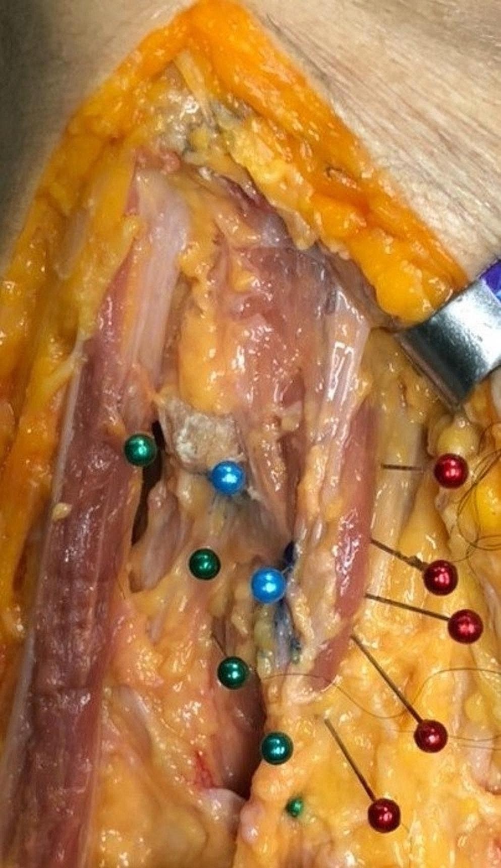
Anatomical dissection featuring the Basilar tip in the pre-pontine cistern at the vicinity of the interpeduncular and crural cisterns, and related neurovascular structures such as cranial nerves III and IV, as well as the Posterior Cerebral, Posterior Communicating and Superior Cerebellar Arteries
Fig. 7
Basilar tip and Posterior Cerebral Artery's neural, cranial and cisternal relations; transcranial subtemporal view
The PCA can be divided into four parts, P1 through P4:
P1 segment (pre-communicating)Also referred to as pre-communicating segment, it extends from the basilar tip to the point in which the PCA is joined by the PCommA. An anatomic variant in 1/3 of hemispheres is known as fetal configuration, in which P1 has a smaller diameter than the PCommA (or even more, completely absent) and the PCA emerges from the ICA, hence supplying blood flow as well to the posterior circulation [26].
P2 segment (post-communicating)This portion begins when the PCA is joint by the PCommA, lies within the crural and ambient cisterns, and terminates lateral to the midbrain. This segment is subdivided in two parts, according to its relationship with the cisterns: P2-A or crural segment, because it travels around the cerebral peduncle in the crural cistern, and P2-P or ambiens segment, because it courses lateral to the midbrain in the ambient cistern. The P2 segment runs through the crural cistern, relating itself to the cerebral peduncle, uncus, the optic tract and basal vein ambient cisterns, and then courses along the ambiens cistern, between the lateral midbrain and the parahippocampal and dentate gyri, in the vicinity of the optic tract, basal vein, geniculate bodies, the trochlear nerve and tentorial edge [13, 48,49,50].
P3 segment (quadrigeminal)P3 courses dorsally from the posterolateral aspect of the midbrain and ambiens cistern to reach the quadrigeminal cistern and then terminates at the anterior limit of the calcarine fissure. The PCA here often divides into its major terminal branches, the calcarine and parieto-occipital arteries. The quadrigeminal segments from both sides approach each other posterior to the cerebral colliculi and the point where both PCAs are nearest is known as the quadrigeminal or collicular point [8].
P4 segment (cortical-calcarine)It is majorly comprised of the branches distributed to the parieto-occipital and calcarine cortical surface. The anterior end of the calcarine sulcus marks the beginning of P4.
The PCA gives off three kind of branches, including central perforating branches, ventricular branches and cerebral branches, providing not only the posterior portion of the cerebral hemispheres, as its denomination suggests, but also sends critical branches to diencephalon, mesencephalon and ventricular system [2, 7, 8, 38, 51]. Central perforating branches divided into direct and circumflex perforating arteries, PCA gives off branches to the diencephalon and midbrain (Tectum, cerebral peduncles, and three nuclei including Edinger-Westphal, oculomotor and trochlear). Among these perforating arteries, one can commonly identify, Interpeduncular thalamoperforators, Mesencephalic perforating arteries, Thalamogeniculate arteries, Quadrigeminal and geniculate branches and Artery of Percheron which is a seldom anatomic variant that nourishes the paramedian thalami and the rostral midbrain bilaterally. It has also been identified a meningeal artery supplying the medial tentorium, arising from the posterior cerebral artery (PCA), the artery of Davidoff and Schechter (ADS), a vessel named in homage to its mentors in neuroradiology [27]. It appears to originate distal to the confluence of the PCA and posterior communicating artery (PCommA), suggesting that it arises from the P2 segment.
Ventricular branches destined for the choroid plexus and walls of the lateral and third ventricles as well as adjacent structures. Very often one or both of these branches join the Anterior choroidal artery in the proximity of the choroid plexus of the lateral ventricles. There are two constant branches including medial posterior choroidal, that arising mostly from P1 or P2 and lateral posterior choroidal, that arising from P2 in most cases.
Cerebral branches are headed to and held accountable for the supply of the cerebral cortex and splenium of the corpus callosum. A wide cortical vascular territory is supplied by the numerous branches that emerge from the cortical segment. Amongst the constant collaterals, the following can be listed [28]: Anterior, middle and posterior temporal branches, in which the anterior branch frequently anastomoses with anterior temporal branch of MCA, Hippocampal branch, Common temporal branch, Parieto-occipital branch, Calcarine branch, and Posterior pericallosal artery (Splenial artery) which often anastomoses with pericallosal artery of ACA (Fig. 8).
Fig. 8
Posterior cerebral artery main branches
留言 (0)