
記住我


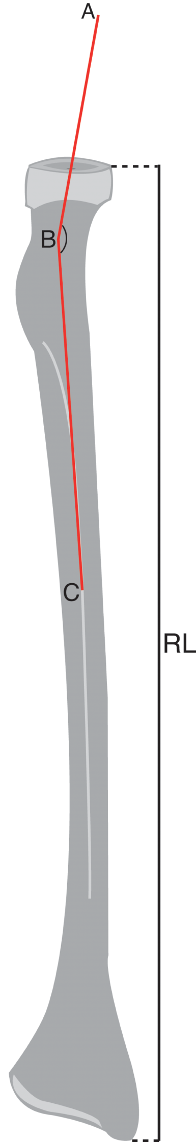
The ICA has been the subject of a multitude of studies and has been one of the most relevant and crucial vessels in the field of neuroanatomy, neurosurgery and neuroradiology. Several classifications exist that divide ICA in a certain number of segments. Keller, Rhoton, Bouthillier, are amongst the most prominent scholars that analyzed its intrinsic anatomy, relation with surrounding structures, microsurgical anatomy, histology, and in this fashion contributed to shape the most accepted and used classifications, of which Rhoton’s is the more widely spread one [25, 43]. According to it, the ICA can be divided in four segments, from C1 to C4, that go along with blood flow from the heart, from proximal to distal. These segments are: the Cervical ICA (C1), the Petrous ICA (C2), the Cavernous ICA (C3) and the Supraclinoidal ICA (C4) [43]. Moreover, C4 is further subdivided into Ophthalmic, Communicating and Choroidal segments. A handful of variant arterial pathways can be found throughout its course towards the intracranial compartment (retrojugular and/or retropharyngeal) and abnormal curvature (kinks, loops, and/or coils) might be encountered along its progress. Several collateral branches exist, often various either embryological persistent or inconstant amongst them [2]. Bouthillier’s classification is also widely spread and divides the ICA into 7 segments, them being: C1, cervical; C2, petrous; C3, lacerum; C4 cavernous; C5, clinoid; C6, ophthalmic; and C7, communicating. [3] For practical purposes, the authors will make use of Rhoton’s classification.
Cervical ICA–C1The Cervical ICA (C1), is the first and longest segment. It commences at the bifurcation of the Common Carotid Artery’s (CCA). Usually at the levelof the fourth cervical vertebrae at Farabeuf’s triangle, which is delineated by the internal jugular vein (IJV) posteriorly, the thyrolinguofacial venous trunk anteriorly, and the hypoglossal nerve superiorly. It lies deep to the medial border of the sternocleidomastoid muscle, and it relates laterally with the internal jugular vein and posteriorly with the X cranial nerve, wrapped along the other structures in the carotid sheath. Along with it, postganglionic sympathetic fibers (PGSNs) ascend to form the Internal Carotid Plexus [4, 12].
It contains the carotid sinus, a crucial baroreceptor that senses changes in systemic blood pressure and is located in the adventitia of the carotid bulb of the internal carotid artery, which is intimately related but a distinct organ from the carotid body, a chemoreceptor found at the CCA’s bifurcation. It arises towards the carotid canal of the petrous bone at the exocranial skull base and on its way, the ICA passes through the parapharyngeal space which is divided into pre-and post-styloid compartments and bordered laterally by the posterior belly of the digastric muscle. Often overlooked, this segment holds utmost significance, particularly when addressing lesions such as Glomus Jugulare. The surgical implications can indeed be dreadful. Consequently, it can be localized along the exocranial paramedian surgical corridor (one of three skull-base surgical corridors postulated as modular compartments: median, paramedian and lateral), as meticulously described by impeccable works [10, 13].
Petrous ICA–C2The Petrous segment, still surrounded by PGSNs, begins when the ICA enters the carotid canal of the petrous bone and ends in the internal orifice of the carotid canal. Three subdivisions can be identified: (a) a shorter one (vertical) that ascends and then bends in a rostral direction; (b) a posterior loop, anterior to the cochlea; (c) and a horizontal longer segment (each segment connected by genu, the posterior and the anterior genu). The latter is intimately related with the V cranial nerve, lying inferomedial to Meckel’s cave [23], where the Gasserian ganglion can be found, in the proximity of the VI nerve’s entrance to Dorello’s canal (below the posterior clinoid process, superior to the petroclival venous confluence [8, 21, 22]); afterwards it traverses in an antero-medial direction, heading to the apex of the petrous bone, parallel to its sagittal axis, to finally reach the aforementioned internal carotid canal orifice, and enter the cavernous sinus anteriorly to petrosphenoidal ligament or “Grubert’s ligament”, which extends from the petrous apex to the petrosal process of the sphenoid bone. Therefore, the horizontal portion, also related to the greater and lesser superficial petrosal nerves, surpasses the foramen lacerum (comprised by the union between the petrous apex, the lateral aspect of the dorsum sellae, and the sphenoid body) forming the anterior genu and ascends with a parasellar trajectory, emerging from the internal carotid canal, piercing the dura as it passes the petrolingual ligament, which bonds the petrous apex and the lingual process of the sphenoid bone, to become the next carotid segment [5, 9, 43].
In the petrous ICA it has also been described a thick “carotid cuff” [44], formed by connective tissue around the artery and a venous plexus. The origin of two branches can be identified from this portion, the caroticotympanic artery (which is inconsistent) and the pterygoid branch (present in approximately 30%) [30]. Additionally, it is worth noting that roughly in more than 80% of cases, there is a dorsal (endocranial) dehiscence of the petrous carotid canal, which can have critical clinical implications [16, 34].
Cavernous ICA–C3Once the ICA emerges from the carotid canal and traverses below the petrolingual ligament, which extends from the petrous apex to the lingual apophysis of the sphenoid bone, it enters the cavernous sinus running through the carotid sulcus, hence becoming the intracavernous ICA. This segment, from proximal to distal, can be divided into four segments: (1) the short vertical segment (a continuation of the paraclival ICA); (2) the posterior genu; (3) the horizontal segment; and (4) the anterior genu, which continues with the paraclinoidal ICA as it emerges from the CS. The intracavernous ICA is covered by a vascular membrane lining the sinus, still surrounded by PGSNs [43]. This portion of the ICA is intimately related to the medial wall of the cavernous sinus, of utmost clinical importance: In the nearest vicinity lies the significant carotico-clinoid, inferior parasellar and posterior parasellar ligament.
The cavernous carotid segment is directed anteriorly and then supero-medially, to bend posteriorly forming in this manner the posterior loop of ICA. The ICA continues horizontally and afterwards bends towards a rostral direction (part of anterior loop of ICA) heading to the anterior clinoid process. Afterwards, it traverses the proximal dural ring—namely the “carotid-oculomotor membrane” (which incompletely encircles the ICA), given the strict adjacency between the ICA and the CN III or oculomotor nerve-, and pierces the dura at the distal dural ring (which completely encircles ICA) where the artery becomes intradural. The segment of the ICA that is found between the proximal and distal dural rings, these also defining the limits of the carotid collar, is additionally known as the clinoid segment. The clinoid segment is bounded medially by the carotid sulcus, anteriorly by the optic strut, and medially by the anterior clinoid process. The distal dural ring is tightly adherent to the anterior and lateral aspect of the clinoid segment, but not the medial and posterior aspect. The posteromedial intradural area around the distal clinoid segment is known as the carotid cave [4, 5, 12, 18, 43, 53].
The cavernous carotid artery has many branches, usually grouped as trunks. The main trunks and branches are the following: (A) Meningohypophyseal trunk, which the largest & most proximal, emerging at the level of the posterior loop and present in 100% of cases. Meningohypophyseal trunk has three branches: The posterolateral branch, tentorial artery (or artery of Bernasconi & Cassinari): provides the blood flow to the proximal III CN, proximal IV CN and Gasserian ganglion [28]. (This branch can often supply petroclival meningiomas.) The posteromedial branch, dorsal meningeal artery (or dorsal clival a.) supplies the proximal VI CN and, seldom, the III CN. Finally, a medial branch giving rise to the inferior hypophyseal artery which supplies the posterior lobe of the hypophysis. It is worth noting that this trunk’s branches can emerge directly from the Internal Carotid Artery. (B) Anterior meningeal artery (C) Inferolateral trunk (ILT), another constant collateral (present in > 90%), which provides supply to the cranial nerves that travel through the inner layer of the cavernous sinus’ lateral wall [6, 8, 18, 28, 47, 50, 53]. All of its branches terminate on the inferomedial aspects of the intracavernous CNs. Extensive anastomoses have been found between the ILT branches and the branches arising from external carotid artery. These anastomoses supply the III CN, proximal IV CN, distal VI CN and proximal V1 and V2 CN [28]. (D) At last, a medial branch is found in less than 10% of cases, the capsular artery of McConnell, which supplies the capsule of the pituitary gland [8, 15, 24, 29, 40, 47].
Supraclinoidal ICA–C4Once the ICA traverses the distal dural ring, it is known as Supraclinoidal ICA. It turns upwards, posteriorly and laterally, toward the lateral aspect of the optic chiasm, then up to the anterior perforated substance to end up bifurcating just below the anterior perforated substance into two terminal branches. One branch extends anteromedially, the anterior cerebral artery. The second branch extends laterally, forming the middle cerebral artery. According to Dr. Rhoton’s classification, C4 Supraclinoidal segment is subdivided into three divisions, namely the ophthalmic, the communicating and the choroidal division [4, 12, 43].
Ophthalmic segmentThe longest portion of C4 commences immediately distal to the dural ring and ends just proximal to the origin of the Posterior Communicating Artery (p-comm). Important perforating arteries arise from this portion, supplying the optic nerve and other chiasm related structures. Two main collaterals are typically present, including the Ophthalmic Artery and Superior Hypophyseal Artery. In more than 96% of cases, the Ophthalmic Artery emerges from the ventral aspect of the Supraclinoidal ICA just inferior to the optic nerve and the anterior clinoid, distal to the cavernous sinus. In the remaining 4%, the ophthalmic artery arises from the cavernous segment. In general, the origin of the ophthalmic artery if often taken to mark the location of the carotid artery entrance into the subarachnoid space, and serves as an important landmark in angiography. The origin of the ophthalmic artery from the ICA can vary by 5 mm anterior to 7 mm posterior to the anterior clinoid process, of which it keeps intimate vicinity. The ophthalmic artery then runs through the optic canal into the orbit, superomedial to the optic strut, along with the II cranial nerve, inferolateral to the latter. This artery has a conspicuous “kink” that can be seen on lateral angiograms [31]. Regarding the Superior Hypophyseal Artery, in the medial wall of the ICA several of the aforementioned perforator arteries emerge, destined to the pituitary stalk and anterior lobe of the pituitary gland. One of the branches is usually and also supplies the dura of the cavernous sinus.
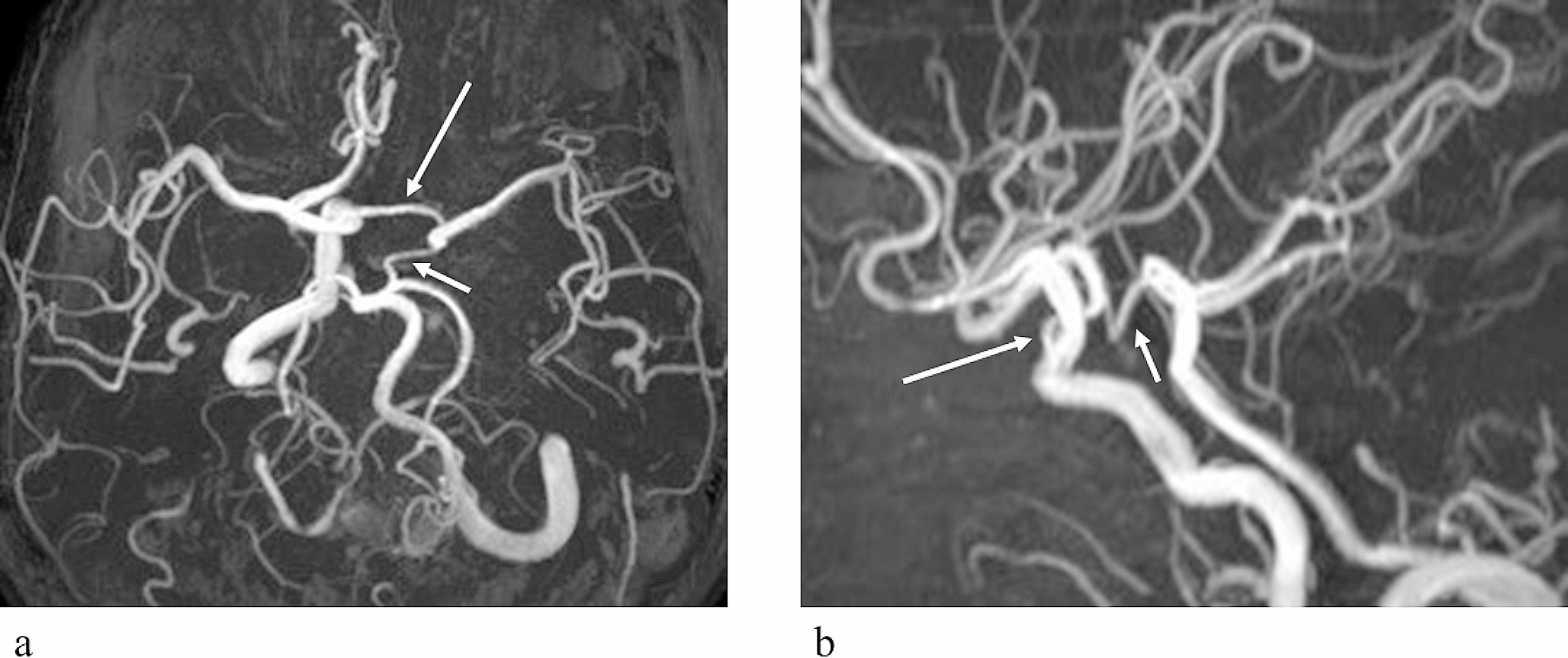
Communicating segmentThe Communicating Segment begins immediately proximal to the Posterior Communicating Artery origin and traverses between II and III cranial nerves, to terminate at the origin of the Anterior Choroidal Artery. Its branches include the PCommA and the Anterior Thalamoperforator Arteries. The PCommA arises from the posterior-medial wall of the ICA, and from there it extends dorsal and slightly medial to encounter Posterior Cerebral Artery, lateral to the optic tract and the mamillary bodies, rostral and dorsal respectively.
The PCommA can also be found as a variant in nearly 18% of cases, known as Fetal p-comm, which has a greater diameter than the proximal P1 segment of the ipsilateral posterior cerebral artery thus providing the majority of blood flow to its respective territory. Several perforators emerge from the PCommA and can be divided into two groups: Anterior perforating group and Posterior perforating group, which are destined to the hypothalamus, ventral thalamus, posterior perforated substance and subthalamic nucleus. Anterior Thalamoperforator Arteries irrigate the optic tract, the optic chiasm, the posterior hypothalamus, the posterior limb of the internal capsule and the subthalamus. Also referred to as premamillary arteries, they often arise from the PCommA. A number of these structures are featured in anatomical dissections and figures all along the text of this article.
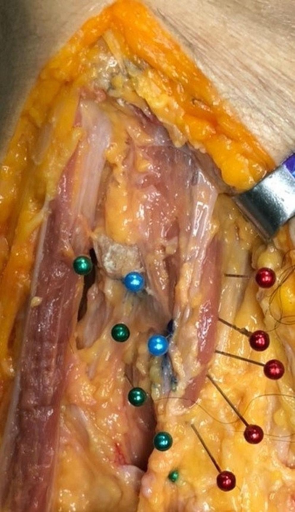
Choroidal segmentThe Choroidal segment spans from the Anterior Choroidal Artery (A.Ch.A.) origin to the bifurcation of the ICA into the ACA and MCA. The Anterior Choroidal Artery originates from the posterior-lateral wall of the ICA. It takes off is usually 2 to 4 mm distal to PCommA and courses posteriorly toward the lateral aspect of the lateral geniculate body. It then directs itself laterally, inferior to the optic tract, to reach the inferior choroidal point, to pierce the temporal horn and anastomose which branches from the posterolateral choroidal artery. One can recognize two portions, the cisternal and plexal segments. The cisternal segment, extends from its origin to the inferior choroidal point and runs through the crural cistern, parallel to the optic tract within it. Limited anterolaterally by the uncus and posteromedially by the cerebral peduncle, it is further divided into proximal and distal cisternal A.Ch.A., according to the relation with the lateral geniculate body. The plexal segment enters the supracornual recess of temporal horn, penetrating the choroidal point. It mainly but not exclusively supplies crucial structures of the diencephalon, mesencephalon, central core and mesial temporal lobe, such as: portion of optic tract, medial globus pallidus, genu of the internal capsule, the inferior half of posterior limb of IC, uncus, retrolenticular fibers (optic radiation), lateral geniculate body, dentate gyrus, fornix, globus pallidus, red nucleus, locus niger, amygdala, temporal horn, lateral geniculate body, hippocampus, and anterior perforated substance. Figures 2, 3 and 4.
Fig. 2
Posterior view of the Circle of Willis’ anterior half. Acom ACommA, anterior communicating artery, AChoA anterior choroidal artery, LICA left ICA, MCA middle cerebral artery, PCA posterior cerebral artery, PCP posterior clinoid process, RAH recurrent artery of Heubner, RICA right ICA
Fig. 3
Terminal ICA bifurcation into A1 and M1, deep in the carotid cistern. It is shown the utilized direction of dissection of the Silvian Cistern's arachnoid
Fig. 4
Schematically illustrated territories and deep brain structures supplied by the anterior choroidal artery and the main blood supply territories and structures nurtured by perforating arteries arising from A1 and M1. Recurrent Artery of Heubner not depicted, although crucial for midline supply of Central Core structures. SN substantia nigra, RN red nucleus, LGB lateral geniculate body
留言 (0)