
記住我


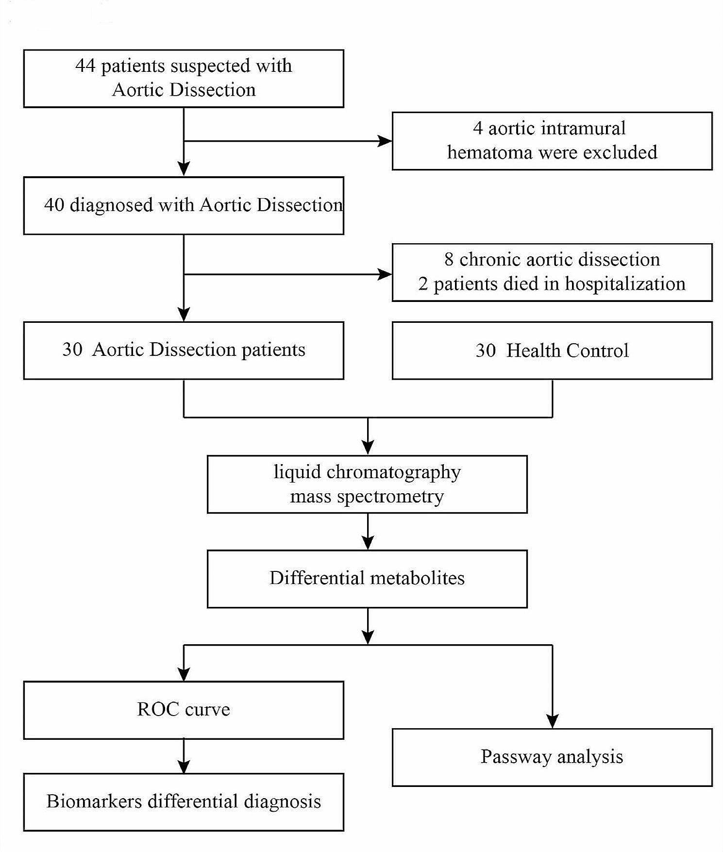


The study protocol conformed to the Regulations for the Administration of Affairs Concerning Experimental Animals and had been approved by the Institutional Animal Care and Use Committee of Sun Yat-Sen University (No. LAEC-2013-0603). After one week of adaptation, 80 male New Zealand white rabbits weighing 2.0–3.0 kg were randomly divided into eight groups (n = 10 per group): normal (N), high cholesterol (H), three different intensities of RTLI groups [six cycles of RTLI once daily (R6qd), three cycles of RTLI once daily (R3qd), and three cycles of RTLI once every other day (R3qod), each cycle of RTLI included 5 min of limb ischemia followed by 5 min limb reperfusion], and three correlated sham RTLI groups [sham ischemia for 60 min once daily (S6qd), sham ischemia for 30 min once daily (S3qd), and sham ischemia for 30 min once every other day (S3qod)] (Fig. 1). The N group was fed a normal diet, while the other groups were fed a 1% cholesterol diet for 12 weeks as described previously [16]. A total of 150 g food was available for all the animals daily; food intake was recorded daily, and body weight was measured weekly.
Fig. 1
Diagram of the experimental grouping. Rabbits in group N were fed only ordinary fodder without intervention. Rabbits in group H were fed only high-cholesterol fodder without intervention. Rabbits in the R3qod, R3qd, and R6qd groups were fed with high-cholesterol fodder and received three cycles of RTLI once every other day, three cycles of RTLI once daily, and six cycles of RTLI once daily, respectively. Each cycle of RTLI included 5 min of limb ischemia followed by 5 min limb reperfusion. Rabbits in the S3qod, S3qd, and S6qd groups were fed with high-cholesterol fodder and received sham ischemia for 30 min once every other day, 30 min once daily, and 60 min once daily, respectively. RTLI, regular transient limb ischemia. n = 10 per group
RTLI administrationThe rabbits in the three ischemia groups were kept in rabbit hutches and received three intensities of RTLI. RTLI were achieved by a blood pressure cuff placed on the left hind limb. Each cycle of RTLI was conducted with 5 min ischemia administered by inflating blood pressure cuff to 200 mmHg, followed by 5 min reperfusion with the cuff deflated. These RTLI procedures were conducted at the beginning of high-cholesterol diet feeding, lasting for 12 weeks. The rabbits in the sham ischemia groups received the same RTLI procedures by a deflated cuff without any pressure.
All the RTLI and sham ischemia procedures were performed without anesthesia, and the rabbits were placed in a prone position and calmed by caressing. The reliability of this RTLI procedure has been demonstrated in conscious rabbits in a previous study [17]. The rabbits in groups N and H received neither RTLI nor the corresponding sham RTLI procedures.
Serum lipid determinationAt the beginning of the experiment (week 0) and the end of the 6th (week 6) and 12th weeks (week 12), blood was collected from the central ear artery the morning following a 14-h fast. Serum samples were separated by centrifugation at 4 °C, 1500 × g for 15 min and stored at -80 °C until analysis. Serum concentrations of total cholesterol (TC), low-density lipoprotein cholesterol (LDL-C) and high-density lipoprotein cholesterol (HDL-C) were determined using an autoanalyzer (ADVIA-2400, Bayer, Germany) [17].
Tissue preparationRabbits were anesthetized by administering sodium pentobarbital (30 mg/kg) intravenously at the end of the 12th week. The left common carotid artery was carefully dissected after ligation and immediately rinsed in freshly prepared 37 °C Krebs buffer for isometric tension recording. The abdominal segment of the aorta was separated by a reference diaphragm and iliac artery bifurcation and immediately rinsed in heparin solution for the apoptotic analysis of endothelial cells. All the above procedures were performed under sterile conditions. The thoracic aorta was dissected from the beginning of the aortic arch to the diaphragm and fixed in 10% neutral formalin for 24 h for the quantification of intimal plaque area.
Plaque area quantification in the thoracic aortaThe percentage of plaque area in the thoracic aorta was determined according to a previous study [18]. The fixed thoracic aorta was dissected free of adhering fat and connective tissue and cut open longitudinally. The aorta was first washed in distilled water and rinsed in 60% isopropanol for 2 min, and then stained with 0.5% isopropanol solution of oil red O (Sigma, USA O0625) for 15 min, differentiated in 60% isopropanol three times (5 min each time), and finally washed in distilled water. Thereafter, the aorta was splayed, pinned plat on a white plate, and photographed using a digital camera (Canon EOS550D, Japan). The plaque area stained with oil red O (red stained) and total aortic surface area were measured using Image J (National Institutes of Health, USA). The extent of AS was assess by the rate of plaque area and the total intimal area (PA/IA).
Vascular ring relaxation assayThe left common carotid artery was carefully dissected free of adhering fat and connective tissue and cut into four rings of 4 mm in length. Two rings with endothelial integrity were used for endothelium-dependent tension recording, and the other two endothelium-denuded rings were subjected to endothelium destruction by gently rubbing the intima with a toothpick for endothelium-independent tension recording. All the rings were carefully suspended between the bases of two triangular-shaped wires and placed into baths filled with oxygenated [95% oxygen (O2), 5% carbon dioxide (CO2)] modified Krebs solution (37 °C, pH 7.4), the Krebs solution contains the following composition (mM): sodium chloride (NaCl) 118.3, kalium chloratum (KCl) 4.7, potassium dihydrogen phosphate (KH2PO4) 1.2, magnesium sulphate (MgSO4) 1.2, sodium bicarbonate (NaHCO3) 24.9, calcium chloride (CaCl2) 2.5, Glucose 11.1. One end of each wire was connected to a force transducer, and the contractive signals passing through the transducer were recorded using the Powerlab system (AD instruments, USA). All the rings were then equilibrated at a resting tension of 1.5 g for at least 2 h. In all the procedures, care was taken to avoid any mechanical damage to the vascular rings. The bathing solution was exchanged with fresh Krebs solution every 15 min.
For endothelium-dependent tension recording, the intact rings were precontracted with phenylephrine (1 µM) and then relaxed by acetylcholine with a series of cumulative concentrations (1 × 10− 9, 3 × 10− 9, 1 × 10− 8, 3 × 10− 8, 1 × 10− 7, 3 × 10− 7, 1 × 10− 6, 3 × 10− 6, and 1 × 10− 5 M). For endothelium-independent tension recording, the endothelium-denuded rings were precontracted with phenylephrine (1 µM), and then relaxed by sodium nitroprusside with a series of cumulative concentration (1 × 10− 9, 3 × 10− 9, 1 × 10− 8, 3 × 10− 8, 1 × 10− 7, 3 × 10− 7, 1 × 10− 6, 3 × 10− 6, and 1 × 10− 5 M). For both endothelium-dependent and -independent tension recording, the rings exhibiting < 1 g tension when precontracted with phenylephrine (1 µM) were considered to be severely damaged and were discarded from the experiments. Relaxation of the rings was expressed as the percent reduction from the amount of phenylephrine-induced tension. Then cumulative concentration-response curves to acetylcholine or sodium nitroprusside were obtained.
Aortic endothelial cell isolation and identificationThe abdominal aorta was separated as described in the “Tissue Preparation” section and then put into sterile 4 °C RPMI 1640 medium (Gibco, USA) before being dissected free of adhering fat and connective tissue, cut open longitudinally, and washed with phosphate buffered saline (PBS) three times. Endothelial cells were isolated using modified methods described by a previous study [19]. The aorta intima was put downward and digested with 3.5 ml 0.2% collagenase (Sigma) at 37 °C for 15 min. Then 3.5 ml endothelial growth medium-2 (Lonza, Switzerland) supplemented with 10% fetal bovine serum (Gibco) was added to terminate the digestion. The solution was then transferred to a centrifuge tube, and cells were obtained after centrifugation [1000 revolutions per minute (rpm), 10 min]. The cells were seeded in a 24-well culture dish precoated with 0.1% gelatin and cultured with endothelial growth medium-2 supplemented with 10% fetal bovine serum.
The cultured cells were identified as endothelial cells based on their characteristic cobblestone morphology and positive staining for endothelial marker CD31. After 7 days, cultured cells were observed under an inverted fluorescent microscope (Leica, Wetzlar, Germany DMI4000B). For CD31 staining, cells with 80% confluency were fixed with 2% paraformaldehyde for 15 min, washed with PBS-tween (PBS-T), and incubated with 5% bovine serum albumin for 1 h to block nonspecific binding. The cells were then incubated with anti-CD31 antibody (clone JC/70A, Abcam, USA; dilution 1:10) for 20 h at 4 °C. After three washes with PBS-T, the cells were incubated with goat anti-mouse lgG complex (code ab96879, Abcam; dilution 1:20) for 55 min. Nucleus counterstaining was performed for 5 min using 4′,6-diamidino-2-phenylindole (DAPI) (5 µg/ml). The cells were then washed with PBS-T and mounted using 50% glycerin. Finally, the cells were viewed and photographed with the inverted fluorescent microscope. Cells demonstrating positive staining of CD31 in the cell membrane were identified as endothelial cells.
Aortic endothelial cell apoptosis assayThe apoptosis of isolated endothelial cells of the abdominal aorta were evaluated. Cell apoptosis was assessed using an Annexin V-fluoresceine isothiocyanate (FITC) apoptosis detection kit (KeyGEN Biotech, China) according to the manufacturer’s instructions. Cells were washed twice with PBS and suspended in 0.5 ml binding buffer. Then the cell suspension was filtered to avoid possible impurity with larger mass. Subsequently, 5 µL of annexin V-FITC and 5 µL of propidium iodide (PI) were mixed with the cell suspension. After incubation for 15 min, mixtures were analyzed by flow cytometry (BD FACSCantoII, USA) within 30 min. Annexin V-negative and PI-negative cells were regarded as live cells, annexin V-positive and PI-negative cells were regarded as early apoptotic cells, while annexin V-positive and PI-positive cells were regarded as late apoptotic or secondary necrotic cells [20].
Statistical analysisThis study was designed as three sub studies. The first sub study was designed to compare the normal and high-cholesterol groups and determine the effect of a hypercholesterolemic diet on AS. The second sub study was designed to compare the effects of different intensities of RTLI on AS and determine the minimum effective intensity of RTLI. The third sub study was designed to explore the mechanisms of the minimum effective intensity of RTLI on AS.
Data are presented as mean values ± standard deviation (SD) or median (interquartile range, IQR) as appropriate. Repeated values were analyzed with the analysis of variance (ANOVA) for repeated measures. The normal (N) and high-cholesterol (H) groups were compared with Student’s t-test (normal distribution) or Mann–Whitney U test (non-normal distribution). In the other sub studies, multiple comparisons for continuous normal distributed variables with homogeneity variance were done with one-way ANOVA followed by the least significant difference test for the differences between two groups. Normally distributed variables with heterogeneity variance or non-normal distributed variables were analyzed using compared using Kruskal Wallis test, resorting to a Mann–Whitney U test for comparisons between two groups. All analyses were performed using SPSS 16.0 statistical software package (SPSS Inc., Chicago, IL, USA). P < 0.05 was considered to indicate significance.
留言 (0)