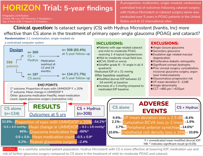
Corneal neuropathic pain (CNP) is being increasingly recognised, particularly in patients with a diagnosis of dry eye disease (DED) [1], for its impact on a patient’s quality of life [2, 3]. The impacts can be mild with minimal effects on activities of daily living to severe with the patient experiencing debilitating symptoms that can lead to a deterioration in their physical and social well-being [4]. Reports have emerged of its occurrence and burden following cataract and refractive surgery [5,6,7] and in those with neurotrophic keratopathy [8], chronic pain syndromes [9, 10] and autoimmune diseases [11, 12]. The overarching feature of CNP is a heightened experience of pain without commensurate clinical signs [4, 13, 14]. Other terminology used to describe the condition include ocular neuropathic pain, corneal neuralgia, neuropathic corneal pain, ocular pain syndrome, corneal pain syndrome, keratoneuralgia, corneal neuropathic disease, phantom cornea, corneal neuropathy and corneal allodynia [13]. Central and peripheral CNP have been defined, with the former responding to topical analgesia [15]. Subtypes of CNP have been identified as; associated with (1) specific ocular disease (2) ocular surface disease without keratitis (3) systemic pain syndrome (4) psychiatric disease (especially depression) and (5) idiopathic [16]. More recently, a new disease association has been noted between CNP and Long COVID [17].
A range of symptoms can be produced by CNP with many overlapping those of DED, such that it is frequently misdiagnosed as dry eye [13, 18]. With corneal vital dye staining - a mainstay in identifying ocular surface damage for the diagnosis of DED [19], CNP has been referred to as ‘pain without stain’. It has been proposed that CNP may be a subtype of Sjogren International Collaborative Clinical Alliance (SICCA) dry eye [20], such that CNP may be the extreme end of the dry eye spectrum [3]. Increased pain sensitivity may influence perceptions of ocular discomfort and dryness and has been reported by contact lens wearers [21].
There remains a lack of epidemiological, long-term and high-quality clinical trial data on CNP, and many clinicians are unfamiliar with the existence of the condition and how to manage it. Registry data is lacking and needed to provide long-term outcomes and disease natural history [22]. Further, current diagnosis of CNP is by exclusion as there is no ‘gold standard’ and patient phenotypes are poorly understood. Evidence is emerging on tools for diagnosis including questionnaires. Technologies such as in vivo confocal microscopy (IVCM) and esthesiometry, have been used to support a diagnosis of CNP but their utility in everyday clinical practice is unknown [15, 23]. Limited clinical studies and trials have provided some data on potential topical, oral, adjuvant and surgical therapies for CNP. Further research is needed to inform the development of evidence-based guidelines for the diagnosis and management of CNP [14].
What is the underlying pathophysiology of corneal neuropathic pain?
The International Association for the Study of Pain defined neuropathic pain as ‘pain caused by a lesion or disease of the somatosensory system’ [4, 24]. CNP can be considered a part of this disorder as it is associated with injury to the corneal nerves, terminal endings of the ophthalmic division of the trigeminal somatosensory system [1, 4, 13, 14]. Corneal nerves can be damaged by a variety of peripheral and systemic aetiologies [1, 4, 13, 14]. Peripheral nerve injuries can result from ocular surface diseases such as DED, contact lens wear, infections, surgery, trauma, toxins, and radiation [1, 4, 13, 14]. In comparison, systemic diseases damage corneal nerves through chronic inflammation and can include disorders such as Systemic Lupus Erythematosus, sarcoidosis, fibromyalgia, diabetes and small-fibre polyneuropathies [1, 4, 13, 14]. In CNP there are two neurobiological processes—peripheral and central sensitisation [1, 14]. Sensitisation can occur after an initial insult with sub-threshold noxious stimuli (hyperalgesia) [4, 13, 14, 18, 25] or even non-noxious stimuli (allodynia, photoallodynia) [18, 26, 27]. Genetic factors may likely contribute to the occurrence of CNP. A variety of genetic polymorphisms have been identified on genome wide association studies in a cohort of veterans CNP [28]. The protein products of the implicated genes may have a role in sensory perception and potentially have links to DED [28].
Peripheral sensitisation occurs when injury to peripheral axons results in the release of pro-inflammatory mediators such as cytokines, prostaglandins and substance P, which decrease the threshold potentials of nociceptors, leading the axons to be triggered by previously non-painful stimuli [1, 4, 13, 14]. Over time, increased peripheral sensitisation results in central neurons becoming highly responsive to non-painful stimuli, leading to an increased response to overall pain, known as central sensitisation [1, 4, 13, 14]. Central CNP is due to abnormal function of the pain cortex in the brain reacting to stimuli that are unpleasant or noxious. Whereas in peripheral CNP, the peripheral sensory nerves are overly sensitive and respond to stimuli that are subthreshold (allodynia), light/non-noxious (photoallodynia) or suprathreshold (hyperalgesia) [15]. These neurobiological processes ultimately produce a wide range of symptoms including hyperalgesia, allodynia, photoallodynia, itching, irritation, burning, dryness, foreign body sensation and a feeling of pressure [1, 4, 13, 14]. Further, neuropathic ocular itch and pain can occur together; with both a result of ocular surface nerve damage and dysfunction [29]. Underlying itch and pain is likely due to inflammation and immune system upregulation [29].
Implications for practice
CNP is a subtype of neuropathic pain arising from damage to the corneal nerves.
Stimuli that usually do not elicit pain may produce CNP.
Symptoms of CNP include itching, irritation, burning, dryness and foreign body sensation along with feelings of pressure.
Who gets corneal neuropathic pain?
CNP may be associated with systemic diseases, with a higher prevalence of females affected, such as autoimmune conditions and fibromyalgia [1, 4, 13, 14]. CNP may also occur with other ocular conditions or following trauma or surgery (e.g. cataract and refractive surgery). Associated ocular conditions include DED, infectious keratitis, herpes simplex keratitis, herpes zoster ophthalmicus, recurrent corneal erosion, radiation keratopathy [4, 14, 25, 30]. Refractive surgery has been known to induce DED [31] but is increasingly being reported to induce ocular pain [4, 6, 14, 25, 30]. Patients with CNP due to refractive surgery and herpes simplex keratitis may have similar clinical characteristics and report moderate to severe pain levels [7]. Both conditions have moderate impacts on quality of life and a significant reduction in total nerve density compared to healthy controls on IVCM [7].
High index of suspicion for CNP
A history of ocular and/or systemic disease should be sought in patients who are suspected to have CNP.
Persistent pain following ocular surgery or infection should raise a high index of suspicion for CNP.
Dry eye disease and corneal neuropathic pain
DED and CNP may occur in the same patient and CNP can exacerbate the symptoms of DED. Indeed, CNP has been associated with more severe dry eye symptoms in an ophthalmology clinic patient population [9]. In DED, there may be various causes of ocular surface damage including infection, inflammation, trauma, adverse environmental conditions, abnormal ocular anatomy and high tear osmolarity [1, 25]. If this damage persists, or if the vicious cycle of DED is not broken, peripheral and central sensitisation can occur, leading to neuropathic pain [1, 25]. As such, some patients with DED may report symptoms that are out of proportion to their ocular surface findings including allodynia, hyperalgesia and hyperaesthesia [1, 25]. Indeed, an overlap exists between CNP and more severe and chronic forms of DED [1, 20, 25]. The DEWS TFOS definition of DED includes the role of neurosensory abnormalities in disease aetiology [32] and peripheral CNP is characterised by sensitisation of sensory and/or nociceptive processing at some level of the trigeminal system. One way to distinguish DED from CNP, there should be a therapeutic failure of conventional treatment for DED and a lack of therapeutic response to topical anaesthetics if the CNP is peripheral [1]. In peripheral neuropathic pain, there may be cutaneous allodynia i.e. pain to light touch around the eye [29]. Indeed as the presence of neuropathic pain is not routinely sought in dry eye patients [25], it has been proposed that such patients should be screened for CNP [33].
Implications for practice
In chronic DED, particularly if it is severe, if pain is out of proportion to the clinical signs, particularly with associated allodynia, consider CNP.
When the patient fails standard dry eye therapy, consider CNP.
Diagnosis of corneal neuropathic pain
There are no standardised diagnostic criteria for CNP. Further, the variability in CNP symptoms often makes it challenging to establish a diagnosis, especially considering the significant overlap with DED, and the lack of clinical signs on examination [1, 4, 13, 14]. Characteristic symptoms include pain, dryness, and itch along with burning, sensitivity to wind, light and temperature [9]. Patients may report of indistinct sensations of pressure [4] and episodes of spontaneous pain [25]. Such symptoms can be present in ocular surface diseases including DED [1].
Validated questionnaires can assist in the diagnosis of CNP [16] as the overlap between severe and chronic DED and CNP led to a variety of DED questionnaires being used to screen for and assess the impact on visual function and quality of life (Table 1) [25, 34,35,36,37,38,39,40,41]. However, such questionnaires were specific for DED and not CNP [25] as most DED questionnaires were not able to differentiate between nociceptive and neuropathic symptoms [1, 25].
Table 1 Questionnaires that assess corneal neuropathic pain [92].Questionnaires specific for ocular pain are now available such as the Ocular Pain Assessment Survey, which is a quantitative, multidimensional questionnaire used to assess corneal and ocular surface pain and the Neuropathic Pain Symptom Inventory—Eye (NPSI-Eye), which is a modified version of the Neuropathic Pain Symptom Inventory (NPSI) that specifically assesses neuropathic-like ocular pain [42, 43]. These questionnaires are valid and reliable, but further studies are needed to validate preliminary findings on their use in CNP [42,


留言 (0)