
記住我

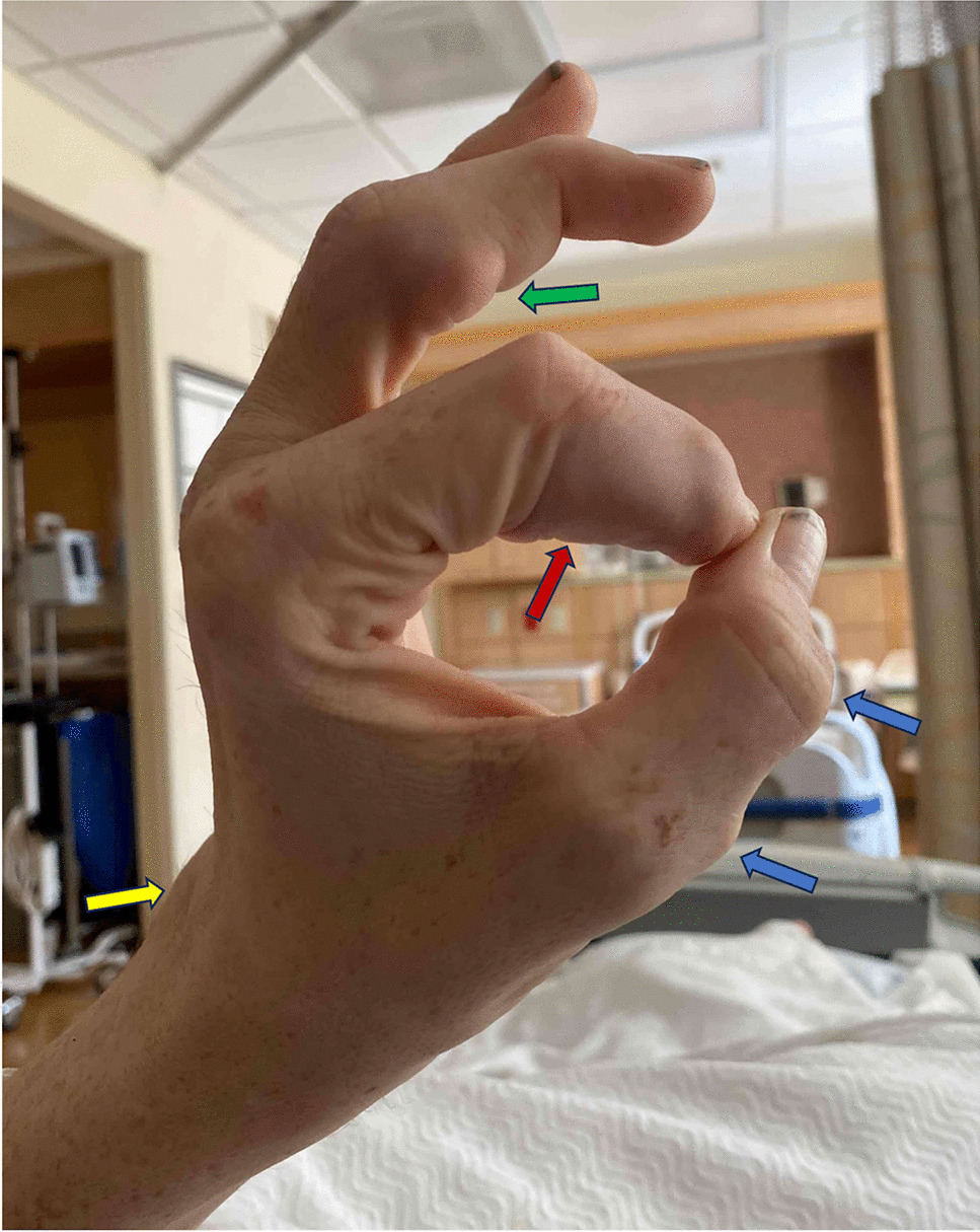


A 42-year-old man with end-stage renal disease presented with progressively enlarging nodules over his left fingers. Examination revealed firm, non-tender nodules in a periarticular distribution (panel A, arrows). Serum labs were notable for elevated phosphorus, parathyroid hormone, normal calcium, and an elevated calcium-phosphate product (CaxPO4). The X-ray showed multiple lobular, soft tissue calcifications (panel B, arrows). He was diagnosed with metastatic calcinosis cutis (MCC). Several months later, his lesions significantly decreased in size with correction of his hyperphosphatemia and dietary restriction.
Calcinosis cutis, of which MCC is one of five subtypes, is characterized by insoluble calcium salt deposition in subcutaneous and cutaneous tissues.1 MCC is most commonly seen in patients with chronic renal failure. It is characterized by benign nodules commonly distributed in a periarticular distribution. It tends to affect large joints, such as hips and shoulders, but can also appear in smaller joints, including fingers.2 Clinical manifestations can range from pain to infection but sometimes can be asymptomatic, as with our patient. It is caused by abnormal calcium and phosphorus metabolism, which includes hyperphosphatemia, normal or elevated serum calcium levels, hyperparathyroidism, and elevated CaxPO4.2 Treatment ranges from lowering CaxPO4 through dietary restriction and the use of phosphate binders to surgery.3


留言 (0)